Download

1 / 25
990 likes | 3.08k Views
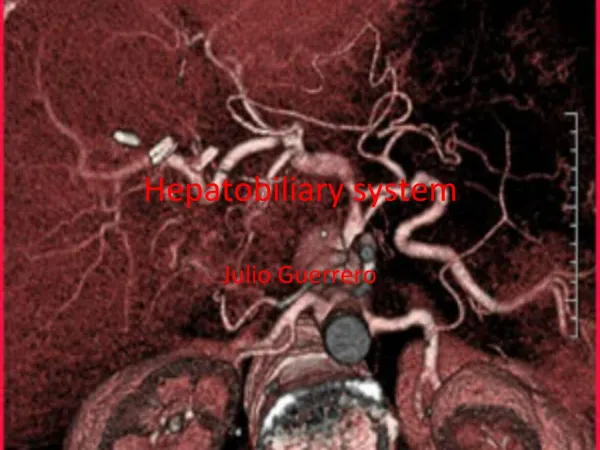
Hepatobiliary system structure. Hepatobiliary System components:. Liver Gallbladder Bile ducts Pancreas. Liver. Four lobes anatomically 1) right (right lobe is the largest lobe-accounting for approximately 3/5 th-2/3rd of the liver size) 2) left 3) caudate 4) quadrate

E N D
Hepatobiliary Systemcomponents: • Liver • Gallbladder • Bile ducts • Pancreas
Liver • Four lobes anatomically 1) right (right lobe is the largest lobe-accounting for approximately 3/5 th-2/3rd of the liver size) 2) left 3) caudate 4) quadrate • surgeons tend to separate the lobes into 8 segments by vascularity • receives blood from an artery (hepatic artery) and also a vein (portal vein)
located in the upper right abdomen • largest of the visceral (internal) organs (weighs approximately 3 lbs.) • unoxygenated blood enters the liver from the portal vein and oxygenated feeds the liver through the hepatic artery • network of tubes called the biliary tree carry bile from the liver • small ducts connect to larger ducts then to a hepatic duct from the right and left lobes which unite to form the common bile duct • connected by ligaments to the diaphragm on its upper section, and on the left, to the stomach
Gall bladder • pear-shaped sac, which stores the bile produced by the liver • Thin walled green muscular sac • 10 cm long • tucked into the ventral fossa of the liver • stores and concentrates bile • expels bile into the cystic duct • sphincter of oddi – regulates the flow of bile back the liver • cholecystectomy – gallbladder removal (doesn’t really effect the patient to have the gall bladder removed)
Pancreas • Three parts head, body and tail. • small organ located behind the stomach. .The bile duct travels through the pancreas immediately before it enters the small intestine. Thus any problem in the pancreas (such as cancer, pancreatitis and cysts) that is adjacent to the bile duct often causes blockage of the bile duct and jaundice.