Download
1 / 4
40 likes | 49 Views
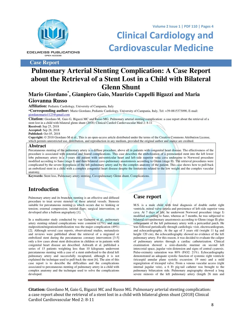
Percutaneous stenting of the pulmonary artery is a diffuse procedure, above all in patients with congenital heart disease. The effectiveness of the procedure is associated with potential and feared complications. This case describes the embolization of a premounted stent into the left lower lobe pulmonary artery in a 3-years old patient with univentricular heart and left-side superior vena cava undergone to Norwood procedure modified according to Sano (stage I) and then bilateral cavo-pulmonary anastomosis according to Glenn (stage II). The retrieval procedures were complicated by the severe hypoplasia of the left pulmonary artery and by the complex anatomy of the patient. This case shows how to pull-back an embolized stent in a child with a complex congenital heart disease despite the limitations related to the low weight and the complex vascular anatomy.<br>Clinical Cardiology and Cardiovascular medicine welcomes all the Hypertension articles, heart transplantation case reports, Coronary angioplasty risk and many more.<br>

E N D
Volume 2 Issue 1 | PDF 110 | Pages 4 Volume 1 . Issue 1 | PDF 101 | Page 1 of x Clinical Cardiology and Cardiovascular Medicine Case Report Pulmonary Arterial Stenting Complication: A Case Report about the Retrieval of a Stent Lost in a Child with Bilateral Glenn Shunt Mario Giordano*, Gianpiero Gaio, Maurizio Cappelli Bigazzi and Maria Giovanna Russo Affiliation: Pediatric Cardiology, University of Campania, Italy *Corresponding author: Mario Giordano,Pediatric Cardiology, University of Campania, Italy, Tel: +39-0815373090, E-mail: giordanomario1123@gmail.com Citation: Giordano M,Gaio G,Bigazzi MC and Russo MG.Pulmonary arterial stenting complication: a case report about the retrieval of a stent lost in a child with bilateral glenn shunt (2018)Clinical Cardiol Cardiovascular Med 2: 8-11 Received: Sep 25, 2018 Accepted: Sep 28, 2018 Published: Oct 05, 2018 Copyright: © 2018 Giordano M et al., This is an open-access article distributed under the terms of the Creative Commons Attribution License, which permits unrestricted use, distribution, and reproduction in any medium, provided the original author and source are credited. Abstract Percutaneous stenting of the pulmonary artery is a diffuse procedure, above all in patients with congenital heart disease. The effectiveness of the procedure is associated with potential and feared complications. This case describes the embolization of a premounted stent into the left lower lobe pulmonary artery in a 3-years old patient with univentricular heart and left-side superior vena cava undergone to Norwood procedure modified according to Sano (stage I) and then bilateral cavo-pulmonary anastomosis according to Glenn (stage II). The retrieval procedures were complicated by the severe hypoplasia of the left pulmonary artery and by the complex anatomy of the patient. This case shows how to pull-back an embolized stent in a child with a complex congenital heart disease despite the limitations related to the low weight and the complex vascular anatomy. Keywords: Stent loss, Pulmonary artery stenting, Cavopulmonary Glenn shunt, Complications. Introduction Pulmonary artery and its branches stenting is an effective and diffused procedure to treat severe stenosis of these arterial vessels. Stenosis suitable for percutaneous stenting is which occurs due to: kinking or tension; external compression; intimal flaps; surgical intervention; or developed after a balloon angioplasty [1]. In a multicenter study conducted by van Gameren et al., pulmonary artery stenting–related complications were common (17%), and stent malposition/migration/embolization was the major complication (49%) [2]. Although several case reports, observational studies, metanalysis and reviews were published about the retrieval of a migrated or embolized stent during the percutaneous coronary intervention [3-5] only a few cases about stent dislocation in children or in patients with congenital heart disease are described. Ashwath et al. published a series of 15 patients weighting less than 10 kilograms underwent percutaneous stenting with a case of a stent embolized to the distal left pulmonary artery and successfully recaptured, although it is not explained the technique used to pull-back the stent [6]. The aim of this case report is to describe the difficulties and the complications associated to percutaneous stenting of pulmonary artery in a child with complex anatomy and the technique used to solve the complications developed. Case report M.S. is a male child with fetal diagnosis of double outlet right ventricle, mitral valve atresia and persistence of left-side superior vena cava. At 7 days of life, he underwent Norwood procedure (stage I) modified according to Sano, whereas at 7 months, he was subjected to bilateral cavopulmonary anastomosis according to Glenn (stage II) plus enlargement of the left pulmonary artery with a pericardial patch. He was followed periodically through cardiologic visit, electrocardiogram, and echocardiography. At the age of 3 years old (weight 11 kg and height 120 cm), the echocardiography showed no evidence of the left pulmonary artery. For this reason, it was decided to evaluate the caliper of pulmonary arteries through a cardiac catheterization. Clinical examination showed: a sisto-diastolic murmur on second left intercostal space, jugular vein distension and signs of central cyanosis. Pulse-oximetry saturation was 80% (FiO2: 21%). Echocardiography demonstrated an adequate systolic function of systemic right ventricle (tricuspid annular plane systolic excursion: 19 mm) and a mild regurgitation of tricuspid valve. From a venous vascular access (right internal jugular vein), a 6 Fr pig-tail catheter was brought to the pulmonary bifurcation side. Pulmonary angiography showed a long severe stenosis of the left pulmonary artery (length 26 mm and Citation: Giordano M, Gaio G, Bigazzi MC and Russo MG. Pulmonary arterial stenting complication: a case report about the retrieval of a stent lost in a child with bilateral glenn shunt (2018) Clinical Cardiol Cardiovascular Med 2: 8-11 8
Giordano M et al. Clinical Cardiology and Cardiovascular, 2018 PDF: 110, 2:1 diameter 2 mm, z-score -8, 54) at the site of the left-side superior vena cava anastomosis (Figure 1). The loss stent was crossed by a 0.014” wire and a progressive inflation of small coronary balloons (Tazuna® semi-compliant balloon catheter 1.5 x 20 mm, 2.0 x 20 mm and 2.25 x 20 mm) was achieved to anchor and pull-back the stent. The balloons were inflated until to achieve a pressure of 10-12 atm (rated burst pressure of the balloon: 14 atm). No balloon bursted during these manoeuvres. This technique of retrieval could expose the patient to a high risk of vessel dissection, but in this case the stenotic tract was a fibrous cord with a great endurance to parietal stress and for this reason the dissection was avoided despite so rough margins. The retrieval of stent failed since it wasn’t able to cross the narrowest tract of pulmonary artery (Figure 4). Luckily, the end-to-side anastomosis of the left-side superior vena cava to the left pulmonary artery was beyond the narrowest tract (Figure 5). Figure 1: Pulmonary angiography in right anterior oblique view (A) and left anterior oblique view (B). Asterisk indicates the stenosis of left pulmonary artery. At hilum side, the left pulmonary artery was measured around 8 mm. The lesion was crossed by a 0.014” wire, and then a more supportive wire (Amplatzer super stiff 0.035”) was placed into the left inferior lobar artery. It was chosen to implant a stainless steel, pre-mounted, open-cell stent: Valeo® balloon expandable vascular stent 36 x 8 mm (Bard Peripheral Vascular, Tempe, Arizona, United States of America), to cover the left pulmonary artery completely. When the stent was brought into the stenotic segment, it seemed to be too much long since it protruded into the inferior lobar artery (Figure 2); and for this reason, it was decided to pull-back its. Nevertheless, despite a large delivery system (Mullins sheath 8 Fr), during the retrieval, the stent slipped from the balloon by positioning beyond the closer segment of the pulmonary artery and by engaging the left inferior lobar artery (Figure 3). Figure 4: Left anterior oblique view. The balloon (Tazuna® semi- compliant balloon catheter 2.25 x 20 mm) inflated into the proximal- end of stent (asterisk) is not able to cross the narrowest segment of pulmonary artery with a diameter measured around 2 mm (arrow). Figure 2: Valeo® balloon-expandable vascular stent 36 x 8 mm appears too much long both in right anterior oblique view (A) and left anterior oblique view (B). Figure 5: Left anterior oblique view. The end-to-side anastomosis of left superior vena cava (circle) to left pulmonary artery is beyond the narrowest tract. It was decided to achieve one more vascular access on the left side (left internal jugular vein) and to insert a large introducer (11 Fr) to pull- back the stent into itself. The various inflation and deflation of different balloons had caused a progressive enlargement of proximal- end of the stent and the balloon wasn’t able to anchor the stent; for this reason, it was used an Amplatz GooseNeck® Snare to tighten the enlarged tract of the stent and to allow the anchor of the balloon. The stent was adequately pulled-back into the introducer that was removed with half stent into and half stent outside of itself (Figure 6). An adequate haemostasis was achieved through a manual compression of left internal jugular vein. Despite the complication, by using the right- side vascular access, the stenosis was crossed by a wire and a pre- mounted Valeo® balloon expandable vascular stent 26 x 8 mm was Figure 3: Left anterior oblique view of the stent (asterisk) slipped from the balloon and engaging the lobar left inferior pulmonary artery. Citation: Giordano M, Gaio G, Bigazzi MC and Russo MG. Pulmonary arterial stenting complication: a case report about the retrieval of a stent lost in a child with bilateral glenn shunt (2018) Clinical Cardiol Cardiovascular Med 2: 8-11 9
Giordano M et al. Clinical Cardiology and Cardiovascular, 2018 PDF: 110, 2:1 implanted and post-dilated until a diameter of 10 mm, with a good angiographic result (Figure 7). to Fontan operation [7], since an adequate function of Fontan circuit requires low pressure in the pulmonary circulation and a balanced blood flow through the pulmonary arteries [8]. In this case, the severe stenosis of left pulmonary artery would have compromised a right function of Fontan circulation since the venous blood flow draining from the inferior vena cava would be distributed to right pulmonary artery predominantly. It was needed to stent the hypoplasic vessel to achieve a balanced pulmonary circulation. The bad choice of stent length has compromised and complicated the procedure. Probably the turbulent flow in the site of left-side superior vena cava anastomosis favoured the migration of stent from the balloon despite a large delivery system used (Mullins sheath 8 Fr, while the premounted vascular stent required a 6 Fr sheath). Kakisis et al. describe a case of a stent dislodged from the original position (in the left brachiocephalic vein) into the left lower lobe pulmonary artery, where he adopted a successful strategy of “wait and see” (the patient was anticoagulated and the stent left “in situ”). In our case, it was impossible to adopt the same strategy because the unexpanded and dislodged stent did not make possible the enlargement of the stenotic left pulmonary artery. The choice to retrieve the stent percutaneously was complicated by the severe stenosis and by the inability of the anchored stent to cross over the narrowest segment. The presence of the left-side superior vena cava and its anastomosis site beyond the narrowest tract allowed the retrieval of loss stent. The procedure was completed by releasing an adequate stent into the left pulmonary artery with good hemodynamic and angiographic result. In literature, several cases about the retrieval of stents migrated into a pulmonary artery are described, but all these ones concern adult patients, and the stent dislodged from a central o peripheral vein to the pulmonary artery [9-11]. Furthermore, in these cases, the pulmonary arteries showed a good diameter without stenotic or hypoplasic segment. The only case about the retrieval of a stent in an infant is described by Kobayashi et al. and it is about a transcatheter retrieval of a stent embolized into the right ventricle [12]. In our experience, this is the first case described about a stent migration into the left lower lobe pulmonary artery in a child with a complex congenital heart disease (an univentricular heart physiology), where the dislodgement of stent beyond the narrowest tract of vessel and complex anatomy of patients complicated the strategies of retrieval. Conclusion The angioplasty and stenting of pulmonary arteries is a diffuse percutaneous procedure although not free from complications, above all in patients with complex congenital heart disease where the different anatomy, the previous surgical operations and the sites of turbulent flow can compromise the results of procedure. The knowledge and the choice of right materials and strategies are necessary to avoid and to solve dangerous complications that can arise during the interventional catheterization. References 1. Trivedi KR and Benson LN. Interventional strategies in the management of peripheral pulmonary artery stenosis (2003) J Intervent Cardiol 16: 171-188. 2. Van Gameren M, Witsenburg M, Takkenberg JJM, Boshoff D, Mertens L, et al. Early complications of stenting in patients with congenital heart disease: a multicentre study (2006) Eur Heart J 27: 2709-2715. 3. Brilakis ES, Best PJM, Elesber AA, Barsness GW, Lennon RJ, et al. Incidence, retrieval methods, and outcomes of stent loss during Figure 6: Right anterior oblique view. The proximal-end of stent is into the introducer whereas the distal-end is outside of its. Figure 7: Left anterior oblique view. The stent (asterisk) is placed rightly (A) and the pulmonary angiography shows an adequate opacification of the left pulmonary artery that appears with an adequate diameter. Final pulmonary angiography showed a balanced flow through both pulmonary arteries and no signs of dissection or thrombo-embolic complications. In the catheterization laboratory, a dose of acetylsalicylic acid (100 mg) was administered intravenously. The patient was treated with an intravenous infusion of sodium heparin in the first 24 hours (15 UI/kg/h), then he assumed acetylsalicylic acid (5 mg/kg) per os. The procedure lasted 300 minutes. Fluoroscopic time was 203 minutes. Absorbed dose and total dose area product were 1076 mGy and 6385 cGy/cm2, respectively. The patient was discharged 72 hours after the procedure, in good clinical condition. The jugular vein distension disappeared and pulse-oximetry saturation increased until 88% (FiO2: 21%). Post-operative echocardiogram showed an adequate blood flow into both the right and the left superior vena cava and into the stent placed in the left pulmonary artery. Discussion Stenosis of pulmonary arteries occurs in 2–3% of patients with congenital heart disease [1] and it can be discrete or associated with long hypoplasic arterial tracts. The development of scars at the site of trans-pulmonary patches or at the anastomotic sites on the pulmonary arteries (in the cases of systemic-to-pulmonary shunts or Glenn shunts) is the most common causes of post-surgical pulmonary arterial stenosis. Complex surgical interventions (as an arterial switch, Norwood operation, Damus–Kaye–Stansel procedure) may determine stretch or distortion of pulmonary branches by developing a hemodynamically significant stenosis. A more aggressive approach is necessary to pulmonary arterial stenosis in patients who are undergoing Citation: Giordano M, Gaio G, Bigazzi MC and Russo MG. Pulmonary arterial stenting complication: a case report about the retrieval of a stent lost in a child with bilateral glenn shunt (2018) Clinical Cardiol Cardiovascular Med 2: 8-11 10
Giordano M et al. Clinical Cardiology and Cardiovascular, 2018 PDF: 110, 2:1 percutaneous coronary intervention: a large single-center experience (2005) Cathet Cardiovasc Intervent 65: 333-340. 4. Alomar ME, Michael TT, Patel VG, Altomare CG, Rangan BV et al. Stent loss and retrieval during percutaneous coronary interventions: a systematic review and meta-analysis (2013) J Invasive Cardiol 25: 637-641. 5. Eggebrecht H, Haude M, Von Birgelen C, Oldenburg O, Baumgart D, et al. Nonsurgical Retrieval of Embolized Coronary Stents (2000) Cathet Cardiovasc Intervent 51: 432-440. 6. Ashwath R, Gruenstein D and Siwik E. Percutaneous stent placement in children weighing less than 10 kilograms (2008) Pediatr Cardiol 29: 562-567. 7. Franco E, Domingo EJB, Del Val VA, Silva LGG, Del Cerro Marín MJ, et al. Percutaneous interventions in Fontan circulation (2015) Int J Cardiol Heart Vasc 8: 138-146. 8. Gewillig M. The Fontan Circulation (2005) Heart 91: 839-846. 9. Kakisis JD, Vassilas K, Antonopoulos C, Sfyroeras G, Moulakakis K, et al. Wandering stent within the pulmonary circulation (1932) Ann Vasc Surg 28: 9-12. 10. Balasubramaniyam N, Garg J, Rawat N, Chugh S, Mittal V, et al . Dual stent migration to the heart and pulmonary artery (2014) Am J Ther 21: 199-203. 11. Dashkoff N, Blessios GA and Cox MR. Migration of covered stents from hemodialysis A-V access to the pulmonary artery: percutaneous stent retrieval and procedural trends (2010) Catheter Cardiovasc Interv 76: 595-601. 12. Kobayashi D, Singh HR, Turner DR, Forbes TJ and Gowda ST. Transcatheter retrieval and repositioning of embolized stent from the right ventricle in an infant (2012) Tex Heart Inst J 39: 639-643. Citation: Giordano M, Gaio G, Bigazzi MC and Russo MG. Pulmonary arterial stenting complication: a case report about the retrieval of a stent lost in a child with bilateral glenn shunt (2018) Clinical Cardiol Cardiovascular Med 2: 8-11 11