Download
1 / 15
170 likes | 247 Views
Gaucher’s Disease – A glycolipid storage disorder characterized by the accumulation of glucosylceramide in the spleen, liver, lungs, bone marrow and brain. The disease is caused by a deficiency of lysosomal acid beta-glucosidase, which hydrolyzes glucosylceramide to ceramide and glucose.

E N D
Gaucher’s Disease – A glycolipid storage disorder characterized by the accumulation of glucosylceramide in the spleen, liver, lungs, bone marrow and brain • The disease is caused by a deficiency of lysosomal acid beta-glucosidase, which hydrolyzes glucosylceramide to ceramide and glucose • Ceramides are the biological building blocks of Sphingolipids Introduction
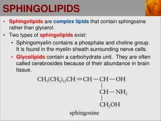
Sphingolipids – are essential components of the plasma membrane of eukaryotic cells. • Sphingolipids differ from phospholipids in being based on a lipophilic amino alcohol (sphingosine) rather than glycerol. • Sphingolipids play important roles in signal transduction Introduction cont.
Topic Application of delayed extraction–matrix-assisted laser desorption ionization time-of-flight mass spectrometry for analysis of sphingolipids in pericardial fluid, peritoneal fluid and serum from Gaucher disease patients Takehisa Fujiwaki, , a, Seiji Yamaguchia, Masaru Tasakaa, Nobuo Sakurab and Tamotsu Taketomic a Department of Pediatrics, Shimane Medical University, 89-1 Enya-cho, Izumo 693-8501, Japanb Department of Pediatrics, Hiroshima University School of Medicine, 1-2-3 Kasumi, Minami-ku, Hiroshima 734-8551, Japanc Department of Biochemistry, Research Center on Aging and Adaptation, Shinshu University School of Medicine, 3-1-1 Asahi-machi, Matsumoto 390-8621, Japan
So, what’s the point???? • The point is……Delayed Extraction – Matrix Assisted Laser Desorption Ionization - Time Of Flight – Mass Spectrometry can be used to evaluate patients with Gaucher’s disease using small amounts of body fluids.
The Basics…..Matrix Assisted Laser Desorption Ionization – Time Of Flight (MALDI-TOF)
The matrix is an organic acid that holds the sample • Absorbs photon energy and transfers it • Reduces intermolecular forces and aggregation by serving as a solvent The basics cont……What is the Matrix???????
UV laser is used to generate ions • The matrix absorbs the energy and transfers the energy to the sample • The sample becomes ionized into a gas phase (proton transfer) The basics cont…Laser Desorption Ionization???
The basics cont……Time of Flight???? • Ions enter the mass spectrometer or time-of flight (TOF) • They are accelerated by high voltage and separated based on the time it takes them to travel to the detector • All ions gain same kinetic energy • A spectrum of ion intensity as a function of travel time is recorded • The ion’s passage through the drift tube can be described as: qE = 1/2 m v2 = 1/2 m (l/t) q= ion charge v= velocity through drift tube l= length of tube t= time of flight through tube m= ion mass E= electric field voltage • Time of flight can be converted to molecular mass: • t ~ (m/q)1/2
Each sample was mixed with a matrix composed of 2,5 dihydroxybenzoic acid (DHB). • Advantages of DHB: • ions undergo little metastable decay • insensitive to contaminations • produces minimal interference in the low molecular weight range Methods • Sphingolipid fractions from body fluids and serum were collected • Samples were loaded into a Voyager DE-RP (2.0 m flight length, reflector mode) and mass spectra of the samples was obtained in the positive ion mode with an N2 laser (337nm) and a scan average of 256. Two point external calibration was performed each time.
Basically – it groups ions and provides better resolution • A time delay between ionization of the sample and extraction from the ion source • Allows ions with identical mass/charge values to arrive at the detector at the same time Delayed Extraction?
Internal and external calibration • External involves acquisition from two samples • 1st sample – two standards derive calibration equation • 2nd sample – contains unknowns and is calibrated with standards • Advantage of External calibration – no risk of concentration/dynamic range or ionization suppression External Calibration?
Basically – it gives you better resolution • Ion reflector – slows down incoming ions and reverses flight path to detector • This results in focusing the ion packets in space and time at the detector. Reflector mode?
Table 1. Measured mass-to-charge ratios (m/z), and proposed molecular species associated with sphingolipids *Ceramide monohexoside includes glucosylceramide. "d" indicates dihydroxy-sphingosine.
Conclusions • Control patients produce sufficient lysosomal acid B-glucosidase and can properly hydrolyze CMH to ceramide, which is a sphingomyelin precursor. Lysosomal acid B-glucosidase HYDROLYSIS Ceramide monohexoside Glucose Ceramide Sphingomyelin • Normal ceramide monohexoside/sphingomyelin (CMH/SM) peak • intensity ratios circled in red.
HYDROLYSIS Conclusions Patients with Gaucher’s disease are deficient in lysosomal acid B-glucosidase and therefore cannot properly hydrolyze CMH to ceramide, resulting in less sphingomyelin. Lysosamal acid B-glucosidase Ceramide monohexoside Ceramide Glucose Sphingomyelin • Ceramide monohexoside/sphingomyelin (CMH/SM) peak intensity • ratios are increased in different body fluids, circled in blue.