Download
1 / 27
470 likes | 1.15k Views
Diastolic Dysfunction . Dr. S. Parthasarathy MD., DA., DNB, MD ( Acu ), Dip. Diab.DCA, Dip. Software statistics PhD ( physio ). Diastole . Isovolumetric Relaxation Phase Rapid Filling Phase Diastasis Atrial systoly. Diastoly . Cardiac cycle . Isovolumetric Relaxation Phase

E N D
Diastolic Dysfunction Dr. S. Parthasarathy MD., DA., DNB, MD (Acu), Dip. Diab.DCA, Dip. Software statistics PhD (physio)
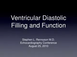
Diastole • Isovolumetric Relaxation Phase • Rapid Filling Phase • Diastasis • Atrialsystoly
Cardiac cycle • Isovolumetric Relaxation Phase • Rapid Filling Phase • Diastasis • Atrialsystoly
Definition • Diastolic Dysfunction refers to abnormalities of active myocardial relaxation and passive ventricular filling. • Condition that includes classic CHF findings and abnormal diastolic and normal systolic function at rest
Why to know ?? • common discharge diagnosis for patients older than 65 years. • a patient cannot have pure systolic heart failure • 40 percent of patients with heart failure have preserved systolic function. • It can be attributed to one of the four underlying mechanisms.
Four causes • Slow/incomplete myocardial Relaxation: the most common cause of this is myocardial ischaemia, which causes the reduced rate of LV pressure decline Impaired peak LV filling rAte: Pericardial constriction: Altered elasticity: What is the net effect??
Cellular level • EC couple • repolarization – relaxation coupling • the calcium transient is prolonged as a result of dysfunction of any of the processes mentioned above. • Lusitropy
Symptoms • decreased exercise capacity; • Neurohumoral activation with sodium and water retention; • paroxysmal nocturnal dyspnoea ; and orthopnoea
Common precipitating factors • volume overload; • tachycardia; exercise; hypertension; • ischemia; • systemic stressors (e.g., anemia, • fever, infection, thyrotoxicosis); • arrhythmia • increased salt intake; • Use of NSAIDs.
Diagnosis – ECHO • EA Under normal conditions, E is greater than A and the E/A ratio is approximately 1.5. Decrease initial to become one the back to 1.5
Pulmonary venous flow (PVF): • During atrial systole, there is normally a small • amount of retrograde PVF. • In DD, PVF reversal associated with atrial contraction becomes progressively more pronounced
Isovolumetric relaxation time • IRT normal 70 ms • DD it becomes 110 ms • Deceleration time (DT): the rate of dissipation of the transmitral pressure gradient is also a function of LV compliance • Normal 180 – 240 ms • Abnormal > 240 → → 180 ms
Diagnosis • Tissue Doppler: • this uses Doppler shifts of ultrasound waves to calculate the velocity of myocardial tissue movement in a similar way to that of blood flow • The serum brain natriuretic peptide (BNP) test can accurately differentiate heart failure from noncardiac conditions in dyspnea, but it cannot distinguish diastolic from systolic heart failure
Treatment -Primary prevention • smoking cessation • aggressive control of hypertension, • Hypercholesterolemia, coronary artery disease. • Lifestyle modifications such as weight loss, dietary changes, limiting alcohol intake, exercise are equally effective in preventing diastolic and systolic heart failure
Treatment • Regress left ventricular hypertrophy (decrease wall thickness and remove excess collagen). • Beta blockers, ACE inhibitors and ARBs • Aldosterone antagonists • Calcium channel blockers • Maintain atrioventricular synchrony by managing tachycardia • Beta blockers (preferred) • Calcium channel blockers (second-line agents) • Digoxin (controversial)
Carvidolol • both agents improve cardiac remodeling in patients with congestive heart failure, carvedilol provides superior resolution of left ventricular fraction. • Patients who do not respond to metoprolol may improve when switched to carvedilol. • carvedilol exhibits more favorable effects on LV function than does nebivolol.
Treatment principles • Optimize circulating volume (hemodynamics). • ACE inhibitors • Aldosterone antagonists (theoretical benefit) • Salt and water restriction • Diuresis, • Improve survival. • Beta blockers • ACE inhibitors
SHF and DHF ?? • Diuretics ?? • Digoxin ?? • Venodilators ?? • Beta blockers !!
summary • Phases of diastoly • Dysfunction • Causes • Symptoms • Diagnosis • Treatment