Download
1 / 26
310 likes | 1.36k Views
Clostridium. - Microscopic appearance of different species. - Differentiation between species according to biochemical reactions. Clostridium perfringens. Clostridium perfringens.

E N D
Clostridium - Microscopic appearance of different species. - Differentiation between species according to biochemical reactions.
On Blood Agar C. perfringensproduceslarge beta-haemolytic coloniesare produced. Some strains produce a double zone of haemolysis.
1- Prepare a plate of lactose egg yolk milk agar 2- Turn the plate over, and using a wax pencil, draw a line across the centre of the plate 3- Using a sterile swab, cover one half of the medium with C. perfringensantitoxin. Allow to dry
4- Inoculate the test organism at right angles to the centre line (inoculum passes from the antitoxin-free half of the plate to the antitoxin-covered half. 6- Incubate the plate anaerobically at 35–37 ºC overnight. 5- Inoculate also a non-toxin producing control organism that will grow anaerobically. 7- Look for an opacity around the inoculum in the half of the plate containing no antitoxin and no opacity in the half containing the antitoxin.
Litmus Milk Reduction Test A heavy inoculum of the test organism is incubated for up to 4 hours in a tube containing litmus milk.Reduction of the litmus milkis indicated by a change in colour of the medium frommauveto white orpale yellow
Nitrate Reduction Test C.perfringens can reduce nitrate to nitrite which detected by addition of sulfanilic acid which react with nitrite to form diazonium salt which react with added alpha-naphthylamine to form redcolour.
Lecithinase C activity: Seen as an opacity in the medium due to the breakdown of lecithin in the egg yolk. Lipase hydrolysis: Seen as (fatty) layer covering colonies and sometimes extending into the medium. Lactose fermentation: There is a reddening in the medium. The colonies become red on exposure to air. Proteinase activity (proteolysis): Shown by an area of clearing around the colonies due to the breakdown of casein in the milk by the enzyme proteinase.
On lactose egg yolk medium, C. perfringens: ● Produces lecithinase C (alphatoxin) ● Ferments lactose
Gelatin Hydrolysis C.perfringensproduce proteolytic enzyme (gelatinase) that liquefy gelatin.
On Robertson’s cooked meat medium C. perfringensis saccharolytic and slightly proteolytic.
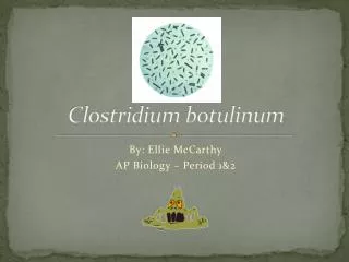
On Blood Agar C. botulinumproduceslarge semi-transparent colonies with a wavy outline. Most strains are beta-haemolytic
On Robertson’s cooked meat medium C. botulinumis proteolytic.
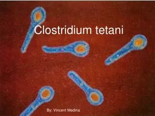
On Blood Agar C. tetaniproduces a fine film of growth. Use a hand lens to examine the plate. On fresh blood agar C. tetaniis haemolytic (alpha first followed by betahaemolysis).
Indole Production When C.tetani is cultured in a medium which contains tryptophan. Indole production is detected by Kovac’s reagent which contains 4 (p)-dimethylaminobenzaldehyde which reacts with the indole to produce a red coloured compound.
On Blood Agar C. difficileproduces large non-haemolytic colonies.
Esculin Hydrolysis C. difficilecan grow in bile esculin agar and turns the indicator ferric ammonium citrate to a dark brown color results from combination of esculetinend product of esculin hydrolysis with ferric ions to form a phenolic iron complex.