Download
1 / 1
10 likes | 161 Views
An Ion Channel Database – Annotation via Homology Modelling. Hyun Ji Kim, John Tate, Charlotte E. Capener, Richard J.Law, David B. Sattelle 1 , Frances M. Ashcroft 2 , Sarah Butcher 3 and Mark S. P. Sansom

E N D
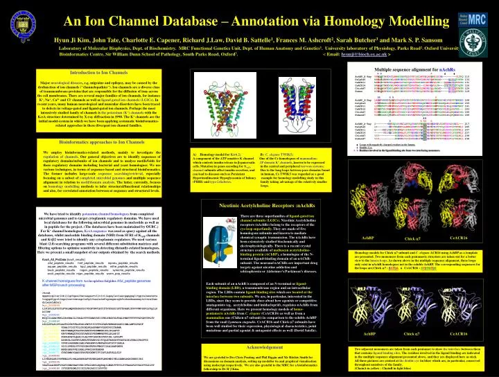
An Ion Channel Database – Annotation via Homology Modelling Hyun Ji Kim, John Tate, Charlotte E. Capener, Richard J.Law, David B. Sattelle1, Frances M. Ashcroft2, Sarah Butcher3 and Mark S. P. Sansom Laboratory of Molecular Biophysics, Dept. of Biochemistry. MRC Functional Genetics Unit, Dept. of Human Anatomy and Genetics1. University laboratory of Physiology, Parks Road2. Oxford University Bioinformatics Centre, Sir William Dunn School of Pathology, South Parks Road, Oxford3. < Email: hyunji@bioch.ox.ac.uk > • Multiple sequence alignment for nAchRs • B B A • AchBP_X-Ray VFWQQTTWSDRTLAWNSSHSPDQVSVPISSLWVPDLAAYNAISKPE---------VLTPQ 110 • Dalpha6 NAWLNLEWNDYNLRWNETEYGGVKDLRITPNKLWKPDVLMYNSAD-----EGFDGTYHTN 133 • CeACR11 NAWLSYTWFDHKLQWEPKKYGGIQDIRFPGSSDHIWKPDVLLYN---SAAEDFDSTFKSN 131 • CeACR16 NAWLDYTWNDYNLVWDKAEYGNITDVRFPAG--KIWKPDVLLYN---SVDTNFDSTYQTN 126 • ChickA7 NIWLQMYWTDHYLQWNVSEYPGVKNVRFPDGLIWKPDILLYNSAD-----ERFDATFHTN 129 • Human NIWLQMSWTDHYLQWNVSEYPGVKTVRFPDGQIWKPDILLYNSAD-----ERFDATFHTN 129 • BB B BA • AchBP_X-Ray LARVVSDGEVLYMPSIRQRFSCDVSGVDTESGATCRIKIGSWTHHSREISVDPTTENSDD 168 • Dalpha6 IVVKHNGSCLYVPPGIFKSTCKIDITWFPFDDQHCEMKFGSWTYDGNQLDLVLNSEDGGD 193 • CeACR11 LLTYHTGTVVWIPPGVLKFVCQLDVTWFPFDDQVCEMKFGSWTFHGYAIDLQIDDDTNGT 191 • CeACR16 MIVYSTGLVHWVPPGIFKISCKIDIQWFPFDEQKCFFKFGSWTYDGYKLDLQPATGG--- 183 • ChickA7 VLVNSSGHCQYLPPGIFKSSCYIDVRWFPFDVQKCNLKFGSWTYGGWSLDLQMQEADISG 189 • HumanA7 VLVNSSGHCQYLPPGIFKSSCYIDVRWFPFDVQHCKLKFGSWSYGGWSLDLQMQEADISG 189 • BA AA A • AchBP_X-Ray S--------EYFSQYSRFEILDVTQKKNSVTYSCCPEAYEDVEVSLNFRKKGRSEIL--- 217 • Dalpha6 LS--------DFITNGEWYLLAMPGKKNTIVYACCPEPYVDITFTIQIRRRTLYYFFNLI 245 • CeACR11 Q----SMDLSTYLVNGEWQVISTNAKRVVSYYKCCPEPYPTVNYYLHIRRRTLYYGFNLI 247 • CeACR16 ------FDISEYISNGEWALPLTTVERNEKFYDCCPEPYPDVHFYLHMRRRTLYYGFNLI 237 • ChickA7 YIS-----------NGEWDLVGIPGKRTESFYECCKEPYPDITFTVTMRRRTLYYGLNLL 238 • HumanA7 YIP-----------NGEWDLVGIPGKRSERFYECCKEPYPDVTFTVTMRRRTLYYGLNLL 238 • Loops with negatively charged residues in the lumen. • Double Cys. • ABResidues involved in the ligand-binding site from two interfacing monomers. Introduction to Ion Channels Major neurological diseases, e.g. migraine and epilepsy, may be caused by the dysfunction of ion channels ("channelopathies"). Ion channels are a diverse class of transmembrane proteins that are responsible for the diffusion of ions across the cell membranes. There are several major families of ion channels, for instance K+, Na+, Ca2+ and Cl- channels as well as ligand gated ion channels (LGICs).In recent years, many human neurological and muscular disorders have been traced to defects in voltage-gated and ligand-gated ion channels. Perhaps the most intensively studied family of channels is the potassium (K+) channels with the KcsA structure determined by X-ray diffraction in 1998. The K+channels are the initial model-system in which we have been applying systematic bioinformatics-related approches in these divergent ion channel families. • General overviews : • bioinofrmatics related approaches • database searching (microbial genomics) • 2) sequence alignments and domain identification • 3) further annotation via homolgy modelling • (structural-functional relationship) Activity (%) • Bioinformatics approaches to Ion Channels • We employ bioinformatics-related methods, mainly to investigate the regulation of channels. Our general objectives are to identify sequences of regulatory domains/subunits of ion channels and to analyse motifs/folds for these regulatory domains including bacterial and yeast homologues. We use various techniques, in terms of sequence-based and structural bioinformatics. The former includes large-scale sequence searching/retrieval, especially focusing on a subset of completed microbial genomes and multiple sequence alignment in relation to motif/domain analysis. The latter, currently, focuses on homology modelling methods to infer structural/functional relationships and also, for correlated annotation between at sequence and structural levels. • Homology model for Kir6.2; B) C. elegansTWIK5; • A component of the ATP sensitive K channel One of the Ce homologues of mammalian- • which controls insulin release in -pancreatic 2P domain K+ channels, known to be expressed • cells. Mutation in genes encoding for KATP in the central and peripheral nervous systems. • channel subunits affect insulin secretion, and Due to the long loops between pore domains found • can lead to diseases such as Persistent in human, Ce TWIK5 was regarded as a good • Hyperinsulinaemic Hypoglycaemia of Infancy example for homology modelling study in this • (PHHI) and type-2diabetes. family taking advantage of the relatively smaller • loops. • General overviews : • bioinofrmatics related approaches • database searching (microbial genomics) • 2) sequence alignments and domain identification • 3) further annotation via homolgy modelling • (structural-functional relationship) NaCL (mM) • General overviewNicotinic Acetylcholine • : • bioinofrmatics related approaches • database searching (microbial genomics) • 2) sequence alignments and domain identification • 3) further annotation via homolgy modelling • (structural-functional relationship) Nicotinic Acetylcholine Receptors :nAchRs • We have tried to identify potassium channel homologues from completed • microbial genomes and to target cytoplasmic regulatory domains. We have used local databases for the following miocrobial genomes in nucleotide as well as in peptide for the project. (The databases have been maintained by OUBC.) • For K+ channel homologues, KcsA sequence was used as query against all the • databases, whilst nucleotide binding domain (NBD) from SUR1 of KATP channels • and Kv2 were tried to identify any cytoplasmic regulators. We used various • blast (2.0) searching programs with several different substitution matrices and • filtering options to optimise sensitivity in detecting distantly-related homologues. • Here we present a small snapshot of our outputs obtained by the search methods. • KcsA_All_ProData [kcsA_results] • aful_peptide_results hinf_peptide_results mpneu_peptide_results aquae_peptide_results hpyl_peptide_results mthe_peptide_results bsub_peptide_results mgen_peptide_results synecho_peptide_results ecoli_peptide_results mjan_peptide_results worm_pep_results • K channel homolgues from Archeoplobus fulgidus Aful_peptide genomes • after MSPcrunch processing • >kcsA • mppmlsgllarlvklllgrhgsalhwraagaatvllvivllagsylavlaergapgaqlitypralwwsveta • tvgygdlypvtlwgrlvavvvmvagitsfglvtaalatwfvgreqerrghfvrhsekaaeeaytrttralher • drlermlddnrr • >gi_2649899 • LIFIFLILFYSTLFVALMQNYEGRSYSIVDGFYWVIATITTVGYGDIYFTTPAGKIFSVVVMVSGVLYFglLP • AIIPW • >gi_2649341 • MIQIILLRAVKKLIRrRRAILIYAGLYTVTLWAVSSIIFHLYEGVDLFDALYWAVTTTTTVGYGDVTPLTV • >gi_2648884 • LKIGIFLSLVVLAGTVGYHFVEwFDSLYMTVITITTTGYGEVKPMGPGGRVISMLLMFVGVGTF • >gi_2649447 VDAAIVIGIVYLYPSELWLAGFMAVVGSFGVSITRAMA • >gi_2649648 KAVYWAMQTFGGYGYAKEYDVERWWREINLIRLAPVT • >gi_2650443 KAVYWAMQTFGGYGYAKEYDVERWWREVNLLRLAPIT • >gi_2648660 FGDSAVATLQGAIILALTFFIARINIFGVIPA-LGVGFV • >gi_2648753 ASGRGLIASPKVLRWLTETAEKYGIPYQLEVAEGGTTDATAIHLTKAGIPAGVVS • >gi_2649912 YSVKLSGERERIARLVGEGERVLVMFAGVGPYPIVIAKLA • >gi_2648225 YGILYPEELVTVVGYGRSFEFAVFMGVIIAAIAAFGMVS • >gi_2648607 MSESSKSFVEISEALSVWIDSFEGERR • >gi_2648771 GVATAMAVGAAVGYAVESGKWFITVIAVLAGVALLSLV • >gi_2649942 • LIVHGRLDEIVPFENGLTLYNLAREPKAFVEVEKGDHFLRHDNRIVELIAEWLDGKIKEKIIRI • >gi_2650635 • YAGVLAAIMSTVSGFISMGTAAVVRDIPTAIGKSLSQRGQILWGRVVTVILTVVAALFGYYGGYVVGILVT • >gi_2649580 IVYGEVSGWLTIIGILVAIAGIIIGVVTG There are three superfamilies of ligand-gated ion channel subunits (LGICs). Nicotinic Acetylcholine receptors (nAchRs) belong to the receptors of the cys-loop superfamily. They are made of five homologous subunits and known to mediate chemical synaptic transmission. The nAchRs have been extensively studied biochemically and electrophysiologically. There is a recent crystal structure available of molluscan acetylcholine-binding protein (AChBP), a homologue of the N-terminal ligand-binding domain of an nAChR-subunit. The neuronal nAChRs are important drug targets against nicotine addiction and schizophrenia or Alzheimer's/Parkinson's diseases. pmol cAMP / mg protein AchBP CeACR16 Chick7 • Homology models for Chick 7 subunit and C. elegans ACR16 using AchBP as a template • are presented. Two monomers from each pentameric structure are taken out for a better • view to the lumen loops. As shown above in the multiple sequence alignment, these loops • only exist in nAchR homologues not in the soluble AchBP. The corresponding sequences for • the loops are;Chick7 : ERFDA& CeACR16 : SVDTNFDS • General overviews : • bioinofrmatics related approaches • database searching (microbial genomics) • 2) sequence alignments and domain identification • 3) further annotation via homolgy modelling • (structural-functional relationship) Each subunit of an nAchR is composed of an N-terminal or ligand-binding domain (LBD), a transmembrane region and an intracellular region. The LBDs contain ligand-binding sites which are located at the interface between two subunits. We are, in particular, interested in the LBDs, since they seem to provide clues about how agonists or competitive anatagonists (eg, acetylcholine and imidacloprid), regulates nAchRs from different organisms. Here we present homology models of homo-pentameric nAchRs from C. elegans (CeACR16) as well as from a mammalian one (Chiken 7 subunit) in comparison to the soluble AchBP from the snail Lymnaea stagnalis. CeACR16 and Chick7 subunits have been well studied for their expression, physiological characteristics, point mutations and partial agonist & antagonist effects as well (David Satelle). AchBP Chick7 CeACR16 • Acknowledgement • We are grateful to Drs Chris Ponting and Phil Biggin and Mr Ritchie Smith for • discussions on domain analysis, setting up modeller 6a and graphical visualisation • using molscript respectively. We are also grateful to the MRC for a bioinformatics • fellowship to Dr H J Kim. • Two adjacent monomers are taken from each pentamer to show the interface between them • that contains ligand binding sites. The residues involved in the ligand binding are indicated • in the multiple sequence alignment presented above, and they are displayed here as stick. • All three pictures are centred at the double cys (in blue) which are, in particular, conserved • throughout members of the family. • (ChainA in yellow : ChainB in light blue)