Download
1 / 21
310 likes | 1.08k Views
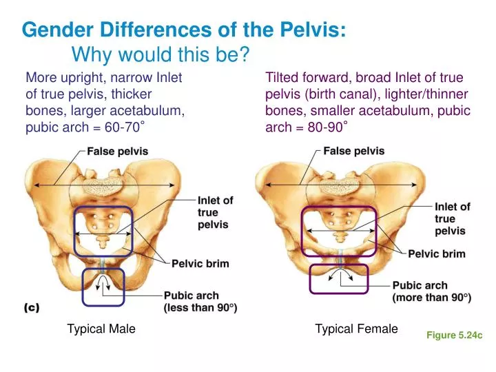
Gender Differences of the Pelvis: Why would this be?. More upright, narrow Inlet of true pelvis, thicker bones, larger acetabulum, pubic arch = 60-70°. Tilted forward, broad Inlet of true pelvis (birth canal), lighter/thinner bones, smaller acetabulum, pubic arch = 80-90°. Typical Male.

E N D
Gender Differences of the Pelvis: Why would this be? More upright, narrow Inlet of true pelvis, thicker bones, larger acetabulum, pubic arch = 60-70° Tilted forward, broad Inlet of true pelvis (birth canal), lighter/thinner bones, smaller acetabulum, pubic arch = 80-90° Typical Male Typical Female Figure 5.24c
You DO have to know these bones… Figure 5.7
…I’m getting dizzy… Figure 5.9
…that’s anatomy for you…it is possible! Figure 5.11
Orbits – what 7 bones? Figure 7.9b
Nasal Cavity Frontal Ethmoid Nasal Sphenoid Inferior Nasal Concha Palatine Maxilla Figure 7.10a
Nasal Septum Figure 7.10b
Paranasal Sinuses – hollow portions of skull Lighten the skull Amplify voice Continuity w/ respiratory tract leads to sinus infections What 5 bones?
The Hyoid Bone • Only bone with no bone articulations • Moveable base for tongue • Aids in swallowing and speech
The Fetal Skull is not fully formed • Fontanels—fibrous membranes connecting the cranial bones (a.k.a. soft spots) • Allows brain growth, slight compression during birth • Convert to bone by 24 months
The Vertebral Column Vertebrae named by location separated by intervertebral discs 7 cervical (neck) 12 thoracic (articulate w/ ribs) 5 lumbar (lower back) 7am breakfast, 12 noon lunch, 5pm dinner? Sacrum = 5 fused vertebrae Coccyx = 3-5 fused vertebrae
The Vertebral Column – changes shape between birth and walking Secondary curvatures of cervical and lumbar regions develop after birth Secondary curves Primary curves Lordosis = exaggerated secondary lumbar curvature (often seen in pregnant women) Kyphosis = exaggerated primary thoracic curvature (humpback) Scoliosis = abnormal lateral curvatures
Vertebral Column: Ligaments Figure 7.14a
Vertebral Column: Intervertebral Discs Figure 7.14b
Atlas (1st Cervical Vertebrae) Atlas = C1, Articulation with occipital condyle = “YES” Figure 5.18a
Axis (Second Cervical Vertebrae) Axis = C2, Dens articulation with Atlas = “NO”; also prevents hyperextension of neck a.k.a. odontoid process Figure 5.18a
Cervical vertebrae have small openings for important nerves and vessels of the head Only in Cervical Vertebrae Present in all vertebrae Figure 5.18b
Thoracic vertebrae have extra facets for rib articulation(look like giraffes?) Figure 5.18c
Lumbar Vertebrae have massive bodies to bear weight (Look like moose?) Figure 5.18d
The Bony Thorax protects major organs Consists of three parts • Sternum • Ribs • True ribs (pairs 1–7) • False ribs (pairs 8–12) • Floating ribs (pairs 11–12) • Thoracic vertebrae