Download
1 / 66
660 likes | 883 Views
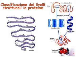
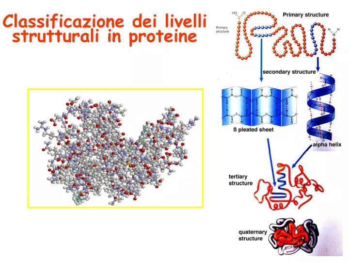
Classificazione dei livelli strutturali in proteine. Classificazione dei livelli strutturali in proteine. http://en.wikipedia.org/wiki/Protein_structure. Proteine: differenti funzioni ed architetture. Proteine: differenti funzioni ed architetture. Proteine:

E N D
Proteine: differenti funzioni ed architetture
Proteine: differenti funzioni ed architetture
Proteine: non tutte le strutture sono possibili
GopalasamudramNarayanaIyer Ramachandran φ ψ
GopalasamudramNarayanaIyer Ramachandran 180 φ 0 ψ ψ φ 0 180 -180
Conformazione delle catene laterali Torsion Axes and Dihedral Angles of the side chain of LysineThe sample amino acid Lysine has four torsion axes within its side chain. The torsion axes are symbolized as arrows, the dihedral angles are labeled chi1 to chi4.
eliche α in α-helix hydrogen bonds between carbonyl i and amide i+4
eliche α Folding mechanism of alpha helices
eliche α 180 ….manon solo 0 ψ φ 0 180 -180
Strutture supersecondarie o motivi (elementi di struttura secondaria uguali o diversi si combinano) (e)
Domini strutturali (subunità avente stabilità strutturale ed una propria funzione) dominio che lega il gliceraldeide-3-fosfato dominio che lega il NAD+ gliceraldeide-3-fosfato deidrogenasi
Enzima bifunzionale Piruvato chinasi PRA-isomerasi IGP-sintetasi
Struttura quaternaria Eteromultimeri Omomultimeri
mettere ordine alle conoscenze strutturali acquisite
La tassonomia è la scienza che si occupa genericamente dei modi di classificazione
La tassonomia è la scienza che si occupa genericamente dei modi di classificazione
La tassonomia è la scienza che si occupa genericamente dei modi di classificazione
La tassonomia è la scienza che si occupa genericamente dei modi di classificazione
La tassonomia è la scienza che si occupa genericamente dei modi di classificazione Rosso: principalmente α Verde: principalmente β Giallo: αβ Blue: basso contenuto di strutture secondarie
La tassonomia è la scienza che si occupa genericamente dei modi di classificazione I possibili modi in cui la catena proteica si ripiega non sono infiniti. Ad oggi SCOP ne suggerisce circa 1.200. Probabilmente, il loro numero non è superiore a 2.000 Dal momento che abbiamo un’ampia conoscenza di come le proteine si strutturano, possiamo fare delle predizioni sulla base della loro sequenza con tecniche di Bioinformatica
Bioinformatica genoma + algoritmo* • Allineamento di sequenze • Individuazione di geni • Ricostruzione di genomi da frammenti • Allineamenti di strutture proteiche • Predizione di strutture proteiche • Predizione di espressione genica • Predizione di interazioni tra proteine *algoritmo= procedimento che risolve un determinato problema attraverso un numero finito di passi
microscopy Il potere risolutivo di un microscopio è la distanza minima alla quale due punti risultano distinti. Nel caso lo strumento si basi sull'utilizzo di radiazione con una propria lunghezza d'onda associata, come i tradizionali microscopi ottici, risoluzione e lunghezza d'onda utilizzata sono parametri tra loro strettamente correlati. Microscopi che si basino su diverse tecnologie, come ad esempio l'AFM, ovviamente rispondono a considerazioni differenti. In prima approssimazione, e non tenendo conto di effetti aberrativi, possiamo considerare che la relazione che lega il potere risolutivo (d o distanza tra due punti tra loro risolti), l'apertura numerica di un sistema ottico (tutto il sistema) e la lunghezza d'onda della radiazione utilizzata sia Questa relazione è generalmente nota come principio di Abbe. Per un microscopio ottico in luce visibile d raggiunge i 0,2 micron, il microscopio elettronico giunge a 0,1nm.
Electron cryomicroscopy (cryo-EM or cryo-electron microscopy) a form of electron microscopy (EM) where the sample is studied at cryogenic temperatures (generally liquid nitrogen temperatures (77 K or −196 °C) Cryo-electron tomography (cryo-ET) is a type of electron cryomicroscopy where tomography is used to obtain a 3D reconstruction of a sample from tilted 2D images at cryogenic temperatures.
Cryo Electron-Tomografy (cryo-ET) Schematic diagram illustrating the sequence of steps involved in obtaining a three-dimensional image of a plunge-frozen cell using cryo-electron tomography. a| A small volume (typically 3–5 l) of a suspension of cells in culture is deposited on a holey carbon grid and then plunge-frozen in liquid ethane cooled to temperatures of 100 K by liquid nitrogen. b | The grid is then transferred into the column of an electron microscope for collection of a series of images at varying tilts. c | The series of tilted images is then converted to a three-dimensional volume (tomogram) that provides a representation of the distribution of densities of cellular components.
Cryo Electron-Tomografy (cryo-ET) http://emnavi.protein.osaka-u.ac.jp/emnavi_gallery.php
Cryo Electron-Tomografy (cryo-ET)
Spectroscopy was originally the study of the interaction between light and matter as a function of wavelength λ
absorption absorption spectroscopy Spectroscopy studies the interaction between energy and matter emission emission spectroscopy ΔΕ=hν Spectrometry is the measurement of these responses and an instrument which performs such measurements is a spectrometer or spectrograph, although the latter term is more limited in use to the original field of optics. Normally, the quantity that is measured is an intensity, either of energy absorbed or produced.
Nuclear Magnetic Resonance NMR Earthmagneticfield: 30-60 µT NMR spectrometer in Siena: 14.2 T
1000 MHz NMR spectrometer (23.5 Tesla superconducting NMR magnet) was delivered to the new ‘Centre de RMN à Très Hauts Champs’ in Lyon, France in July 2009
Main Features World’s first, standard-bore, high homogeneity 1 GHz NMR magnet Persistent superconducting magnet UltraStabilized™ sub-cooling technology Magnetic field strength of 23.5 Tesla Proton NMR frequency of 1000 MHz Standard bore size of 54 mm delivered to the new ‘Centre de RMN à Très Hauts Champs’ in Lyon, France in July 2009