Download
1 / 7
70 likes | 211 Views
Nerve Stimulus Excites the Muscle Cell. A muscle cell must receive a stimulus to begin the excitation-contraction coupling Series of events linking electrical signal to muscle contraction Muscle cells can be stimulated by ACh ACh- Acetylcholine- neurotransmitter

E N D
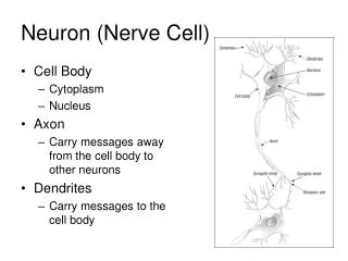
Nerve Stimulus Excites the Muscle Cell • A muscle cell must receive a stimulus to begin the excitation-contraction coupling • Series of events linking electrical signal to muscle contraction • Muscle cells can be stimulated by ACh • ACh- Acetylcholine- neurotransmitter • Nerve impulse reaches axon terminal • Axon- long extension of nerve cell, relays stimulus • Neuromuscular Junction- axon branches as it enters muscle, each branch goes to 1 muscle fiber • Synaptic cleft- small space between axon terminal & muscle fiber 2. Voltage-gated Ca2+ channels on axon terminal open Ca2+ goes in synaptic vesicles fuse with membrane • Synaptic vesicles- sacs filled with neurotransmitter 3. Exocytosis of ACh • Motor end plate- folded part of sacrolemma with millions of ACh receptors Animated Neurotransmission
Resting Potential- Polarized • Partial negative charge inside a neuron or muscle cell at rest • More K+ inside, more Na+ outside • Both K+ & Na+ diffuse through cell membrane, K+ can get out easier than Na+ can get in • Polarized- difference in charge inside & outside the cell Resting membrane potential K+ Outside the cell K+ Na+ K+ K+ K+ - Na+ Na+ Na+ Na+ K+ K+ Na+ - - - - Membrane Cytoplasm K+ Na+ Na+ K+ K+ - Na+ - K+ Na+ - K+ - - - K+ K+ K+ - -
Action Potential (AP)- Depolarized • When muscle cell is stimulated by ACh, chemically gated ion (Na+ & K+) channels open • Na+ flows in faster than K+ flows out Depolarization- change of charge (action potential) • Causes a ripple effect along sarcolemma, voltage gated Na+ gates open • Also causes slower K+ gate to open, K+ rushes out Repolarization- return to resting charge • Active transport is used to move Na+ back outside & K+ back inside • Refactory period- cell cannot be stimulated again until repolarization & active transport of ions is complete Animated Neurotransmission Na+ K+ Outside the cell K+ K+ Na+ K+ K+ - Na+ Na+ Action Potential Na+ Na+ K+ K+ Na+ Na+ - - - - Membrane Cytoplasm K+ Na+ Na+ K+ K+ - Na+ - Na+ K+ - K+ - - - K+ Na+ K+ Na+ Na+ Na+ K+ - -
Excitation-Contraction Coupling • AP ends before signs of contraction are obvious • AP goes along sacrolemma & down T tubules • AP in T tubules causes release of Ca2+ from adjacent terminal cisternae • Ca2+ binds to troponin, causing it to move myotroponin away for actin active site • Mysosin heads form cross bridges with active sites on actin & pull thin filaments toward center of sacromere (power stroke) Excitation-Contraction Coupling Excitation-Contraction Coupling 2 Actin Myosin Bridge
ATP and the Power Stroke • Myosin heads have ATP attached to them, used for E to “cock” heads back • Release ADP & P • Myosin attaches to active sites to form “cross-bridges” • Myosin head returns to its lower E position once cross bridge is formed, moving the thin filament (power stroke) • ATP binds to myosin head, actin filament is released Actin Myosin Bridge
Contraction • Full contraction of the muscle cell requires 30+ repeats of power stroke action • Process repeats until Ca2+ is no longer available • Acetylcholinesterase • enzyme that digests acetylcholine to ensure contraction does not persist without nervous stimulation • No more acetylcholine Ca2+ is reabsorbed by SR by active transport (uses more ATP) Actin Myosin Bridge
Rigor Mortis • When breathing stops, no more O2 can’t make ATP • Dying cells cannot keep extracellular Ca2+ out • Ca2+ goes into muscle cells and promotes myosin-actin cross-bridges • ATP is still being consumed at the cross bridge, when it runs out, detachment becomes impossible stiffness • Usually starts to set in 3-4hrs postmortem, peaks about 12 hrs postmortem • As muscle protein begin to break down, rigor mortis gradually goes away