Download
1 / 16
160 likes | 240 Views
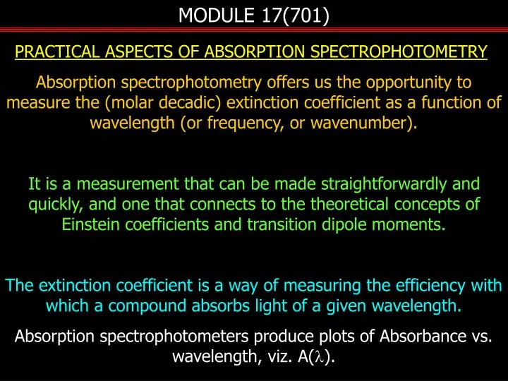
MODULE 17(701). PRACTICAL ASPECTS OF ABSORPTION SPECTROPHOTOMETRY Absorption spectrophotometry offers us the opportunity to measure the (molar decadic) extinction coefficient as a function of wavelength (or frequency, or wavenumber).

E N D
MODULE 17(701) PRACTICAL ASPECTS OF ABSORPTION SPECTROPHOTOMETRY Absorption spectrophotometry offers us the opportunity to measure the (molar decadic) extinction coefficient as a function of wavelength (or frequency, or wavenumber). It is a measurement that can be made straightforwardly and quickly, and one that connects to the theoretical concepts of Einstein coefficients and transition dipole moments. The extinction coefficient is a way of measuring the efficiency with which a compound absorbs light of a given wavelength. Absorption spectrophotometers produce plots of Absorbance vs. wavelength, viz. A(l).
MODULE 17(701) • Since • Other good reasons for measuring absorption spectra are • contains information about the vibrational progression in excited states. • provide information as to where compounds have a high value of e and thereby at which wavelength photoexcitation can be optimally carried out. • lead to knowledge of the number/rate of photons absorbed for a given incident photon flux.
MODULE 17(701) detector mono- chromator lamp sample reference Instrumentation Two principal types of instruments are available on the commercial market, scanning and diode array. The vibrating reflector and the positioning and orientation of the intervening mirrors ensures that the beam striking the detector is one that has been transmitted by either reference or sample cells, alternately.
MODULE 17(701) Thus, the PMT output is a square-wave voltage signal that oscillates between Vil and Vtl. These relate linearly to the transmitted intensities (I values) in the two cuvettes. The voltages are converted into digital format by an analog-to-digital converter (ADC), the output of which is sent to a computer that calculates the absorbance at the operating wavelength according to The monochromator grating is moved to another wavelength and the process repeated over and over for the wavelength range chosen. Typically the scan is continuous
MODULE 17(701) . The processor then generates a plot of A versus l - an absorption spectrum of the sample.
MODULE 17(701) In the array-based device the whole of the wavelength range of the lamp is passed through the sample after which it is dispersed in a spectrograph. This is a monochromator without an exit slit. An array detector (e.g. CCD) is placed at the focal plane of the spectrograph such that the dispersed white light spectrum covers the whole of the array. Each pixel (measuring unit) of the detector (typically 512 or 1064 pixels) records a different wavelength of the dispersed light and the array records intensity of transmitted light at all wavelengths simultaneously. This is called the Felgett (or multiplex) advantage and it represents a major increase in the speed of measurement.
MODULE 17(701) S light source to computer CCD spectrograph The stored charge on each of the pixels is read into a computer, which calculates the absorbance at each one. Each pixel represents a narrow band of wavelengths Placing a cuvette filled with solvent in the sample position and repeating the measurement records the reference spectrum.
MODULE 17(701) 100 %T l 0 white = reference (Vi); cyan = sample (Vt) Errors in Absorptiometry In the double beam scanning instrument the detector alternately sees light transmitted by the reference and the sample channels as the wavelength is changed. In reality the ADC samples the voltage amplitude several times while the instrument is “paused” at a particular wavelength, as indicated by the red arrows in the next sketch.
MODULE 17(701) Here we are taking 5 samples at each wavelength step. the average of the five is calculated and stored as a Vi(l) or a Vt(l) reading from which A(l) is computed. What governs how precisely the device can measure A(l)? How accurately can it measure A(l) at the low end and the high end of the A(l) range?
MODULE 17(701) The first is a signal-to-noise problem and the second is a dynamic range problem. Dynamic Range How the intrinsic error (dV) in the measurement of V propagates. Let the intrinsic error in our ADC be ±1 mV, and let the reference measurement show 1000 mV. In the following Table the % error in A is calculated at different values of A, neglecting all errors except that in Vt .
MODULE 17(701) * the lower value is zero, but a small positive value has been entered so an A value can be calculated. for optimum precision arrange to measure A ~ 1. Change the path length to bring A into the best range.
MODULE 17(701) Signal-to-noise The Table shows that the error in A improves as A decreases, implying that the nearer A is to zero, the more the precision. This is misleading because as the absorbance approaches zero, the ADC is being required to take the ratio of two voltages which are very close to the same value (1000 mV in our example) with ±1 mV error on both. For this reason the errors in A increase again at A < 0.1. [At A = 0.01 the error is 8%, and at A = 0.001 it is ~ 40%.] Thus the intrinsic noise in the ADC leads to a lack of precision.
MODULE 17(701) We can improve the situation by recognizing that if we increase Vi to a value significantly greater than 1000 mV, e.g. to 10 V then our signal-to-noise ratio will also increase by a factor of 10, because the ADC noise remains at 1 mV. Remember that the absorbance measurement is a relative one; we measure ratios of voltages (Vi/Vt) to obtain A, so the absolute values of the voltages are unimportant. Thus if we increase Vi to pull it out of the noise, then we do the same to Vt . A first thought would be to add a voltage amplifier after the detector and before the ADC. This provides only marginal improvement because the amplifier will amplify the noise in addition to the signal.
MODULE 17(701) A better solution is to arrange for the ADC to take more samples of the voltage on each half cycle at every wavelength. Signal-to-noise improves as the square root of the number of samples (for random noise). In our example we used five samples per half cycle. Increasing this to 50 decreases the noise amplitude by a factor of ~3, and so on. The clock rate of an ADC has limits and in order to achieve the goal it may be that you need to increase the time that the instrument dwells at a particular wavelength, viz., scan more slowly.
MODULE 17(701) A lock-in amplifier can be useful in these circumstances. A lock-in amplifies signals that have a particular frequency. To employ such a device you “chop” the optical beam at a given frequency (say 500 Hz) and then send a synchronous electrical output from the chopper to the reference channel of the lock-in. This instructs the lock-in to amplify only those signals that have a 500 Hz repetition rate. Noise typically has random frequencies and only a fraction of the noise will be amplified, hence improving the signal-to-noise ratio.
MODULE 17(701) V saturation curve I Another way to enhance Viand Vt is to increase Ii ,the optical power of the incident beam (increase lamp power; open monochromator exit slit). This works because Ii/It is fixed for a given value of A, and increasing Ii leads to a corresponding increase in It. This is a limited approach because PMT detectors saturate at high values of cathode illumination.


![700-701 Exam Dumps - Real 700-701 Dumps PDF [2018]](https://cdn4.slideserve.com/7919108/cisco-700-701-exam-cisco-specialist-dt.jpg)