Download
1 / 4
40 likes | 173 Views
Supplementary Figure S1-S2. Suppl. Fig . S2. Suppl. Fig . S1. Cd. Control. Fig. S2B. Fig. S2A. MRP1. MK. Prob. MK. Prob. Cd - + - + - +. Cd - + - + - +. MRP1. p-Ser GSK3 αβ. Procaspase-3. p-Tyr GSK3 αβ. Cleaved caspase-3. Hoechst. GSK3 αβ.

E N D
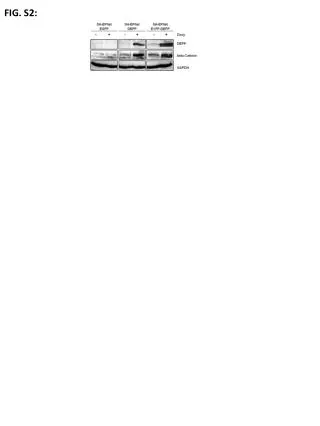
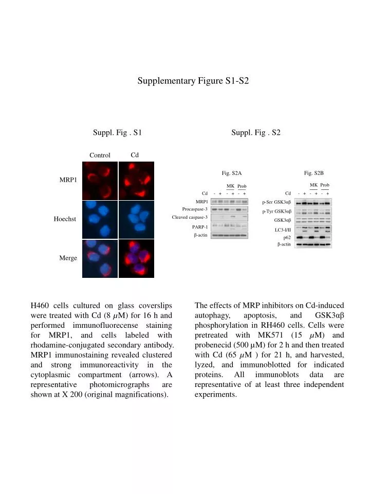
Supplementary Figure S1-S2 Suppl. Fig . S2 Suppl. Fig . S1 Cd Control Fig. S2B Fig. S2A MRP1 MK Prob MK Prob Cd - + - + - + Cd - + - + - + MRP1 p-Ser GSK3αβ Procaspase-3 p-Tyr GSK3αβ Cleaved caspase-3 Hoechst GSK3αβ PARP-1 LC3-I/II β-actin p62 β-actin Merge The effects of MRP inhibitors on Cd-induced autophagy, apoptosis, and GSK3αβ phosphorylation in RH460 cells. Cells were pretreated with MK571 (15 µM) and probenecid (500 µM) for 2 h and then treated with Cd (65 µM ) for 21 h, and harvested, lyzed, and immunoblotted for indicated proteins. All immunoblots data are representative of at least three independent experiments. H460 cells cultured on glass coverslips were treated with Cd (8 µM) for 16 h and performed immunofluorecense staining for MRP1, and cells labeled with rhodamine-conjugated secondary antibody. MRP1 immunostaining revealed clustered and strong immunoreactivity in the cytoplasmic compartment (arrows). A representative photomicrographs are shown at X 200 (original magnifications).
Supplementary Figure S3-S4 Suppl. Fig. S4 Hoechst Merge MRP1 Suppl. Fig. S3 N Control MRP * * * Mortality (% of control) N Cd → → MRP → → → → → → OA / Cd → → → Vanadate / Cd For Trypan blue assays, after treating cells with OA and Cd, floating and adherent cells were collected, centrifuged, and stained with 0.4% trypan blue for 5 min at room temperature. Numbers of trypan blue-positive (dead) and negative (alive) cells were counted on a hemocytometer under a microscope. Cell viabilities are expressed relative to those of untreated controls. Data were statistically analyzed by one-way ANOVA test. P < 0.05 Modulation ofMRP1 by p-Ser or p-Tyr GSK3αβ in Cd-treated RH460 cells. Cells cultured on glass coverslips were treated OA (5 µM), vanadate (300 µM) for 2 h and then further treated with Cd (65 µM) for 18 h, and performed immunofluorescence staining for MRP1 and labeled with FITC- conjugated secondary antibodies. Nucleus was stained with Hoechst 33342. White arrows indicate MRP1 localized in the cytoplasmic compartment. A representative photomicrographs are shown at X 200, original magnifications.
Supplementary Figure S5 Suppl. Fig. S5 Vector GSK3β Cd - + - + - + MRP1 Endogenous GSK3α → GSK3β-HA → → Endogenous GSK3β HA LC3- I/II β-actin H460 cells transduced with pcDNA3.1 and GSK3β-HA plasmid DNA (1 ug, each) were cultured for 48 h, and further treated with Cd (8 μM) for 12 h, harvested, lysed, and immunoblotted for indicated proteins. Data shown are representative of two independent experiments.
Supplementary Figure S6-S7 Suppl. Fig. S7 Suppl. Fig. S6 Control Cd Control Cd CathepsinD MRP1 Lamp-2 CathepsinB Hoechst Hoechst Merge Merge Colocalization of cathepsinB and Lamp-2 in Cd-treated RH460 cells. Cells cultured on glass coverslips were treated with Cd (65 µM) for 12 h, and performed immunofluorescence staining for cathepsinD and Lamp-2 (S6), or cathepsinB and MRP1(S7), and labeled with FITC (cathepsinD, MRP1)- or rhodamine (Lamp-2, cathepsinB) -conjugated secondary antibodies. Nucleus was stained with Hoechst 33342. A representative photomicrographs are shown at X 200, original magnifications.