Download
1 / 33
350 likes | 369 Views
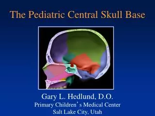
EE 137: MR Imaging Review of the Skull Base Foramina and Their Lesions. Brandon W. Sur MD, MSE Department of Imaging Sciences University of Rochester Medical Center. Disclaimer. The author has no financial disclosure or conflicts of interest with the presented material in this presentation.

E N D
EE 137: MR Imaging Review of the Skull Base Foramina and Their Lesions Brandon W. Sur MD, MSE Department of Imaging Sciences University of Rochester Medical Center
Disclaimer • The author has no financial disclosure or conflicts of interest with the presented material in this presentation.
Objectives • Review anatomical location of the skull base foramina with CT images • Review contents of each foramen • Learn various lesions affecting each foramen with exemplary MR images.
Cribriform Plate A C B Esthesioneuroblastoma: Axial (A), coronal (B), and sagittal (C) T1+C images show a heterogeneously enhancing mass in the nasal cavity destroying the cribriform plate and extending intracranially
Cribriform Plate D E F Olfactory groove meningioma: Sagittal (D), axial (E) , and coronal (F) T1+C images show an intensely enhancing dural based mass within the olfactory groove.
Optic Canal A Optic Glioma: Axial T2 (A) shows diffusely thickened right optic nerve in a patient with neurofibromatosis type I. Axial T1+C (B) shows no significant enhancement. B
Optic Canal C Optic nerve sheath meningioma: Coronal (C) and axial (D) T1+C images show enhancement along the left optic nerve and extending into the optic canal. D
Foramen Rotundum B A Nasopharyngeal Carcinoma: Coronal (A) and axial (B) T1+C images show an ill-defined, enhancing sinonasal mass extending through the right foramen rotundum.
Foramen Rotundum D C Skin Squamous Cell Carcinoma Metastasis: Coronal (C) and axial (D) T1+C images show enhancement along the left foramen rotundum.
Foramen Ovale A B Meningioma: Sagittal (A) and axial (B) T1+C images show a dural based enhancing mass extending through the right foramen ovale.
Stylomastoid Foramen A C B Adenoid cystic carcinoma: Coronal T1+C (C) image shows an enhancing right parotid gland mass extending intracranially through the stylomastoid foramen and destroying the temporal bone. Schwannoma: Axial (A) and coronal (B) T1+C images show enhancement along the facial canal and the stylomastoid foramen.
Internal Auditory Canal B A Schwannoma: Axial (A) and coronal (B) T1+C images show a heterogeneous, enhancing mass in the left cerebellopontine angle involving the left internal auditory canal.
Internal Auditory Canal D C Bilateral Acoustic Neuromas: Axial (C) and coronal (D) T1+C images of neurofibromatosis type II patient show bilateral enhancing masses in the bilateral internal auditory canals.
Jugular Foramen B A Glomus Tumor: Coronal (A) and axial (B) T1+C images show a heterogeneous, enhancing mass within the left jugular foramen.
Jugular Foramen E C Chondrosarcoma: Axial (C) and coronal (D) T2 images demonstrate a T2 hyperintense lesion in the left jugular foramen. Axial T1+C image (E) shows heterogeneous enhancement. D
Hypoglossal Canal Meningioma: Coronal T1+C (A) and axial T1+C (B) images show an enhancing, dural based mass occupying the foramen magnum and extending into the right hypoglossal canal. C A Schwannoma: Coronal T1+C (C) and axial T1+C (D) images show a dumbell-shpaed (not fully seen in these two images), enhancing mass within the right hypoglossal canal. D B
Hypoglossal Canal G E Persistent Hypoglossal Artery:Axial (E) and coronal (F) images of CT angiogram of the head show persistent hypoglossal artery within expanded left hypoglossal canal. F Dissection of Persistent Hypoglossal Artery: Axial image of CT angiogram of the head/neck (G) shows dissection of the persistent hypoglossal artery within the left hypoglossal canal.
Foramen Magnum D A B Meningioma Coronal (A), sagittal (B) and axial T1+C (C) images show an intensely enhancing, dural based mass occupying the foramen magnum. Schwannoma: Coronal T1+C (D) image of a patient with NF2 shows an enhancing mass within the foramen magnum extending down to the spinal canal. C
Conclusion • Understanding of the anatomy of the skull base foramina, knowledge of the characteristic and differential imaging findings of disorders affecting them, and reviewing pertinent clinical history will allow radiologists to formulate accurate diagnoses and present appropriate differential diagnostic considerations.
References • Boulton MR, Cusimano MD. Foramen magnum meningiomas: concepts, classifications, and nuances. Neurosurg Focus. 2006;14 (6): e10. • Fujita N, Shimada N, Takimoto H et-al. MR appearance of the persistent hypoglossal artery. AJNR Am J Neuroradiol. 1995;16 (4): 990-2. • Snyder WE, Shah MV, Weisberger EC et-al. Presentation and patterns of late recurrence of olfactory groove meningiomas. Skull Base Surg. 2011;10 (3): 131-9. • Vogl T, Brüning R, Schedel H et-al. Paragangliomas of the jugular bulb and carotid body: MR imaging with short sequences and Gd-DTPA enhancement. AJR Am J Roentgenol. 1989;153 (3): 583-7. • Vogl TJ. Differential Diagnosis in Head and Neck Imaging. Thieme. 1999