Download
1 / 85
970 likes | 1.64k Views
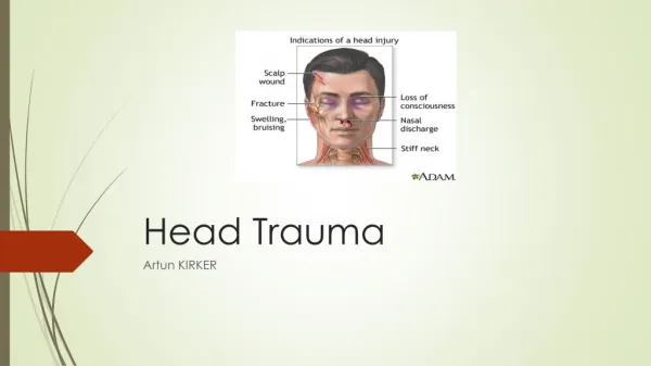
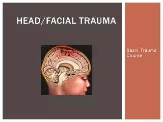
Basic Knowledge and Film Practices: Head Trauma. Nuttha Sanghan, M.D. Radiology Department Prince of Songkla University. Head Trauma. Imaging modalities Primary traumatic lesions Secondary effects. Imaging Modalities. Skull radiography Calvarial fractures Penetrating injuries

E N D
Basic Knowledge and Film Practices: Head Trauma Nuttha Sanghan, M.D. Radiology Department Prince of Songkla University
Head Trauma • Imaging modalities • Primary traumatic lesions • Secondary effects
Imaging Modalities Skull radiography • Calvarial fractures • Penetrating injuries • Radiopaque foreign bodies • Shift away from skull radiography toward CT scan AJNR 2007: 28; 1619-21.
CT Scan Faster Wide availablility Lack contraindication High accuracy Treatable lesions MRI Longer time Lesser availability Difficult monitoring More sensitive Parenchyma Extraparenchyma Lower accuracy Skull fracture Imaging Modalities AJNR 2007: 28; 1619-21.
Primary Traumatic Lesions • Skull and scalp lesions • Extraaxial hemorrhage Epidural hematoma (EDH) Subdural hematoma (SDH) Subarachnoid hemorrhage (SAH) • Intraaxial lesions Diffuse axonal injury (DAI) Cortical contusion Deep gray matter/brainstem injury
Skull and Scalp Lesions • Skull fractures Linear fracture Comminuted fracture Depressed fracture Compound fracture Diastatic fracture http://radiopaedia.org
Skull and Scalp Lesions “SCALP” • S : skin • C : connective tissue • A : [galea] aponeurosis • L : loose connective tissue • P : periosteum http://radiopaedia.org
Skull and Scalp Lesions Bilateral subgaleal hematoma http://radiopaedia.org
Skull and Scalp Lesions Acute and chronic cephalohematoma; a crescent-shaped soft-tissue mass in the periosteum of the left parietal and a partially calcified mass in the periosteum of the left parietal bone. The mass originated from a cephalohematoma. Radiographics2004; 24:1655–1674
Primary Traumatic Lesions Skull and scalp lesions Extraaxial hemorrhage Epidural hematoma (EDH) Subdural hematoma (SDH) Subarachnoid hemorrhage (SAH) Intraaxial lesions Diffuse axonal injury (DAI) Cortical contusion Deep gray matter/brainstem injury
Primary Traumatic Lesions • Extraaxial hemorrhage Epidural hematoma (EDH) Subdural hematoma (SDH) Subarachnoid hemorrhage (SAH)
Epidural Hematoma (EDH) • 1-4% craniocerebral trauma • Lucid interval 50% • Delay 10-30% 24-48 hours
Epidural Hematoma (EDH) Imaging • Displaced dura matter Certain diagnosis • Focal biconvex/lentiform configuration • Not cross suture • Cross dural attachments Falx cerebri Tentorium cerebelli
Epidural Hematoma (EDH) http://emedicine.medscape.com
Epidural Hematoma (EDH) • Arterial origin Laceration: fracture 85-90% Stretching Temporal/temporoparietal region: MMA • Venous origin Lacerated dural sinus Posterior cranial fossa
Epidural Hematoma (EDH) Classic arterial epidural hematoma; a lentiform, high attenuation collection adjacent to the right temporal lobe, caused by skull fracture with middle meningeal artery injury. Neurotherapeutics 2011; 8: 39-53.
Epidural Hematoma (EDH) Venous epidural hematoma; scalp soft tissue swelling with lentiform EDH and pneumocephalus indicated an associated skull fracture. CT venogram was obtained as the fracture line extended over the expected location of the right transverse sinus. The opacified transverse sinuses are patent with compression and displacement from the inner table by the EDH caused by injury to the transverse sinus. Neurotherapeutics 2011; 8: 39-53.
Epidural Hematoma (EDH) • Size variable • 2/3 hyperdense • 1/3 mixed hyper/hypodense • Swirl sign Low density: active bleeding Osborn
Epidural Hematoma (EDH) • Coronal reconstructions Vertex EDH http://radiopaedia.org
Epidural Hematoma (EDH) • Small EDH Not always differentiation SDH Classic shape Fracture
Epidural Hematoma (EDH) • Mortality 5% • Poor outcome Delayed diagnosis Posterior cranial fossa
Primary Traumatic Lesions Extraaxial hemorrhage Epidural hematoma (EDH) Subdural hematoma (SDH) Subarachnoid hemorrhage (SAH)
Subdural Hematoma (SDH) • 10-20% craniocerebral trauma • 30% fatal injuries • Lack trauma • Stretching & tearing of bridging veins • Asymptomatic-unconsciousness
Subdural Hematoma (SDH) Imaging • Crescent shape • Cross suture • Not cross dural attachment
Subdural Hematoma (SDH) • Supratentorial convexity • Posterior cranial fossa • Falx-tentorium Nonaccidental trauma http://www.taem.or.th
Subdural Hematoma (SDH) Acute (a few days) • Hyperdense 60% • Mixed 40% • Nearly isodense Coagulopathy Severe anemia http://www.neurosurgery.com.sg
Subdural Hematoma (SDH) Subacute (a few weeks) • Isodense Displaced GW interface Sulci-inner table • Contrast administration Cortical vessels Neomembrane http://radiopaedia.org
Subdural Hematoma (SDH) Chronic (months) • Hypodense • Neomembrane Capillary-rich • Rebleeding 10-40% Mixed density Fluid-fluid levels Septate-loculation • Calcification 1-2%
Subdural Hematoma (SDH) AJR 2011: 196; 166-73.
Subdural Hematoma (SDH) http://www.surgicalneurologyint.com
Epidural hematoma Fracture 85-95% Arterial/venous Biconvex Not cross suture Cross dura Subdural hematoma Stretching/tearing Bridging cortical veins Crescent Cross suture Not cross dura Differentiation
Primary Traumatic Lesions Extraaxial hemorrhage Epidural hematoma (EDH) Subdural hematoma (SDH) Subarachnoid hemorrhage (SAH)
Subarachnoid Hemorrhage (SAH) • Moderate to severe head trauma • Superficial sulci • CSF cisterns http://www.radswiki.net
Subarachnoid Hemorrhage (SAH) Common causes • Head trauma • Intracranial aneurysm • Perimesencephalic hemorrhage Less frequent etiologies • Arteriovenous malformation • Arterial dissection • Extension from intracerebral hemorrhage
Subarachnoid Hemorrhage (SAH) • Pseudo-subarachnoid hemorrhage Severe diffuse brain edema Relatively hyperdense vasculatures A 34-year-old man with cardiac arrest. A, On the first day, no abnormal finding is seen. B, On the 8th day, the brain shows diffuse low attenuation with obliteration of cisterns-sulci and narrowed ventricles. High-attenuation areas along sylvian fissures and tentorium cerebelli. C, On the 129th day, brain edema becomes more severewith more prominent high attenuation areas.
Subarachnoid Hemorrhage (SAH) • Pseudo-empty delta sign: SSS thrombosis Posterior parafalcine/interhemispheric SDH SAH around sinus AJR 2007; 189: 64-75.
Primary Traumatic Lesions Skull and scalp lesions Extraaxial hemorrhage Epidural hematoma (EDH) Subdural hematoma (SDH) Subarachnoid hemorrhage (SAH) Intraaxial lesions Diffuse axonal injury (DAI) Cortical contusion Deep gray matter/brainstem injury
Primary Traumatic Lesions • Intraaxial lesions Diffuse axonal injury (DAI) Cortical contusion Deep gray matter/brainstem injury
Diffuse Axonal Injury (DAI) • Shear-strain deformation • Acceleration/deceleration • Rotational force • Diffuse, bilateral http://www.givengain.com
Diffuse Axonal Injury (DAI) Locations • GW interface • Corpus callosum Posterior body Splenium • Dorsolateral upper brainstem • Sequentially deeper with increasing severity http://www.nursing-lectures.com
Diffuse Axonal Injury (DAI) Imaging • Initial normal/subtle • Nonhemorrhagic 80% Hypodense foci Site of shearing • Petechial hemorrhages 20-50% • Tip of the iceberg • Delayed scans with new lesions http://radiopaedia.org
Diffuse Axonal Injury (DAI) Diffuse axonal injury in a patient who was on anticoagulants: CT scan on day1 and day 2 http://radiopaedia.org
Diffuse Axonal Injury (DAI) Diffuse axonal injury: small hemorrhagic foci in the right parasagittal posterior frontal lobe and in the splenium of the corpus callosum Neurotherapeutics 2011; 8: 39-53.
Primary Traumatic Lesions Intraaxial lesions Diffuse axonal injury (DAI) Cortical contusion Deep gray matter/brainstem injury
Cortical Contusions • Second primary intraaxial lesions • Less initial loss of consciousness • Gray matter More vascularity Likely hemorrhage Variable size http://www.givengain.com
Cortical Contusions Locations Temporal lobe Petrous part Greater sphenoid wing Frontal lobe Cribiform plate-orbit Planum sphenoidale Lesser sphenoid wing http://www.nebraskabraininjurylawyer.com
Cortical Contusions Locations Cerebellum 10% Superior vermis Tonsils Inferior hemisphere Gliding contusion Parasagittal region emedicine.medscape.com
Cortical Contusions http://www.braininjury.com