Download
1 / 1
10 likes | 110 Views
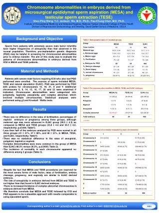
Investigating chromosomal abnormalities in embryos from severe male factor infertility cases undergoing ICSI with MESA, TESE, or EJAC sperm sources. Results show similar rates of aneuploidy but higher complexity in MESA embryos.

E N D
Chromosome abnormalities in embryos derived from microsurgical epididymal sperm aspiration (MESA) and testicular sperm extraction (TESE)Shao-Ping Weng, T.C. Jackson Wu, M.D., Ph.D., Pau-Chung Chen, M.D. Ph.D. Institute of Occupational Medicine and Industrial Hygiene, National Taiwan University College of Public HealthDivision of Reproductive Endocrinology and Infertility, David Geffen School of Medicine, UClLA Sperm from patients with extremely severe male factor infertility have higher frequencies of aneuploidy than that observed in the normal population. Therefore, pre-implantation genetic diagnosis (PGD) may be helpful in screening for chromosomal abnormalities prior to embryo transfer.The aim of this study was to evaluate the patterns of chromosome abnormalities in embryos derived from ICSI in MESA and TESE patients. Background and Objective Conclusions Table 1 Demographic data of 3 studied groups. Patients with severe male factors requiring ICSI who also had PGD performed were enrolled. The sources of sperm included MESA, TESE and natural ejaculation (EJAC). PGD was performed by FISH with probes for chromosomes 13, 18, 21, X and Y. Additional chromosome 8, 9, 14, 15, 16, 17, 20 and 22 were examined if indicated. Chromosome abnormalities were categorized into polyploidy, haploidy, aneuploidy, and complex abnormal, which involved multiple chromosomes. Statistical analyses were performed using χ2 and Kruskall - Wallis tests. Material and Methods Results p<0.001 Table 2 Chromosome abnormalities in MESA, TESE, and EJAC embryos. • There was no difference in the rates of fertilization, percentages of euploid embryos or pregnancy among three groups, although maternal age was more advanced in EJAC group (38.5 ± 4.3) as compared to MESA and TESE groups (33.8 ± 4.4 and 36.5 ± 4.0, respectively; p=0.004; Table1). • Less than half of the embryos analyzed by PGD were normal in all three groups (41 ± 31%, 37 ± 38%, and 48 ± 31%, in MESA, TESA, and EJAC, respectively; Table1) • There was no statistical difference in the rates of aneuploid, polyploid, haploid or euploid. • Complex Abnormalities were more common in the group of MESA than EJAC (48.3% versus 26.5%, p=0.0005; Table 2). • The incidence of normality in each chromosome appeared no difference among 3 groups (Table 3). p=0.004 Table 3 the incidence of normality evaluated by each chromosome. • Despite the fact that MESA and TESE procedures are reserved for the most severe forms of male factor, rates of fertilization, embryo cleavage, pregnancy, and euploidy are similar to EJAC derived embryos. • The rate of aneuploidy in embryos derived from MESA and TESE is not higher than that found in EJAC derived embryos. • There is increased incidence of complex abnormal chromosomes in embryos derived from MESA. • We therefore conclude that MESA and TESE followed by ICSI and PGD appears to be a plausible approach with results comparable to using ejaculated sperm. Corresponding author’s e-mail: pchen@ntu.edu.tw; First author’s e-mail: r95841021@ntu.edu.tw OMIH