Download
1 / 6
60 likes | 65 Views
N-Glycans will be obtained from the free protein of hydraxinolysis or exhaustive digestion with protease. It helps to remove all amino acids. The released N-Glycans can be purified by conventional ion-exchange and High-pressure liquid chromatography (HPLC) method.<br>

E N D
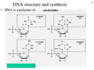
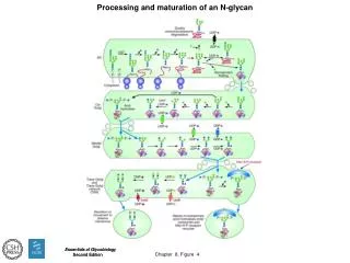
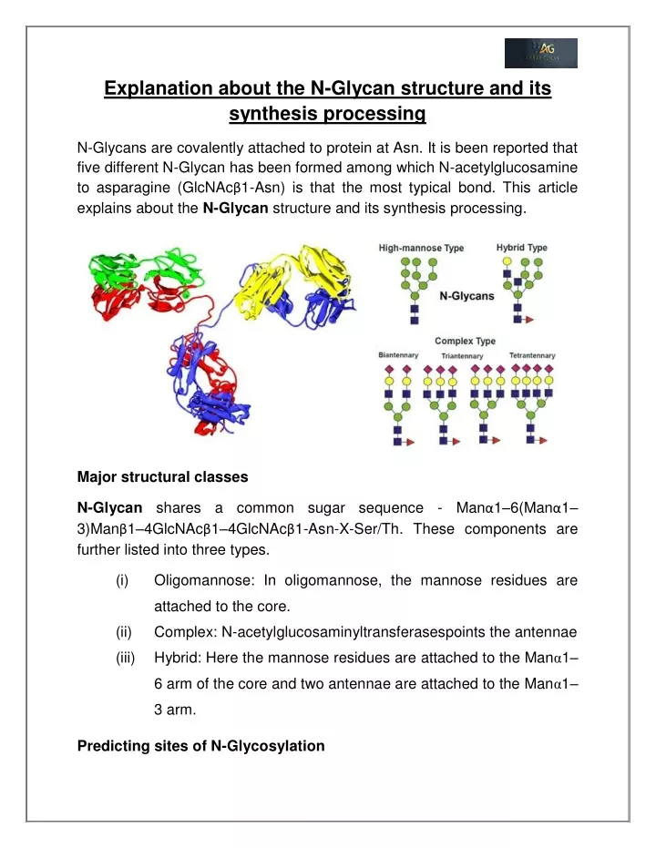
Explanation about the N-Glycan structure and its synthesis processing N-Glycans are covalently attached to protein at Asn. It is been reported that five different N-Glycan has been formed among which N-acetylglucosamine to asparagine (GlcNAcβ1-Asn) is that the most typical bond. This article explains about the N-Glycan structure and its synthesis processing. Major structural classes N-Glycan shares a common sugar sequence - Manα1–6(Manα1– 3)Manβ1–4GlcNAcβ1–4GlcNAcβ1-Asn-X-Ser/Th. These components are further listed into three types. (i) Oligomannose: In oligomannose, the mannose residues are attached to the core. (ii) Complex: N-acetylglucosaminyltransferasespoints the antennae (iii) Hybrid: Here the mannose residues are attached to the Manα1– 6 arm of the core and two antennae are attached to the Manα1– 3 arm. Predicting sites of N-Glycosylation
N-Glycans exist on various secreted membrane-bound glycoprotein at Asn- X Ser/Thr sequins. Analyses haverevealed that two-third of the entries contain consensus sequence in which 2/3 of the sequons are likely to be N- glycosylated. N-Glycans occur neither on cytoplasmic proteins nor on the cytoplasmic portions of membrane proteins. The shift of N-Glycans to Asn-X-Ser/Thr sequins occurs on the luminal side of the endoplasmic reticulum(ER) membrane. The presence of Asn-X-Ser/Thrsequon is necessary for the receipt of N- Glycan. Also, it is identified that “X” may reduce the efficiency of glycosylation when Asn-X-Ser/Thr is present in a deduced amino acid sequence by cDNA. These sequences are referred to as potentialN- Glycan Sites. The membranes of the ER membrane are exposed to the ER lumen and other portions in the membrane and other regions to the cytoplasm. The domains that are accessible to the ER lumen receive an N-Glycan. The glycoprotein lack of Trans membrane will also receive N-Glycan contritely. Purification and Analysis Process
N-Glycans will be released using a bacterial enzyme called peptide from asparagines. This enzyme will remove oligomannose, complex, and hybrid- glycans attached to the asparagine. Bacterial enzymes called PNGase A and PNGase F will remove the structures of N-Glycan. These enzymes further remove nitrogen of asparagines which is attached to N-Glycan. Thereby it converts asparagine to aspartate. N-Glycans will be obtained from the free protein of hydraxinolysis or exhaustive digestion with protease. It helps to remove all amino acids. The released N-Glycans can be purified by conventional ion-exchange and High-pressure liquid chromatography (HPLC) method. Synthesis of N-Glycans The biosynthesis of N-Glycans begins at the cytoplasmic face of the ER membrane with the shift of GlcNAc-P from UDP-GlcNAc to the lipid dolichol phosphate (Dol-P) to generate acetylglucosamine (Dol-P-P-GlcNAc). In this Dol-P process, fourteen sugars are sequentially added to an Asn-X- Ser/Thrsequon in a protein. This is being synthesized and passed through the ER membrane. The N-Glycan protein model is remodelled in the ER and Golgi by a series of glycosyltransferases. dolichol pyrophosphate N-
Most of the enzymes are exquisitely sensitive to the biochemical state of the cell in which glycoprotein is impressed. Thus the cell type during a glycoprotein is solely depended on the population of sugars attached to every glycosylated asparagines in an exceedingly mature glycoprotein. It also depends on the physiological status of the cell during the development of the altered disease. Maturation of N-Glycans The sugar additions occurring in the trans-Golgi convert the limited repertoire of hybrid N-Glycans into an extensive array of mature complex N-Glycans.Thus the bio-synthesis processes are further classified into three components. (i) (ii) Sugar additions to the core Elongation of residues by sugar additions in the branching N- acetylglucosamine Decoration or capping of elongated branches. (iii) Summary of detailed processing
Addition: In the vertebrate, N-Glycans are the main core modification of fucose in α1–6 linkage to the N-acetylglucosamine. In invertebrate Fucosylation, proteins occur on N- acetyl glucosamine but here the fucose is added in α1–3 and/or α1–6 linkages. The acetylglucosamine. It is commonly notable in plant and glycoprotein. In addition to this xylose is linked to the β-mannose of the core. This xylosyltransferase requires the action of GlcNAcT-I where Xylose has not been detected in N-Glycans. fucosyltransferases transfer the fucose to the core N- Elongation: In the elongation residue process, the elongated branches that are made by the addition of β-linked galactose residue produce the ubiquitous building block Galβ1-4GlcNAc by initiating N-acetylglucosamine is referred to as type-2 N-acetylglucosamine. Antennae: The complex N-Glycan has two antenneintiated by the addition of dual N-acetylglucosamine residues. Here the antennae are further extended by the sequential addition of N-acetylglucosamine and galactose residues results in LacNAc (-3Galβ1–4GlcNAcβ1-)n or type-2 N- acetylglucosamine. Additional branches can be initiated at C-4 of the core mannoseα1–3 and C-6 of the core mannose α1–6 are used to yield tri- and tetra-antennary N- glycans.
In the variation, β-linkedN-acetylgalactosamine is added to N- acetylglucosamineinstead of galactose will result in antennae with a GalNAcβ1–4GlcNAc (“LacdiNAc”) extension. Capping: The capping or decoration reaction involves the addition of sialic acid, galactose, fucose, N-acetylglucosamine and sulfate to the branches. Capping sugars are α-linked and are protrude away from the β-linked ribbon-like poly-N-acetylgalactosamine branches to facilitate the presence of terminal sugars to antibodies and lectins. Most of these structures are being shared by N- Oglycans and glycolipids. The various process involved potentially will yield complex N-Glycans that differ in length, composition, capping arrangements, branch number, and core modifications. To know more about: www.arrayglycan.com