Download
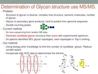
1 / 24
340 likes | 710 Views
N-Glycan Analysis. Jake S. Yang. Center for Biomarker Discovery and Translation. Oct 25, 2013. Glycosylation play crucial roles . Glycosylation is the most abundant posttranslational modification (PTM) and glycans are most structurally diverse;

E N D
N-Glycan Analysis Jake S. Yang Center for Biomarker Discovery and Translation Oct 25, 2013
Glycosylation play crucial roles • Glycosylation is the most abundant posttranslational modification (PTM) and glycans are most structurally diverse; • More than 50% of all proteins have been modified by glycans; • Glycoforms are depending upon many factors which are related to both gene expression and cellular metabolism.
Molecular markers are glycoproteins [D. Sidransky, Nat. Rev. Cancer 2002, 2, 210-219]
Diverse glycosylation • Individual glycosylation sites on the same protein contain different glycan structures • Reflect cell type and status • Same protein have different glycan structures in different organs (e.g., membrane protein Thy-1 in brain vs. lymphocytes, Rudd and Dwek, 1997) • Changes in peptide sequence or structure could alter the types of glycan structures attached • The robust and high-throughput techniques are needed to understand the roles of glycans in biological activities.
Technology Innovation glycans Glycoprotein Immobilization for Glycan Extraction (GIG) Reversible HydrazoneSolid-Phase Extraction (rHSPE) Sialic Acid Modification Glycan profiling by GIG-chipLC Quantitative glycomics (iARTs) glycoproteins Carbohydrates and Glycobiology, Science, Vol.291, No. 5512, pp. 2263-2502. Glycan isolation, tissue imaging Nat. Biotechnol. 2003, 21, 660-666. Anal. Chem. 2012, 84 (5), 2232-2238. Proteomics Clin. Appl. 2012, 6, 596-608. Anal. Chem. 2013, 85, 5555-5561. Anal. Chem. 2013, 85, 3606-3613. Anal. Chem. 2013, 85, 8188-8195. Anal. Chem. 2013, 85, DOI: 10.1021/ac4013013. Glycan chip imaging
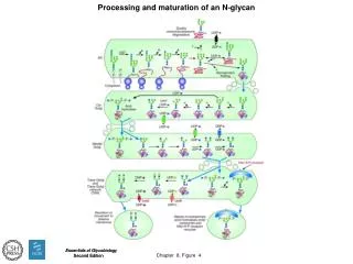
N-glycan workflow Sample (protein extraction from tissue or cell) Buffer exchange (amine-free) rHSPE (glycan reducing-end capture) GIG (solid-phase) (protein immobilization) modification Solid-phase Sialic acid (modification and quantitation) quantitation On beads (glycan capture) On slide (glycan imaging) iARTs (isobaric quantitation) separation Detection (MALDI-MS) chipLC (microchip) detection Detection (MALDI or ESI - MS)
Current methods • Glycan extraction • Potential issues • Non-specific binding • Sample loss (affinity; multiple purification) • Difficulty to removal of reagents after derivatization (sialic acid modification: reagents severely interfere glycan ionization) Carbo Enzyme C18/C8 modify Carbo MS S. Yang and H. Zhang, Proteomics Clin. Appl. 2012, 11-12, 596-608
GIG (chemoselective method) Aldehyde beads Glycoprotein Immobilization for Glycan Extraction (GIG)1 MS immobilize modify2 enzyme wash -elimination3 MS Immobilization on solid-phase: Immobilization in pH 10 on N-terminus and lysine 1S. Yang et al., Anal. Chem. 2013, 85(11), 5555-5561. 2P. Shah et al., Anal. Chem. 2013, 85 (7), 3606-3613. 3G.J. Rademaker et al., Anal. Biochem. 1998, 257, 149-160.
Complex sialic acids About 50 different sialic acids known [Schauer, 2009]
On GIG: glycan modification and extraction N-glycan modification on solid-phase O-glycan -elimination [S. Yang et al., Anal. Chem. 2013, 85(11), 5555-5561.]
On GIG: sialic acid isotope quantitation Demonstration of sialylated N-glycan isotope labeling by mixing 1:1 light to heavy (p-toluidine) P-toluidine amidation EDC @pH 4.5 – 5.5 Sialic acid [P. Shah et al., Anal. Chem. 2013]
GIG integration on a microchip A interface C B [S. Yang, S. ToghiEshighi, H. Chiu, D.L. DeVoe, and H. Zhang, Anal. Chem. 2013, DOI: 10.1021/ac4013013]
Microchip implementation 2). Union and capillary installation 1). Needle insertion 1 2 3 4 3). AminoLink bead packing 4). Graphitized carbon packing
GIG-chipLC operation 1) Protein capture and glycan release 2) Glycan separation • Cap needle C • Inject proteins from needle B • Conjugate proteins to AminoLink beads • Release glycans and wash column • Cap needle B, go to 2) • Cap needle of B and up-cap C • Wash column through needle A • Elute glycans to needle C • Analyze elution by MS
GIG-chipLC: mouse glycan analysis • Experimental procedure • Isolation of glycans using GIG • Modification of sialic acids on beads • Separation of N-glycans using porous graphitized carbon • Profiling of N-glycans by Shimadzu Resonance MALDI-MS • Analyze glycans of mouse heart tissue and blood serum
Identification of glycans without chipLC • Abundant oligomannoses are observed on mouse tissue • Sialylated N-glycans are observed in mouse blood serum • Less number of N-glycans are expected without LC separation ( # of N-glycans < 50)
GIG-chipLC reproducibility • The majority of N-glycans are eluted in respective same fraction. • Isomers of N-glycans are observed by porous graphitized carbon. • Has advantages using microfluidics • High-throughput, low sample and reagent consumption, fast analysis, and flexible interfacing Mouse blood serum, 400 g of serum proteins Mouse blood serum, 200 g of serum proteins
Mouse N-glycan profiling • Detected unique unsialylated N-glycans in tissue only • Observed mature and sialylated structures from tissue and serum • Demonstrated GIG-chipLC as a simple and robust platform for glycomic analysis Mouse tissue Mouse serum 7 65 31 [S. Yang et al., manuscript under review, 2013]
Glycan quantification • Glycan quantification is essential for determination for both fundamental studies of biological activities and biomarker identification [J. Zaia, Chem. Biol. Rev. 2008] • A current challenge in the field of glycomics is to determine how to quantify changes in glycan expression between different cells, tissues, or biological fluids [J.A. Atwood III, R. Orlando et. al, J. Proteome Res. 2007] • MS-based quantification methods include isotope and isobaric labeling • Isotope: pair-wise measurement, increasing MS complexity • Isobaric: concurrent measurement, improving throughput and sensitivity
GIG-iARTs iARTs GIG [S. Yang et al., Anal. Chem., 2013 (accepted)
Improved sensitivity and quantification 15 N-glycans identified, 17 others confirmed as glycans gp120
Summary • A novel method (GIG) is developed for solid-phase glycan isolation and modification. • GIG improves specificity and facilitates glycan modification with minimizing sample loss using covalent immobilization. • Glycan structure can be enzymatically analyzed on GIG. • GIG-chipLC is the high-throughput platform for glycomic analysis from complex biological samples. • Isobaric labeling could quantify glycans for clinical application. GIG: a robust technique for glycomic analysis
Acknowledgements • Dr. Don DeVoe • Dr. Shuwei Li • All members of CBDT • Dr. Hui Zhang • Dr. Daniel Chan • Dr. Lori Sokoll • Dr. Zhen Zhang • Dr. Scott Kuzdzal • Brian Field • Dr. Jennifer Van Eyk • Sarah Parker Funding National Institute of Health National Heart, Lung and Blood Institute (NHLBI) Programs of Excellence in Glycoscience (PEG) With Prof. Hart National Cancer Institute The Early Detection Research Network (EDRN) Clinical Proteomic Tumor analysis Consortium (CPTAC) • Dr. David Graham • David Colquhoun • Dr. Kevin Yarema