Download
1 / 27
270 likes | 428 Views
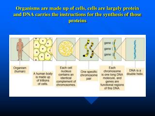
Synthesis and Degradation of Nucleotides Part 2: September 2 nd , 2009. Champion CS Deivanayagam Center for Biophysical Sciences and Engineering University of Alabama at Birmingham Birmingham, AL 35294-4400. Recollection’s from yesterday’s lecture.

E N D
Synthesis and Degradation of Nucleotides Part 2: September 2nd, 2009 Champion CS Deivanayagam Center for Biophysical Sciences and Engineering University of Alabama at Birmingham Birmingham, AL 35294-4400
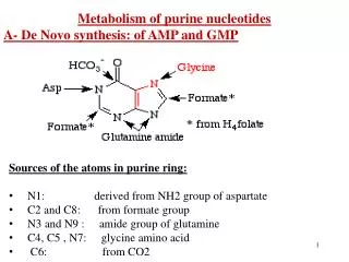
Recollection’s from yesterday’s lecture 1. The purine ring is built on a ribose-5-P foundation through 11 steps to get IMP 2. GMP and AMP are derived from IMP
Today’s lecture will concentrate on Pyrimidine synthesis and catabolism Gylocosidic bond Gylocosidic bond Note that the numbering are slightly different and note where the glycosidic bonds are attached
What do you need to learn from this lecture ? 1. What are the Committed steps that are unique in this synthesis cycle 2. What are the different feed back inhibition steps in this synthesis cycle 3. What steps can be utilized to develop inhibitors in this synthesis cycle 4. What are some of the diseases that are related to this synthesis cycle
De novo pyrimidine synthesis: • In contrast to purines, pyrimidines are not synthesized as nucleotides • Rather, the pyrimidine ring is completed before a ribose-5-P is added • Carbamoyl-phosphate and aspartate are the precursors of the six atoms of the pyrimidine ring • Mammals have two enzymes for carbamoyl phosphate synthesis – carbamoyl phosphate for pyrimidine synthesis is formed by carbamoyl phosphate synthetase II (CPS-II), a cytosolic enzyme
In bacteria, six enzymes catalyze the reactions to form the pyrimidine ring In mammals, these are encoded in three protein: a. CPS-II, aspartatetranscarbomylase and dihydrorotate are in a 210 kDacytosolic polypeptide b. DHO dehydrogenase is a separate enzyme associated with the outer surface of the inner mitochondrial membrane c. Orotatephosphoribosyltranferase and OMP carboxylase are encoded on a single cytosolic polypeptide known as UMP synthase
The advantages of multifunctional enzymes: The enzymatic activities are catalyzed by single polypeptide chains in mammals. The advantages are: The product of one reaction in a pathway is the substrate for the next, and the product remains bound and are channeled directly to the next active site rather than disassociated into the surrounding medium for diffusion to the next active site. Transit time for movement from one active site to the next is shortened Substrates are not diluted into the solvent phase Chemically reactive intermediates are protected from decomposition into aqueous mileu No pools of intermediates accumulate and Intermediates are shielded from interactions with other enzymes that might metabolize them
A comparison of the regulatory circuits that control pyrimidine synthesis in E. coli and animals. E.ColiATCase: Feebackinhibitied by the end product CTP ATP is an allosteric regulator CTP and ATP compete for a common allosteric site. CPS II in mammals:UDP and UTP are feed back inhibitors PPRP and ATP are allosteric regulators
How Are Pyrimidines Degraded? • In some organisms, free pyrimidines are salvaged and recycled to form nucleotides via phosphoribosyltransferase reactions • In humans, however, pyrimidines are recycled from nucleosides, but free pyrimidine bases are not salvaged • Catabolism of cytosine and uracil yields -alanine, ammonium ion, and CO2 • Catabolism of thymine yields -aminoisobutyric acid, ammonium ion, and CO2
How Do Cells Form the Deoxyribonucleotides That Are Necessary for DNA Synthesis? • In most organism NDP’s are the substrates fordeoxyribonucleotide formation. • Reduction at 2'-position commits nucleotides to DNA synthesis • Replacement of 2'-OH with hydride is catalyzed by ribonucleotidereductase • Three classes of ribonucleotidereductases differ in their mechanisms of free radical generation The enzyme system for dNDP formation consists of four proteins: Two constitute the riboneuclotidereductase Other two are Thioredoxin and Thioredoxinreductase
E. Coli RibonucleotideReductase Has Three Different Nucleotide-Binding Sites • An 22-type enzyme - subunits R1 (86 kD) and R2 (43.5 kD) • R1has two regulatory sites, a specificity site and an overall activity site • Activity depends on Cys439, Cys225, and Cys462 on R1 and on Tyr122 on R2 • Cys439 removes 3'-H, and dehydration follows, with disulfide formation between Cys225 and Cys462 • The net result is hydride transfer to C-2' • Thioredoxin and thioredoxinreductase deliver reducing equivalents
R1 homodimer carries two type of regulatory sites in addition to the catalytic site Catalytic site binds substrates: ADP, CDP, GDP and UDP One regulatory site binds: ATP, dATP, dGTP or dTTP Depending on which one of the nucleotides is bound there determines which NDP is bound at the catalytic site Other regulatory site binds: ATP (the activator) or dATP (the negative effector) Overall activity site that determines whether the enzyme is active or inactive The 2 Fe atoms within the single active site formed by the R2 homodimers generate the free radical required for ribonucleotide reduction on a specific R2 residue, Tyr 122. This in turn generates the thiyl free radical (Cys-S·) on Cys439. Cys439-S· initiates ribonucleotide reduction by abstracting the 3’ H from the ribose ring of the nucleoside diphosphate substrate and form s a free radical on C-3’. Subsequent dehydration forms the deoxyribonucleotide product
RibonucleotideReductase Uses a Free Radical Mechanism Cys residues undergo reversible oxidation-reduction between (-S-S-) and (-SH-SH-) In their reduced form serve as electron donors to regenerate the reactive –SH pair in the active site The sulfhydryls of thioredoxinreductase, mediates the NADPH-dependent reduction of thioredoxin.
RibonucleotideReductase is Regulated by Nucleotide Binding Regulation of deoxynucleotide biosynthesis: the rationale for the various affinities displayed by the two nucleotide-binding regulatory sites on ribonucleotidereductase.
How Are Thymine Nucleotides Synthesized? • Cells have no requirement for free thymine ribonucleotides and do not synthesize them • dUDP anddCDP lead to the formation of dUMP the immediate precursor for dTMP synthesis • Interestingly, formation of dUMP from dUDP passes through dUTP, which is then cleaved by dUTPase, a pyrophosphatase that removes Ppi from dUTP. • The action of dUTPase prevents dUTP from serving as a substrate in DNA synthesis. • An alternative route to dUMP formation starts with dCDP, which is dephosphorylated to dCMP, and then deaminated by dCMPdeaminase yielding dUMP.
dCMPDeaminase Provides an Alternative Route to dUMP TrimericdCMPdeaminase. Each chain has a bound dCTP molecule (purple) and a Mg2+ ion (orange). An alternative route to dUMP is provided by dCDP, which is dephosphorylated to dCMP and then deaminated by dCMPdeaminase. It is allosterically activated by dCTP and feedback inhibited by dTTP. Only dCTP does not interact with either regulatory sites on ribonucleotidereductase. Instead it acts upon dCMPdeaminase.
Synthesis of dTMP from dUMP is catalyzed by thymidylatesynthase Thymidylatesynthasedimer. Each monomer has a bound folate analog (green) and dUMP (light blue). • ThymidylatesynthasemethylatesdUMP at 5-position to make dTMP • N5,N10-methylene THF is 1-C donor • Once again folate derivatives are used as inhibitors to disrupt DNA synthesis similar to the purine synthesis.
Fluoro-Substituted Analogs as Therapeutic Agents Carbon-fluorine bonds are extremely rare in nature, and fluorine is not common in nature. Moreover, F is electronegative and relatively unreactive. Thus fluoro-substituted agents are often potentially useful drug candidates. Shown here is the effect of 5-fluoro substitution on the mechanism of action of thymidylatesynthase. The ternary complex is stable and prevents further enzyme turnover. 5-Fluorouracil is a thymine analog. It is converted to 5'-fluorouridylate by a PRPP-dependent phosphoribosyltransferase and passes through the reactions of dNTP synthesis, becoming 2'-deoxy-5-fluorouridylic acid, a potent inhibitor of dTMPsynthase. 5-Fluorocytosine is an antifungal drug, and 5-fluoroorotate is an anti-malarial drug.