Download
1 / 21
230 likes | 427 Views
Somite Derivatives: Muscle and Bone Formation. Gilbert - Chapter 14. Today’s Goals. Become familiar with the mesodermal sub-populations Examine somite maturation, muscle and bone formation. Muscle and Bone Formation. Which compartment of the somite will give rise to muscle?

E N D
Somite Derivatives: Muscle and Bone Formation Gilbert - Chapter 14
Today’s Goals • Become familiar with the mesodermal sub-populations • Examine somite maturation, muscle and bone formation
Muscle and Bone Formation • Which compartment of the somite will give rise to muscle? • Which compartment of the somite will give rise to bone?
Myogenesis • The generation of muscle cells • Come from 2 cell lineages within the myotome • Primaxial - don’t mix with Lateral Plate Mesoderm • Abaxial - Do mix with Lateral Plate Mesoderm • Muscles are induced to form by paracrine factors (ex. WNT’s, BMP’s)
Myogenic bHLH proteins (Basic Helix Loop Helix) • Transcription factors in MRF family (myogenic regulatory factors) • All bind similar sites on DNA, activate muscle genes • Expressed only in muscle cells/precursors • Sufficient to specify/commit a cell to the muscle lineage • A variety of cells types in culture transfected with myf-5 or myoD will become muscles
Muscle cell fusion • Muscle tissue is multinucleate • Myoblasts (muscle precursors) fuse to form multinucleate myotubes • At this point, cells are differentiated • Become organized into a muscle fiber
Differentiation of Myotubes • In order to begin differentiation, myoblasts must stop proliferating (dividing) • Depletion of certain growth factors allows myoblasts to exit the cell cycle and differentiate • If growth factors are present, myoblasts will continue to proliferate
Muscle Cell “Regeneration” in response to injury • Muscle cells are differentiated - cannot divide to replace themselves after injury • As in many tissues, replacement of lost tissue comes from stem cell populations associated with that tissue type • Satellite cells - found by basement membrane of muscle fibers • Not well understood
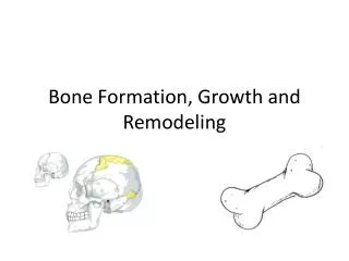
Bone Formation • In vertebrates, 3 systems must form • Craniofacial bones • Axial Skeleton • Appendicular Skeleton
Anatomy of the Musculoskeletal System of Jawed Vertebrates The locomotor anatomy is composed of 2 systems: Axial: Vertebrae, Ribs, Associated musculature Appendicular: Paired appendages, Pelvic and pectoral Girdles, Associated musculature Must work together for an organism to function properly. Skeleton Musculature
Bone formation • 3 lineages of bone structures • Somites: axial structures (vertebrae, ribs) • Scapula? • Lateral plate mesoderm: appendicular structures (limbs) • Scapula • Cranial neural crest: craniofacial bones
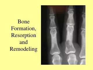
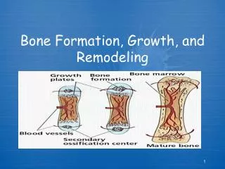
Bone Formation (Osteogenesis) • 2 major modes • Intramembranous (dermal) ossification • Endochondral ossification
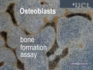
Intramembranous (dermal) ossification • Directly convert mesenchyme into bone • Involves osteoblasts, osteoclasts • Typical of skull formation
Endochondral ossification • Convert mesenchyme to cartilage first, then converts to bone • Typical of the formation of vertebrate ribs, limbs • Involves chondrocytes (make cartilage), osteoblasts (make bone)
Zebrafish Development - Day 2 • Draw 3 embryos, label structures • Use lab printout and poster to identify structures and stage of embryonic development • When finished examine cleared and stained chick embryo - stained for cartilage • Remove marbles from fish tank