Download
1 / 41
420 likes | 631 Views
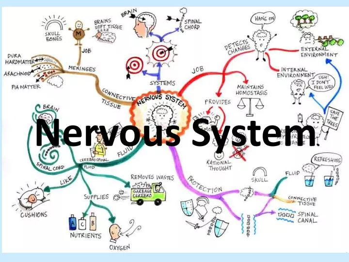
Nervous System. Sodium-Potassium Pump. Resting Potential . The sodium potassium pump pumps out and into the axon in unequal amounts This results in an excess of charge (Na+) outside and an excess of charge (organic ions) inside the axomembrane

E N D
Resting Potential • The sodium potassium pump pumps out and into the axon in unequal amounts • This results in an excess of charge (Na+) outside and an excess of charge (organic ions) inside the axomembrane • An electrical potential difference of - millivolts is generated • To maintain the resting potential of resting neurons, is continually needed to supply to the sodium-potassium pumps as sodium ions and potassium ions tend to or diffuse across the • potassium tends to out faster than the sodium back in, and the axoplasm contains large organic negative that contribute to the negative charge inside the
Action Potential • If nerve is stimulated by electric , pH change, stimulation, a nerve impulse is generated, and there is a in potential • The action potential has three phases: , , and a recovery period
Action Potential - Depolarization • A nerve is • The sodium open to allow Na+ to into axon through a channel so the Na+ ions rush from the outside of the to the inside • This changes the across the neuron from - (resting potential) to + • The is now charged compared to the outside of the neuron. Note: during the depolarization phase, there is of sodium ions
Action Potential - Repolarization • potassium gated channels open and + ions rush to the outside of the axomembrane via the • This makes the of the membrane positively charged relative to the inside once • The across the membrane returns from +40mV back to - Note: during the phase, there is facilitated diffusion of ions
Action Potential – Recovery (Refractory) Period • follows • a neuron can not an action potential during this phase since the sodium cannot open • the sodium-potassium pump is busy the resting potential by pumping sodium ions out and ions back in through the • recovery period the action potential from moving backwards • Resting potential is re-established
Threshold Stimulus • Action potentials occur only when the membrane is stimulated (depolarized) enough so that sodium channels open completely. The minimum stimulus needed to achieve an action potential is called the . • If the membrane potential reaches the (generally 5 - 15 mV less negative than the resting potential), the voltage-regulated sodium channels all open. Sodium ions rapidly diffuse inward, & depolarization occurs. All-or-None Law - action occur maximally or not at all. In other words, there's no such thing as a or action potential. Either the threshold potential is reached and an action potential occurs, or it isn't and no potential occurs.
Transmission of an Impulse from Neuron to Neuron What happens to a nerve impulse once it reaches the end of an axon? How does one nerve communicate with another? The answer lies in the specialized regions at the ends of axons called SYNAPSES. • Synapse: the region end of an axon and the body or dendrite to which it is attached. • Synaptic Endings: swollen knobs on the ends of axon terminal branches. • Presynaptic Membrane: the membrane of the axon synaptic ending. • Postsynaptic Membrane: the membrane of the next neuron just beyond the axon's synaptic membrane. • Synaptic Cleft: the space between the and the postsynaptic membranes • Neurotransmitter Substances ( ): chemicals that transmit the nerve impulses across a synaptic cleft. • Synaptic Vesicles: contain the neurotransmitters. Contained near surface of synaptic endings. • (Ach), Noradrenalin (NA), Serotonin, Adrenalin (epinephrine) are some important neurotransmitters.
Sequence of Events • Nerve (action potential) travels along axon, and a synaptic ending. • Arrival of the action potential causes calcium ion gates on the to open and calcium ions move into the . • The rise in in the axon bulb causes synaptic vesicles containing to move towards the inner surface of the presynaptic membrane • Synaptic vesicles merge the presynaptic membrane and of neurotransmitters (Ach) into the synaptic cleft occurs • Recall that endocytosis requires ATP energy. The axon bulb contains many to produce
Sequence of Events • Neurotransmitters diffuse synaptic cleft to on membrane. The receptors control selective ion channels; binding of a to its specific receptors opens the ion channels. • The resulting ion flux changes the of the postsynaptic membrane. One of the following can occur: • an synapse: moves the membrane voltage closer to the ‘ voltage’ required for an action potential and the excitatory neurotransmitters cause sodium ions to move through receptor depolarizing the membrane • nerve impulse will be down the dendrite of the second neuron • Examples of excitatory neurotransmitters: ACETYLCHOLINE (ACh), (epinephrine) • an : Inhibitory neurotransmitters do not depolarize the postsynaptic membrane • Examples of inhibitory neurotransmitters: GABA (gamma aminobutyricacid)
Sequence of Events • To prevent stimulation or inhibition of the postsynaptic membrane, are broken down by enzymes or are reabsorbed through the presynaptic by (also requires ATP energy) • neurotransmitter is degraded by enzymes e.g., breaks down acetycholine • synaptic ending reabsorbs the neurotransmitter e.g. this is what happens to This results in only a single stimulus and propagation of the impulse
Divisions of the Nervous System • Central Nervous System: (CNS) - includes spinal cord and brain. In the "center" of the body. • Peripheral Nervous System: (PNS) - the rest of the nervous system:
The Central Nervous System • The central nervous system is made up of the and cord. • The brain is protected by the and the spinal cord is protected by the of the column • The brain and spinal cord are by and fluid that protects the CNS • The brain • contains approximately billion neurons (nerve cells) and between 1.3 and 1.4 kgs in the average adult human. • The function of the brain is to information from other areas of the central nervous system and the nervous system via the spinal cord. • Thespinal cord • is about 45 cm long and weighs about 35-40 grams. • The function of the spinal cord is to from the brain to the peripheral nerves and from peripheral to the brain. • The spinal cord also reflex arcs or involuntary responses that the brain
The Peripheral Nervous System • The peripheral nervous system is made up of the and (groups of cell bodies) that are found outside of the CNS • Recall, sensory neurons transmit impulses from the PNS to the and motor neurons transmit impulses from the to the . • Peripheral nerves that communicate directly with the brain are called (12 pairs - connect sensory in nose, eyes, ears, tongue, etc.) • Peripheral nerves that communicate with the brain via the spinal cord are called (31 pair - muscles of the body and various glands and organs) • The PNS is broken down into other divisions. • nervous system and nervous system. • autonomic nervous system (ANS) is further divided into sympathetic and parasympathetic branches.
Somatic Nervous System • peripheral that receive and send to skeletal muscle, skin, and tendons • Sensory in skin, muscle and send information to the CNS about body position and conditions • The CNS relays to motor neurons that control the of skeletal muscle and the movement of the body. • The somatic nerves voluntary movements of the body such as walking, jumping, writing, typing, etc. Somatic nerves also reflex actions that skeletal muscles as the effector, such as when you touch sharp or hot.
Autonomic Nervous System • Controls involuntary to stimuli by the body. • Autonomic nerves serve muscle, smooth muscle, glands, and all of the organs. • The ANS acts on these effectors to maintain homeostasis within the body (parasympathetic branch - neurotransmitter) and respond to stress (sympathetic branch - neurotransmitter).
Reflex Arc • Reflex actions are responses to a stimulus and are a part of the nervous system. • These actions do not involve the cortex (conscious brain) only the cord and are faster than actions that involve the brain. • The of these reflex actions often prevents injury
Parts of the Reflex Arc • Reflex actions are carried out by a path called a arc • Reflex arcs involve five main parts: • sensory • Sensory • Interneurons • effectors
How a Reflex Arc Functions • Sensory Receptor (e.g. in skin) – receive a stimulus and generates a • Sensory Neuron - takes message to CNS. Impulses move along , proceed to (in dorsal root ganglia) and then go from cell body to axon in of cord. • Interneuron - passes message to motor neuron • Motor neuron - takes message away from CNS to axon of spinal nerve • Effector - receives nerve impulses and : glands secrete and muscles contract SUMMARY: The sensory receptors receive a stimulus and generate a nerve impulse. The sensory neurons then carry this impulse to the interneurons of the spinal cord. The interneurons then carry the impulse directly to the motor neurons . The motor neurons then carry the impulse to an effector. The effector, which is normally a muscle or a gland, responds by contracting or releasing a biologically active compound. The action of the muscle or gland often is beneficial to the individual
Sympathetic System • The sympathetic branch of the prepares the body for “ or ". • Involves several responses to a stressful such as: • increases in rate (effector is cardiac muscle) and rate • of the pupils (effector is smooth muscle) • of blood from the digestive organs to make more blood to muscles (effector is smooth muscle of arterioles), • the of hormones such as (effector is adrenal gland)
Adrenalin • also called • is produced in the of the adrenal glands. • located on the top of each . • The sympathetic nervous system the adrenal gland (an effector) to release the hormone adrenalin or epinephrine into the . • The target tissue for adrenalin is mainly and muscle. • Adrenalin increases heart and blood providing more to working muscles. • It also increases blood sugar levels providing more to cardiac and muscles
Parasympathetic System • Acts to conditions in the body and return the body to a relaxed state. • Parasympathetic nerves also cause responses that: • increase function • decrease heart rate and • the pupils
The Brain • The brain is made up of an estimated 100 billion . • Each of these neurons has of connections (synapses) with other . • These neurons or brain cells do not and cannot be if damaged. • It has been estimated that the brain which makes up about of body weight uses % of the body's energy. • Many of the related to the functioning of the brain are still not well . • The brain receives input from sensory neurons throughout the body, the information it receives (this may involve communication between neurons in different regions of the brain), and an appropriate output response using motor .
Meninges • Is the ofmembranes which the central nervous system. • The meninges consist of three layers: • the mater • the mater • the mater • The function of the meninges and of the cerebrospinal fluid is to the central nervous system.
Corpus Callosum • The brain is divided into a and a hemisphere. • Each hemisphere is further divided into lobes. • The left hemisphere of the brain controls the side of the body and the hemisphere controls the side of the body. • The corpus callosum is an area of nervous tissue that connects the two of the brain. It allows the two hemispheres to and information.
Cerebrum • The cerebrum is the part of the human brain • gives us and the ability to think, wonder, ponder, etc. • It is highly in structure. • The cerebrum is responsible for receiving signals (ex. touch) and voluntary responses by the body such as movement. • The billion neurons of the cerebrum also allow for , learned behaviours, of speech, and determine personality and .
Thalamus • is called the “ " for the cerebrum. • All information received by the cerebrum is routed through the (except smell). • Like a switchboard, the thalamus signals entering the cerebrum and sends them to the areas (called sensory processing). • The thalamus also plays a role in the reticular activating system ( ) that makes the cerebrum aware of important (ex: awakening you from sleep) and out stimuli (background noise).
Hypothalamus • lies below the . • maintains in the body • Some of the bodily functions controlled by the hypothalamus are • sleep • Thirst • water • pressure • body (thermostat). • The mechanism by which it controls the above functions often involves released from the pituitary gland that have effects on target organs in the body • Ex: thyroid gland, glands, and kidneys
Medulla Oblongata • located in the brain stem at the of the skull. • It has neurons that with both the brain and the spinal cord. • The medulla oblongata involuntary functions vital to life and is the site of the (breathing) and cardiac (heartbeat) control centers mentioned in the course. • Other reflexes controlled by the medulla oblongata include; , coughing, sneezing, , and swallowing
Cerebellum • Lies behind the stem. • Responsible for coordination and balance. • Receives information from the and related to and of the body and from proprioceptors (sensory receptors that signal the position of body parts and control posture) in and . • It is very well in animals such as birds. • The cerebrum also provides the with information about the normal position of body parts. • The cerebellum uses this information to initiate responses that are coordinated and balanced. • This helps us when preventing a fall or when learning a new motor skill such as walking or .
Neuroendocrine Center • Located in the • The control centerable to maintain or internal balance in the body with the help of the system. • It receives information about the status of things such as temperature, water , and the levels of many hormones within the blood and acts to keep them • The neuroendocrine center interacts with the anterior and posterior pituitary
Posterior Pituitary Gland • The pituitary gland is made up of the pituitaryand pituitary (one lies in front of the other). • cells (neurons that produce hormones) in the hypothalamus produce hormone (ADH) and oxytocin. • ADH helps gauge the water level in blood and tells kidneys to either expel or reabsorb water in urine. • Oxytocin is released during childbirth to stimulate uterine contractions and during breastfeeding to stimulate milk letdown • These hormones travel along axons to terminal bulbs in the posterior pituitary where the hormones are . • The posterior pituitary does not produce these hormones but does act as a gland.
Anterior Pituitary • The anterior pituitary contains cells that produce many hormones. • The anterior pituitary or "master gland" produces hormones that the following: • (thyroid stimulating hormone or TSH) • adrenal (adrenal cortex stimulating hormone or ACTH) • glands (prolactin - for milk production) • bones and (growth hormone or GH) • ovaries and (follicle stimulating hormone or FSH and luteinizing hormone or LH).
Anterior Pituitary and the Hypothalamus • Neurosecretory cells (neurons that produce hormones) in the hypothalamus produce hormones • These hormones are transported directly to the pituitary. • When they reach the anterior pituitary, they cause the of a specific anterior pituitary hormone into the blood which is transported to a specific organ or gland (ex: TSH which stimulates the ) • When pituitary hormones reach the target , target organs produce hormones that perform a within the body (ex: Thyroxin – the hormone by the thyroid gland which increases metabolic rate) • These hormones, produced by target organs, inhibit the of their own releasing hormones in the hypothalamus and their own stimulating hormones in the anterior pituitary • This is to keep hormone levels relatively
Mandatory Vocabulary acetylcholine (ACh), acetylcholinesterase (AChE), action potential, adrenal medulla, adrenalin, “all-ornone” response, autonomic nervous system, axomembrane, axon, axoplasm, calcium ion, cell body, central nervous system, cerebellum, cerebrum, contractile protein, corpus callosum, dendrite, depolarization, effector, excitatory neurotransmitter, hypothalamus, impulse, inhibitory neurotransmitter, interneuron, medulla oblongata, meninges, motor neuron, myelin sheath, myelinated nerve fibre, neuroendocrine control centre, neuron, neurotransmitters, node of Ranvier, norepinephrine, parasympathetic division, peripheral nervous system, pituitary gland, polarity, postsynaptic membrane, potassium gate, presynaptic membrane, receptor, reflex arc, refractory period, repolarization, resting potential, saltatory transmission, Schwann cell, sensory neuron, sodium gate, sodium-potassium pump, somatic nervous system, sympathetic division, synapse, synaptic cleft, synaptic ending, synaptic vesicle, thalamus, threshold value
By the end of this section, you should be able to: • identify and give functions for each of the following: dendrite, cell body, axon, axoplasm, and axomembrane • differentiate among sensory, motor, and interneurons with respect to structure and function • explain the transmission of a nerve impulse through a neuron, using the following terms: – resting and action potential – depolarization and repolarization – refractory period – sodium and potassium gates – sodium-potassium pump – threshold value – “all-or-none” response – polarity • q relate the structure of a myelinated nerve fibre to the speed of impulse conduction, with reference to myelin sheath, Schwann cell, node of Ranvier, and saltatory transmission
identify the major components of a synapse, including – synaptic ending – presynaptic and postsynaptic membranes – synaptic cleft – synaptic vesicle – calcium ions and contractile proteins – excitatory and inhibitory neurotransmitters (e.g., norepinephrine, acetylcholine – ACh) – receptor – acetylcholinesterase (AChE) • explain the process by which impulses travel across a synapse • describe how neurotransmitters are broken down in the synaptic cleft • describe the structure of a reflex arc (receptor, sensory neuron, interneuron, motor neuron, and effector) and relate its structure to how it functions
compare the locations and functions of the central and peripheral nervous systems • identify and give functions for each of the following parts of the brain: – medulla oblongata – cerebrum – thalamus – cerebellum – hypothalamus – pituitary gland – corpus callosum – meninges • explain how the hypothalamus and pituitary gland interact as the neuroendocrine control centre • differentiate between the functions of the autonomic and somatic nervous systems
describe the inter-related functions of the sympathetic and parasympathetic divisions of the autonomic nervous system, with reference to – effect on body functions including heart rate, breathing rate, pupil size, digestion – neurotransmitters involved – overall response (“fight or flight” or relaxed state) • identify the source gland for adrenalin (adrenal medulla) and explain its role in the “fight or flight” response