Download
1 / 49
510 likes | 680 Views
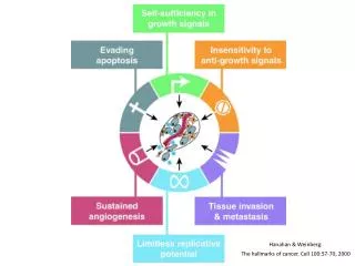
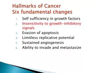
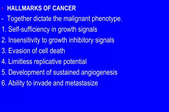
HALLMARKS OF CANCER - Together dictate the malignant phenotype. 1. Self-sufficiency in growth signals 2. Insensitivity to growth inhibitory signals 3. Evasion of cell death 4. Limitless replicative potential 5. Development of sustained angiogenesis 6. Ability to invade and metastasize.

E N D
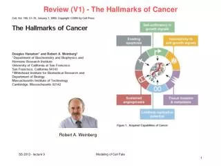
HALLMARKS OF CANCER - Together dictate the malignant phenotype. 1. Self-sufficiency in growth signals 2. Insensitivity to growth inhibitory signals 3. Evasion of cell death 4. Limitless replicative potential 5. Development of sustained angiogenesis 6. Ability to invade and metastasize
Emerging" hallmarks of cancer, a. Reprogramming of energy metabolism b. Evasion of the immune system, Enabling characteristics, a. Genomic instability b. tumor-promoting inflammation Note: gene symbols are italicized but their protein products are not (e.g., RB gene and Rb protein, TP53 and p53, ).
I.Self-Sufficiency in Growth Signals - Cancer cells use a strategies to drive their proliferation and become insensitive to normal growth regulators. In physiologic conditions, cell proliferation has steps: a. The binding of a growth factor to its specific receptor b. Transient activation of the growth factor receptor c. Activation of several signal-transducing proteins d. Transmission of the transduced signal to the nucleus e. Induction and activation of nuclear regulatory factors that initiate and regulate DNA transcription
f. Entry and progression of the cell into the cell cycle, resulting ultimately in cell division - The mechanisms that endow cancer cells with the ability to proliferate can be grouped according to their role in the growth factor-induced signal transduction cascade and cell cycle regulation
A. Growth Factors -All normal cells require stimulation by growth factors to undergo proliferation and most soluble growth factors are made by one cell type and act on a neighboring cell (paracrine action) . - Normally, cells that produce the growth factor do not express the cognate receptor and this specificity prevents the formation of positive feedback within the same cell. 1- Many cancer cells acquire growth self-sufficiency by acquiring the ability to synthesize the same growth factors to which they are responsive.
For Example: Many glioblastomas secrete platelet-derived growth factor (PDGF) and express the PDGF receptor 2. Another mechanism by which cancer cells acquire growth self-sufficiency is by interaction with stroma. - In some cases, tumor cells send signals to activate normal cells in the supporting stroma, which in turn produce growth factors that promote tumor growth.
B. Growth Factor Receptors and Non-Receptor Tyrosine Kinases - Overexpression of growth factor receptors, which can render cancer cells hyperresponsive to levels of the growth factor that would not normally trigger proliferation. - The best-documented examples of overexpression :. a. ERBB1, the EGF receptor, is overexpressed in 80% of squamous cell carcinomas of the lung b. The gene encoding a related receptor, HER2/NEU (ERBB2), is amplified in 25% to 30% of breast cancers.
-High level of HER2/NEU in breast cancer cells a. is a harbinger of poor prognosis b. The significance of HER2/NEU in the pathogenesis of breast cancers is illustrated dramatically by the clinical benefit derived from blocking the extracellular domain of this receptor with anti-HER2/NEU antibodies(Herceptin).
C. Downstream Signal-Transducing Proteins - Cancer cells may acquire growth autonomy is mutations in genes that encode components of signaling pathways I. RAS Protein: is the most commonly mutated proto-oncogene in human tumor andApproximately 30% of all human tumors contain mutated versions of the RAS gene, with higher frequency in colon and pancreatic carcinomas. - RAS is a member of G proteins that bind guanosine nucleotides (guanosinetriphosphate [GTP] and guanosinediphosphate [GDP]),
- Normal RAS proteins flip back and forth between an excited signal-transmitting state and a quiescent state . a- RAS proteins are inactive when bound to GDP b- Stimulation of cells by growth factors such as EGF leads to exchange of GDP for GTP that generate active RAS. - This excited state is short-lived, because the intrinsic guanosine triphosphatase (GTPase) activity of RAS hydrolyzes GTP to GDP, returning RAS to Inactive state. - The GTPase activity of activated RAS protein is magnified by GTPase-activating proteins (GAPs), which act as molecular brakes that prevent uncontrolled RAS
activation by favoring hydrolysis of GTP to GDP. • The RAS protein most commonly is activated by point mutations that interfere with GTP hydrolysis, which is essential to inactivate RAS, and RAS is thus trapped in its activated, GTP-bound form, and the cell is forced into a continuously proliferating state -The consequences of mutations in RAS would be mimicked by loss-of-function mutations in the GAPs with a failure to simulate GTP hydrolysis and thereby restrain normal RAS. - Indeed, disabling mutation of neurofibromin-1 (NF-1), a GAP, is associated with familial neurofibromatosis type 1
II.ABL - Several non-receptor-associated tyrosine kinases function as signal transduction molecules and ABL is the most well defined with respect to carcinogenesis. - The ABL proto-oncogene has tyrosine kinase activity that is dampened by internal negative regulatory domains. - The Philadelphia (Ph) chromosome is found in 90% of chronic myelogenous leukemia, consisting of balanced translocation between chromosomes 22 and 9 in which a part of the ABL gene is translocated from its normal site on chr 9 to chr 22, where it fuses with breakpoint cluster
region (BCR) gene and the BCR-ABL hybrid protein maintains the tyrosine kinase domain - The crucial role of BCR-ABL in transformation has been confirmed by the clinical response of patients with chronic myelogenous leukemia to BCR-ABL kinase inhibitors ( imatinib mesylate (Gleevec), (so-called targeted therapy). - Acquired resistance of tumors to BCR-ABL inhibitors often is due to the outgrowth of a subclone with a mutation in BCR-ABL that prevents binding of the drug to the BCR-ABL protein
D. Nuclear Transcription Factors - Growth autonomy may be a consequence of mutations affecting genes that regulate transcription of DNA such as MYC, MYB, and JUN - MYC gene is involved most commonly in human tumors and its activities include. 1.Activation of cyclin-dependent kinases (CDKs), whose products drive cells into the cell cycle 2. MYC repress the CDK inhibitors (CDKIs).
a-In more than 90% of cases of Burkitt lymphoma the cells have a translocation, usually between chromosomes 8 and 14, which leads to overexpression of the MYC gene on chromosome 8 by juxtaposition with immunoglobulin heavy chain gene regulatory elements on chromosome 14. b. NMYC gene is amplified in neuroblastomas c. LMYC genes is amplified in small cell cancers of lung.
E. Cyclins and Cyclin-Dependent Kinases - The ultimate outcome of all growth-promoting stimuli is the entry of quiescent cells into the cell cycle. CELL CYCLE:- Cancers may become autonomous if the genes that drive cell cycle become mutated or amplified - To achieve DNA replication and division, the cell goes through the cell cycle which consists of G1, (presynthetic), S (DNA synthesis), G2 (premitotic), and M (mitotic) phases - Quiescent cells that have not entered the cell cycle are in the G0 state and to enter the cycle, they must first go through the transition from G0 to G 1 which functions as a
gateway to the cell cycle - Cells can enter G1 either from G0 or after completing mitosis (continuously dividing cells) and progress through the cycle and reach a critical stage at the G1-S known as a restriction point, a rate-limiting step for replication - Progression through the G1-S transition, is regulated by cyclins, and cyclin-dependent kinases (CDKs). - The activity of CDK-cyclin complexes is regulated by CDK inhibitors (CDKIs), which enforce cell cycle checkpoints - CDKs acquire catalytic activity by forming complexes with the cyclins that that phosphorylate crucial target proteins
that drive the cell through the cell cycle and one such protein is retinoblastoma protein (Rb). - cyclins D, E, A, and B appear sequentially during the cycle - Embedded in the cell cycle mechanisms that sense damage to DNA called checkpoints ; a. The G1-S checkpoint monitors the integrity of DNA before DNA replication, b. The G2-M checkpoint checks DNA after replication and monitors whether the cell can safely enter mitosis. - When cells sense DNA damage, checkpoint activation delays the cell cycle and triggers DNA repair mechanisms
. - If DNA damage cant be repaired, the cells undergo apoptosis, or enter a nonreplicative state ( senescence), - Mutations in genes regulating these checkpoints allow cells with damaged DNA to divide, producing daughter cells carrying mutations - There are several families of CDKIs. 1. p21 , p27 , and p57 inhibits the CDKs broadly, 2. p15 , p16 , p18, p19 are called INK4 (A to D) proteins have selective effects on cyclin CDK4 and cyclin CDK6.
Alterations in Cell Cycle Control Proteins in Cancer Cells - cyclins and CDKs mutations favor cell proliferation and all cancers have genetic lesions that disable the G1-S point causing cells to continually reenter the S phase. - Mishaps increasing the expression of cyclin D or CDK4 seem to be a common event in neoplastic transformation. 1. The cyclin D genes are overexpressed in many cancers, including mainly mantle cell lymphomas 2. Somatically acquired deletion or inactivation of CDKN2A is seen in 75% of pancreatic carcinomas
II. Insensitivity to Growth Inhibitory Signals - The products of tumor suppressor genes apply brakes to cell proliferation and Disruption of such genes renders cells refractory to growth inhibition and mimics the growth-promoting effects of oncogenes. 1. RB Gene: Governor of the Cell Cycle - Retinoblastoma gene (RB), the first tumor suppressor gene to be discovered and, the discovery of tumor suppressor genes was accomplished by the study of retinoblastoma, - Approximately 60% of retinoblastomas are sporadic, and the remaining ones are familial,
- The predisposition to develop the tumor being transmitted as an autosomal dominant trait . - two-hit hypothesis, explains the sporadic and familial occurrence of an identical tumor, for example retinoblastoma and these involve the RB gene, on chr 13. - Both of the normal alleles of the RB locus must be inactivated for the development of retinoblastoma 1. In familial cases, children inherit one defective copy of the RB gene in the germline; the other copy is normal and retinoblastoma develops when the normal RB gene is lost in retinoblasts as a result of somatic mutation
- Because in retinoblastoma families only a single somatic mutation is required for expression of the disease, the familial transmission is autosomal dominant 2. In sporadic cases, both normal RB alleles are lost by somatic mutation in one of the retinoblasts. - The end result is the same: a retinal cell that has lost both of the normal copies of the RB gene becomes cancerous. - Although the loss of RB genes initially discovered in retinoblastomas, it is now evident that homozygous loss of this gene is common in breast cancer and small cell cancer of the lung
- Patients with familial retinoblastoma also are at greatly increased risk for development of osteosarcomas . - A cell heterozygous at the RB locus is not neoplastic. - Tumors develop when the cell loses its normal RB gene copy and thus becomes homozygous for the mutant allele Mechanism of action of RB - The RB gene product is a DNA-binding protein that is expressed in every cell type where it exists in an active hypophosphorylated state and inactive hyper-phosphorylated state.
- The Rb regulates G1/S checkpoint. - During development, as cells become terminally differentiated, and enter G0 , and remain there until mitogenic signaling, push them back into the cell cycle - In G1,diverse signals determine whether the cell should progress through the cell cycle, or exit it and differentiate, and Rb integrates mitogenic signals to make this decision - The initiation of DNA replication requires the activity of cyclin E/CDK2 complexes, and expression of cyclin E is dependent on the E2F family of transcription factors. - Early in G1, Rb is in its hypophosphorylated active form
, and it binds to and inhibits the E2F family of transcription factors, preventing transcription of cyclin E. Hypophosphorylated Rb blocks E2F-mediated transcription a. By sequestering E2F, preventing it from interacting with other transcriptional activators. b. Rb recruits chromatin remodeling proteins, histone deacetylases and histone methyltransferases, which bind to the promoters of E2F-responsive genes such as cyclin E and These enzymes modify chromatin at the promoters to make DNA insensitive to transcription factors This situation is changed on mitogenic signaling.
.a- Growth factor signaling leads to cyclin D expression and activation of cyclin D-CDK4/6 complexes which phosphorylate Rb, inactivating it and releasing E2F to induce cyclin E. b. Expression of cyclin E then stimulates DNA replication and progression through the cell cycle and When the cells enter S phase, they are committed to divide without additional growth factor stimulation - During M phase, the phosphate groups removed from Rb by phosphatases, regenerating the hypophosphorylated form of Rb.
why RB is not mutated in every cancer - Mutations in other genes that control Rb phosphorylation can mimic the effect of RB loss and such genes are mutated in many cancers that have normal RB genes 1. For example, mutational activation of CDK4 or overexpression of cyclin D favors cell proliferation by facilitating Rb phosphorylation and inactivation. 2. Mutational inactivation of CDKIs also would drive the cell cycle by unregulated activation of cyclins and CDKs. The CDKN2A gene is an extremely common target of deletion or mutational inactivation in human tumors.
- Loss of normal cell cycle control is central to malignant transformation and that at least one of the four key regulators of the cell cycle CDKN2A, cyclin D, CDK4, Rb is mutated in most cancers 3. the transforming proteins of several oncogenic DNA viruses act, by neutralizing the growth inhibitory activities of Rb, For example, the human papillomavirus (HPV) E7 protein binds to the hypophosphorylated form of Rb, preventing it. from inhibiting the E2F transcription factors - Thus, Rb is functionally deleted, leading to uncontrolled growth
TP53 Gene: Guardian of the Genome - TP53 is one of the most commonly mutated genes in cancers. And The p53 protein thwarts neoplastic transformation by : a. activation of temporary cell cycle arrest (quiescence), b. induction of permanent cell cycle arrest (senescence), c. or triggering of programmed cell death (termed apoptosis) A variety of stresses trigger the p53 response pathways, a. Anoxia, b. Damage to the integrity of DNA
- In healthy cells, p53 has a short half-life (20 minutes) because of its association with MDM2, a protein that targets p53 for destruction. - When the cell is stressed, by an assault on DNA, sensors such as ATM (ataxia telangiectasia mutated) are activated - These activated complexes p53 from MDM2 and increase its half-life and enhance its ability to drive the transcription of target genes that suppress -neoplastic transformation by three mechanisms : a. p53-mediated cell cycle arrest may be considered the primordial response to DNA damage :-
It occurs late in the G1 phase and is caused mainly by p53-dependent transcription of the CDKI gene CDKN1A (p21). - The p21 protein inhibits cyclin-CDK complexes and - prevents phosphorylation of Rb, thereby arresting cells in the G1 phase and such a pause in cell cycling gives the cells "breathing time" to repair DNA damage. 2. The p53 protein also induces expression of DNA damage repair genes.: If DNA damage is repaired successfully, p53 upregulates transcription of MDM2, leading to destruction of p53 and relief of the cell cycle block.
- If the damage cannot be repaired, the cell may enter p53-induced senescence or undergo p53-directed apoptosis.p53-induced senescence is a permanent cell cycle arrest 3. p53-induced apoptosis of cells with irreversible DNA damage is the ultimate protective mechanism against neoplastic transformation and mediated by several pro-apoptotic genes such as BAX and PUMA - " With homozygous loss of the TP53 gene, DNA damage goes unrepaired, mutations become fixed in dividing cells, leading to malignant transformation - More than 70% of human cancers have a defect in this
gene, and the remaining malignant neoplasms have defects in genes upstream or downstream of TP53. - Biallelic loss of the TP53 gene is found in virtually every type of cancer, including carcinomas of the lung, colon, and breast-the three leading causes of cancer deaths. Note: - In most cases, inactivating mutations affecting both TP53 alleles are acquired in somatic cells - Less commonly, some patients inherit a mutant TP53allele; the disease is called the Li-Fraumeni syndrome. inheritance of one mutant allele predisposes
affected persons to develop malignant tumors because only one additional hit is needed to. inactivate the second, normal allele. - Cancers in Li-Fraumeni syndrome are varied; the most common are sarcomas, breast cancer, leukemia, brain tumors, and compared with sporadic tumors, the cancers develop tumors at a younger age and are multiple - Normal p53 also can be rendered nonfunctional by certain DNA viruses, Proteins encoded by oncogenic HPVs , and possibly Epstein-Barr virus (EBV) can bind to normal p53 and nullify its protective function.
3. Transforming Growth Factor-β Pathway - In normal cells, TGF-β is a potent inhibitor of proliferation and it binds to a complex composed of receptors I and II - Binding of TGF-B to its receptors leads to: a.Activation of CDKIs with growth-suppressing activity, b. Repression of growth-promoting genes such as MYC, NOTE - In many forms of cancer, the growth-inhibiting effects of the TGF-β pathways are impaired by mutations affectingTGF-β signaling. a. Mutations affecting the type II receptor are seen in cancers of the colon, stomach, and endometrium
.b. Mutational inactivation of SMAD4, involved in TGF-β signaling, is common in pancreatic cancers. - . In 100% of pancreatic cancers and 83% of colon cancers, at least one component of the TGF-β pathway is mutated c. In many cancers, loss of TGF-β-mediated growth control occurs at a level downstream of the core signaling pathway, for example, persistent expression of MYC. d. in many late-stage tumors, TGF-β signaling activates epithelial-to-mesenchymal transition (EMT), a process that promotes migration, invasion, and metastasis.
4. Contact Inhibition, NF2, and APC - When nontransformed cells are grown in culture, they proliferate until confluent monolayers are generated; cell-cell contacts formed in these monolayers suppress further cell proliferation and "contact inhibition" is abolished in cancer cells, allowing them to pile on top of one another. - Cell-cell contacts in many tissues are mediated by interactions between proteins called cadherins. E-cadherin mediates cell-cell contact in epithelial layers. a. It sustains contact inhibition is mediated by the tumor suppressor gene NF2
- Its product, neurofibromin-2, called merlin, which facilitates E-cadherin mediated contact inhibition and Homozygous loss of NF2 is known to cause neurofibromatosis 2 2. There are other mechanisms of E-cadherin regulation and is illustrated by the rare hereditary disease adenomatous polyposis coli (APC).
adenomatous polyposis coli (APC). a. This disorder is characterized by the development of numerous adenomatous polyps in the colon that have a very high incidence of transformation into colonic cancers. b. They consistently show loss of a tumor suppressor gene called APC (named for the disease)which exerts antiproliferative effects by encoding a cytoplasmic protein whose dominant function is to regulate the intracellular levels of β-catenin, a protein with many functions. a. β-catenin binds to the cytoplasmic portion of E-cadherin b. translocate to the nucleus and activate cell proliferation.
3. -β-Catenin is an important component of the so-called WNT signaling pathway that regulates cell proliferation - WNT is a soluble factor that can induce cellular proliferation by transmitting signals that prevent the degradation of β-catenin, allowing it to translocate to the nucleus , where it acts as a transcriptional activator in conjunction with TcF - In quiescent cells, which are not exposed to WNT, cytoplasmic β-catenin is degraded by a destructioncomplex, of which APC is an integral part -With loss of APC (in malignant cells), β-catenin degradation is prevented, and the WNT signaling response is activated
Inappropriately in the absence of WNT and this leads to a. transcription of cyclin D1 and MYC, b. And transcription of TWIST and SLUG, that repress E-cadherin expression and thus reduce contact inhibition • APC behaves as a typical tumor suppressor gene.and - Persons born with one mutant allele are found to have hundreds to thousands of polyps in the colon by their teens ; these polyps show loss of the other APC allele and invariably, one or more polyps undergo malignant change - APC mutations are seen in 70% to 80% of sporadic colon cancers.