Download
1 / 37
370 likes | 528 Views
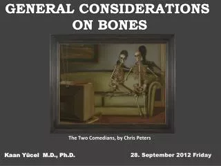
INTRORUCTION TO OSTEOLOGY. The Two Comedians , by Chris Peters. Kaan Yücel M.D., Ph.D . 19. November 201 3 Tuesday. . OSTEOLOGY. Gk , osteon, bone, logos, science. branch of medicine concerned with the development and diseases of bone tissue. 270 bones. 222 bones.

E N D
INTRORUCTION TO OSTEOLOGY TheTwoComedians, byChrisPeters Kaan Yücel M.D., Ph.D. 19. November2013 Tuesday
.OSTEOLOGY Gk, osteon, bone, logos,science branch of medicine concerned with the development and diseases of bone tissue 270 bones 222 bones 206 bones
Skeletalsystemdividedintotwofunctional/anatomicalparts: Axialskeleton bones of the head, neckandtrunk Appendicularskeleton bones of the limbs including those forming the pectoral (shoulder) and pelvic girdles. 80 bones 126 bones
Bone one of the hardest structures of the animal body calcification of its extracellular matrix some elasticity results from the organic matter great rigidity results from their lamellous structures and tubes of inorganic calcium phosphate colorin a fresh state pinkish-whiteexternally, deep red within.
CartilageSand Bones • The skeleton is composed of cartilages and bones. • Cartilage • resilient, semirigid form of connective tissue • forms parts of the skeleton where more flexibility is required. articulating of bones participating in a synovial joint capped with articular cartilage provides smooth, low-friction, gliding surfaces for free movement
CartilageSand Bones • The skeleton is composed of cartilages and bones. • Cartilage • resilient, semirigid form of connective tissue • forms parts of the skeleton where more flexibility is required. articulating of bones participating in a synovial joint capped with articular cartilage provides smooth, low-friction, gliding surfaces for free movement
Blood vessels do not enter cartilage avascular • Diffusion • bone /cartilage in the skeleton • changes as the body grows • younger a person the more cartilage • bones of a newborn are soft and flexible because mostly composed of cartilage.
CartilageSand Bones • The skeleton is composed of cartilages and bones. • The amount and kind of extracellular fibers in the matrix • depends on the type of cartilage. • Heavyweightbearing areas or areas prone to pulling forces • Morecollagenfibers, lessflexiblecartilage.
Functionsof cartilage support soft tissues provide a smooth, gliding surface for bone articulations at joints enable the development and growth of long bones.
Typesof cartilage 1. Hyaline most common,matrix w/ moderate amount of collagen fibers articularsurfaces of bones 2. Elastic large number of elastic fibers external ear 3. Fibrocartilage limited number of cells &ground substance amidst substantial amount of collagen fibers intervertebral discs
Bones function as • supportive structures for the body • protectors of vital organs • reservoirs of calcium and phosphorus • levers on which muscles act to produce movement • containers for blood-producing cells
TYPES OF BONES • according to their shape gross anatomy • Long bones • tubular humerusin the arm • 3)Flat bones • protectivefunctions • flat bones of the cranium protect the brain 2)Short bones cuboidal tarsus (ankle) carpus (wrist)
Classification of Bones 4) Irregular bones various shapes other than long, short, or flat bones of the face
Classification of Bones 5) Sesamoidbones patella or knee cap protect the tendons from excessive wear often change the angle of the tendons as they pass to their attachments.
Long bones develop by replacement of hyaline cartilage plate endochondral ossification • a shaft diaphysis - two ends epiphyses • Metaphysis • a part of the diaphysis adjacent to the epiphyses. • Diaphysisencloses the marrow cavity.
2types of bones according to histological features compact bone &spongy (trabecular) bone relative amount of solid matter #&size of the spaces they contain
All bones have a superficial thin layer of compact bone • around a central mass of spongy bone • except where the spongy boneis replaced by a medullary (marrow) cavity. • Spongy bone • found @ expanded heads of long bones +fills most irregular bones. • Compact bone • forms outer shell of all bones+shafts in long bones.
Spongy (cancellous)bone consists of thin threads of bone trabeculae The orientation of the trabeculae is modelled by the mechanical stress to which the bone is exposed Wolff'slaw
Architecture & proportion of compact and spongy bone vary according to function Compact bone providesstrength for weight bearing. . Fig. 11. (a) Frontal section of the humerus head of a younger person (male, 32 years). The superior border of the medullary cavity is marked by the dotted line. The arrows point to the very thin lamella of compactbone in this region. (b) Frontal section of the humerus head of an aged person (female, 97 years). The superior border of the medullary cavity is marked by the dotted line. Notice that the very thin lamella of compactbone (arrows) is not supported by spongious osseous substance.
Bone Markings and Formations • Bone markings appear wherever tendons, ligaments, and fascias are attached or where arteries lie adjacent to or enter bones. • Other formations occur in relation to the passage of a tendon (often to direct the tendon or improve its leverage) or to control the type of movement occurring at a joint.
TEMPORALIS MUSCLE Arisesfrom the bony floor & overlying temporal fascia attaches superiorly superior temporal line inferiorlylateral &medial surfaces of the zygomaticarch Insertion: Coronoid process of mandible & ramus of mandible Elevation and retraction of mandible
Linear elevations Line (in Latin linea), crest (in Latine crista) Cristagalli (crest of the cock) in the anterior part of the skull Superior temporal line Inferior temporal line in the skull
Round elevations tubercule(small eminence), protuberance (swelling) Tubercle of a rib External occipital protuberance rear side of the head (skull)
Sharp elevations spine, process Spinousprocess of a vertebra Styloidprocess in the skull
Rounded articular areas head, condyle Head of the mandible Condylarprocess
Depressions fossae (small depression), groove (sulcus, long narrow depressions) Submandibularfossa Costalgroove
Foramen Hole Mentalforamen Foramenmagnum in theskull
Canal a foramen having length Optic canal in the skull in the orbita where the eye is located.
Meatus acanal entering a structure Externalauditory meatus
Vasculature and Innervation of Bones • Bones are richly supplied with blood vessels. • Veins accompany arteries. • Nerves accompany blood vessels supplying bones.
Accessory Bones • Accessory (supernumerary) bones develop when additional ossification centers appear and form extra bones. • Many bones develop from several centers of ossification, and the separate parts normally fuse. • Sometimes one of these centers fails to fuse with the main bone, giving the appearance of an extra bone. .
Heterotopic Bones Bones sometimes form in soft tissues where they are not normally present (e.g., in scars). Horse riders often develop heterotopic bones in their thighs (rider's bones), probably because of chronic muscle strain resulting in small hemorrhagic (bloody) areas that undergo calcification and eventual ossification. .
Changes in Bones &Bone Fractures Trauma to a bone may break it. For the fracture to heal properly, the broken ends must be brought together, approximating their normal position. reduction of a fracture. Fractures are more common in children than in adults. .
Changes in Bones &Bone Fractures Immediately after a fracture, the patient suffers severe local pain and is not able to use the injured part. Deformity may be visible if the bone fragments have been displaced relative to each other. .
OSTEOPOROSIS decreases in theorganic & inorganiccomponents of the bone byaging . Bones become brittle, lose their elasticity, and fracture easily. Bone scanning is an imaging method used to assess normal and diminished bone mass.
(BONE) SCINTIGRAPHY metabolic activity of bone and its affinity to uptake a detectable marker image can be captured by a scan a wide range of indications ranging from sports related injuries to detection of metastasis (spreading of cancer) to the bones. .
BONE DENSITOMETRY (DEXA, DXA) enhanced form of x-ray technology used to measure bone loss . most often used to diagnose osteoporosis effective in tracking the effects of treatment for osteoporosis and other conditions that cause bone loss.