Download
1 / 77
770 likes | 784 Views
The Immune System: Innate and Adaptive Body Defenses Part B. 21. Antibodies. Also called immunoglobulins Constitute the gamma globulin portion of blood proteins Are soluble proteins secreted by activated B cells and plasma cells in response to an antigen

E N D
The Immune System: Innate and Adaptive Body Defenses Part B 21
Antibodies • Also called immunoglobulins • Constitute the gamma globulin portion of blood proteins • Are soluble proteins secreted by activated B cells and plasma cells in response to an antigen • Are capable of binding specifically with that antigen • There are five classes of antibodies: IgD, IgM, IgG, IgA, and IgE
Classes of Antibodies • IgD – monomer attached to the surface of B cells, important in B cell activation • IgM – pentamer released by plasma cells during the primary immune response • IgG – monomer that is the most abundant and diverse antibody in primary and secondary response; crosses the placenta and confers passive immunity • IgA – dimer that helps prevent attachment of pathogens to epithelial cell surfaces • IgE – monomer that binds to mast cells and basophils, causing histamine release when activated
Basic Antibody Structure • Consists of four looping polypeptide chains linked together with disulfide bonds • Two identical heavy (H) chains and two identical light (L) chains • The four chains bound together form an antibody monomer • Each chain has a variable (V) region at one end and a constant (C) region at the other • Variable regions of the heavy and light chains combine to form the antigen-binding site
Basic Antibody Structure Figure 21.12a, b
Antibody Structure • Antibodies responding to different antigens have different V regions but the C region is the same for all antibodies in a given class • C regions form the stem of the Y-shaped antibody and: • Determine the class of the antibody • Serve common functions in all antibodies • Dictate the cells and chemicals that the antibody can bind to • Determine how the antibody class will function in elimination of antigens
Mechanisms of Antibody Diversity • Plasma cells make over a billion different types of antibodies • Each cell, however, only contains 100,000 genes that code for these polypeptides • To code for this many antibodies, somatic recombination takes place • Gene segments are shuffled and combined in different ways by each B cell as it becomes immunocompetent • Information of the newly assembled genes is expressed as B cell receptors and as antibodies
Antibody Diversity • Random mixing of gene segments makes unique antibody genes that: • Code for H and L chains • Account for part of the variability in antibodies • V gene segments, called hypervariable regions, mutate and increase antibody variation • Plasma cells can switch H chains, making two or more classes with the same V region
Antibody Targets • Antibodies themselves do not destroy antigen; they inactivate and tag it for destruction • All antibodies form an antigen-antibody (immune) complex • Defensive mechanisms used by antibodies are neutralization, agglutination, precipitation, and complement fixation
Complement Fixation and Activation • Complement fixation is the main mechanism used against cellular antigens • Antibodies bound to cells change shape and expose complement binding sites • This triggers complement fixation and cell lysis • Complement activation: • Enhances the inflammatory response • Uses a positive feedback cycle to promote phagocytosis • Enlists more and more defensive elements
Other Mechanisms of Antibody Action • Neutralization – antibodies bind to and block specific sites on viruses or exotoxins, thus preventing these antigens from binding to receptors on tissue cells
Other Mechanisms of Antibody Action • Agglutination – antibodies bind the same determinant on more than one antigen • Makes antigen-antibody complexes that are cross-linked into large lattices • Cell-bound antigens are cross-linked, causing clumping (agglutination) • Precipitation – soluble molecules are cross-linked into large insoluble complexes
Mechanisms of Antibody Action Figure 21.13
Monoclonal Antibodies • Commercially prepared antibodies are used: • To provide passive immunity • In research, clinical testing, and treatment of certain cancers • Monoclonal antibodies are pure antibody preparations • Specific for a single antigenic determinant • Produced from descendents of a single cell
Monoclonal Antibodies • Hybridomas – cell hybrids made from a fusion of a tumor cell and a B cell • Have desirable properties of both parent cells – indefinite proliferation as well as the ability to produce a single type of antibody
Cell-Mediated Immune Response • Since antibodies are useless against intracellular antigens, cell-mediated immunity is needed • Two major populations of T cells mediate cellular immunity • CD4 cells (T4 cells) are primarily helper T cells (TH) • CD8 cells (T8 cells) are cytotoxic T cells (TC) that destroy cells harboring foreign antigens • Other types of T cells are: • Suppressor T cells (TS) • Memory T cells
Major Types of T Cells Figure 21.14
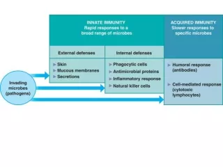
Importance of Humoral Response • Soluble antibodies • The simplest ammunition of the immune response • Interact in extracellular environments such as body secretions, tissue fluid, blood, and lymph
T Cells • T cells mature in the thymus under negative and positive selection pressures • Negative selection – eliminates T cells that are strongly anti-self • Positive selection – selects T cells with a weak response to self-antigens, which thus become both immunocompetent and self-tolerant
Importance of Cellular Response • T cells recognize and respond only to processed fragments of antigen displayed on the surface of body cells • T cells are best suited for cell-to-cell interactions, and target: • Cells infected with viruses, bacteria, or intracellular parasites • Abnormal or cancerous cells • Cells of infused or transplanted foreign tissue
Antigen Recognition and MHC Restriction • Immunocompetent T cells are activated when the V regions of their surface receptors bind to a recognized antigen • T cells must simultaneously recognize: • Nonself (the antigen) • Self (a MHC protein of a body cell)
MHC Proteins • Both types of MHC proteins are important to T cell activation • Class I MHC proteins • Always recognized by CD8 T cells • Display peptides from endogenous antigens
Class I MHC Proteins • Endogenous antigens are: • Degraded by proteases and enter the endoplasmic reticulum • Transported via TAP (transporter associated with antigen processing) • Loaded onto class I MHC molecules • Displayed on the cell surface in association with a class I MHC molecule
Class I MHC Proteins Figure 21.15a
Class II MHC Proteins • Class II MHC proteins are found only on mature B cells, some T cells, and antigen-presenting cells • A phagosome containing pathogens (with exogenous antigens) merges with a lysosome • Invariant protein prevents class II MHC proteins from binding to peptides in the endoplasmic reticulum
Class II MHC Proteins • Class II MHC proteins migrate into the phagosomes where the antigen is degraded and the invariant chain is removed for peptide loading • Loaded Class II MHC molecules then migrate to the cell membrane and display antigenic peptide for recognition by CD4 cells
Class II MHC Proteins Figure 21.15b
Antigen Recognition • Provides the key for the immune system to recognize the presence of intracellular microorganisms • MHC proteins are ignored by T cells if they are complexed with self protein fragments
Antigen Recognition • If MHC proteins are complexed with endogenous or exogenous antigenic peptides, they: • Indicate the presence of intracellular infectious microorganisms • Act as antigen holders • Form the self part of the self-antiself complexes recognized by T cells
T Cell Activation: Step One – Antigen Binding • T cell antigen receptors (TCRs): • Bind to an antigen-MHC protein complex • Have variable and constant regions consisting of two chains (alpha and beta)
T Cell Activation: Step One – Antigen Binding • MHC restriction – TH and TC bind to different classes of MHC proteins • TH cells bind to antigen linked to class II MHC proteins • Mobile APCs (Langerhans’ cells) quickly alert the body to the presence of antigen by migrating to the lymph nodes and presenting antigen
T Cell Activation: Step One – Antigen Binding • TC cells are activated by antigen fragments complexed with class I MHC proteins • APCs produce co-stimulatory molecules that are required for TC activation • TCR that acts to recognize the self-antiself complex is linked to multiple intracellular signaling pathways • Other T cell surface proteins are involved in antigen binding (e.g., CD4 and CD8 help maintain coupling during antigen recognition)
T Cell Activation: Step One – Antigen Binding Figure 21.16
T Cell Activation: Step Two – Co-stimulation • Before a T cell can undergo clonal expansion, it must recognize one or more co-stimulatory signals • This recognition may require binding to other surface receptors on an APC • Macrophages produce surface B7 proteins when nonspecific defenses are mobilized • B7 binding with the CD28 receptor on the surface of T cells is a crucial co-stimulatory signal • Other co-stimulatory signals include cytokines and interleukin 1 and 2
T Cell Activation: Step Two – Co-stimulation • Depending on receptor type, co-stimulators can cause T cells to complete their activation or abort activation • Without co-stimulation, T cells: • Become tolerant to that antigen • Are unable to divide • Do not secrete cytokines
T Cell Activation: Step Two – Co-stimulation • T cells that are activated: • Enlarge, proliferate, and form clones • Differentiate and perform functions according to their T cell class
T Cell Activation: Step Two – Co-stimulation • Primary T cell response peaks within a week after signal exposure • T cells then undergo apoptosis between days 7 and 30 • Effector activity wanes as the amount of antigen declines • The disposal of activated effector cells is a protective mechanism for the body • Memory T cells remain and mediate secondary responses to the same antigen
Cytokines • Mediators involved in cellular immunity, including hormonelike glycoproteins released by activated T cells and macrophages • Some are co-stimulators of T cells and T cell proliferation • Interleukin 1 (IL-1) released by macrophages co-stimulates bound T cells to: • Release interleukin 2 (IL-2) • Synthesize more IL-2 receptors
Cytokines • IL-2 is a key growth factor, which sets up a positive feedback cycle that encourages activated T cells to divide • It is used therapeutically to enhance the body’s defenses against cancer • Other cytokines amplify and regulate immune and nonspecific responses
Cytokines • Examples include: • Perforin and lymphotoxin – cell toxins • Gamma interferon – enhances the killing power of macrophages • Inflammatory factors
Helper T Cells (TH) • Regulatory cells that play a central role in the immune response • Once primed by APC presentation of antigen, they: • Chemically or directly stimulate proliferation of other T cells • Stimulate B cells that have already become bound to antigen • Without TH, there is no immune response
Helper T Cells (TH) Figure 21.17a
Helper T Cell • TH cells interact directly with B cells that have antigen fragments on their surfaces bound to MHC II receptors • TH cells stimulate B cells to divide more rapidly and begin antibody formation • B cells may be activated without TH cells by binding to T cell–independent antigens • Most antigens, however, require TH co-stimulation to activate B cells • Cytokines released by TH amplify nonspecific defenses
Helper T Cells Figure 21.17b
Cytotoxic T Cell (Tc) • TC cells, or killer T cells, are the only T cells that can directly attack and kill other cells • They circulate throughout the body in search of body cells that display the antigen to which they have been sensitized • Their targets include: • Virus-infected cells • Cells with intracellular bacteria or parasites • Cancer cells • Foreign cells from blood transfusions or transplants
Cytotoxic T Cells • Bind to self-antiself complexes on all body cells • Infected or abnormal cells can be destroyed as long as appropriate antigen and co-stimulatory stimuli (e.g., IL-2) are present • Natural killer cells activate their killing machinery when they bind to MICA receptor • MICA receptor – MHC-related cell surface protein in cancer cells, virus-infected cells, and cells of transplanted organs
Mechanisms of Tc Action • In some cases, TC cells: • Bind to the target cell and release perforin into its membrane • In the presence of Ca2+ perforin causes cell lysis by creating transmembrane pores • Other TC cells induce cell death by: • Secreting lymphotoxin, which fragments the target cell’s DNA • Secreting gamma interferon, which stimulates phagocytosis by macrophages
Mechanisms of Tc Action Figure 21.18a, b
Other T Cells • Suppressor T cells (TS) – regulatory cells that release cytokines, which suppress the activity of both T cells and B cells • Gamma delta T cells (Tgd) – 10% of all T cells found in the intestines that are triggered by binding to MICA receptors
Summary of the Primary Immune Response Figure 21.19