Download
1 / 21
230 likes | 267 Views
For more details visit our website at - http://www.cryoviva.in/

E N D
Hindawi Publishing Corporation Advances in Andrology Volume 2014, Article ID 140618, 20 pages http://dx.doi.org/10.1155/2014/140618 Review Article Advances in Stem Cell Therapy for Erectile Dysfunction Ching-Shwun Lin Knuppe Molecular Urology Laboratory, Department of Urology, School of Medicine, University of California, San Francisco, CA 94143-0738, USA Correspondence should be addressed to Ching-Shwun Lin; clin@urology.ucsf.edu Received 21 November 2013; Accepted 20 January 2014; Published 12 March 2014 Academic Editor: Enzo Vicari Copyright © 2014 Ching-Shwun Lin. This is an open access article distributed under the Creative Commons Attribution License, which permits unrestricted use, distribution, and reproduction in any medium, provided the original work is properly cited. Stem cell (SC) therapy for erectile dysfunction (ED) has been investigated in 35 published studies, with one being a small-scale clinical trial. Out of these 35 studies, 19 are concerned with cavernous nerve (CN) injury-associated ED while 10 with diabetes mellitus- (DM-) associated ED. Adipose-derived SCs (ADSCs) were employed in 18 studies while bone marrow SCs (BMSCs) in 9. Transplantation of SCs was done mostly by intracavernous (IC) injection, as seen in 25 studies. Allogeneic and xenogeneic transplantations have increasingly been performed but their immune-incompatibility issues were rarely discussed. More recent studies also tend to use combinatory therapies by modifying or supplementing SCs with angiogenic or neurotrophic genes or proteins. All studies reported better erectile function with SC transplantation, and the majority also reported improved muscle, endothelium, and/or nerve in the erectile tissue. However, differentiation or engraftment of transplanted SCs has rarely been observed; thus, paracrine action is generally believed to be responsible for SC’s therapeutic effects. But still, few studies actually investigated and none proved paracrine action as a therapeutic mechanism. Thus, based exclusively on functional outcome data showninpreclinicalstudies,twoclinicaltrialsarecurrentlyrecruitingpatientsfortreatmentwithICinjectionofADSCandBMSC, respectively. 1. Introduction options treat only the symptoms, not the underlying causes, and most of them need to be taken by or administered to the patient when having an erection which is perceived as necessary. Thus, current research efforts are geared toward findinglong-termsolutionsthatcanreversethepathogenesis of ED and thereby restore the patient’s ability to achieve natural penile erection. One of such research efforts is the investigation of stem cells (SCs) as therapeutic agents, and in this review article I will summarize and discuss all available relevant data in this field of research. Erectile dysfunction (ED) is a term recommended by a panel of experts in 1992 to replace the term “impotence” [1]. These experts also defined ED as an inability of the male to attain and/or maintain penile erection sufficient for satisfactory sexual performance. Although not life threatening by itself, ED is a strong predictor of high-mortality diseases such as coronary artery disease and cardiovascular disease [2– 5]. ED does directly and negatively impact the quality of life of the afflicted men and their spouse [6–11]. In a 1999 report the worldwide prevalence of ED was estimated to be 152 million men in 1995 and predicted to increase to 322 millionmen by 2025 [12]. While the majorityof ED cases can be treated with currently available medications or devices, approximately 20% of the overall ED patient population remainsunresponsivetotreatment[13],andincertainpatient populations, such as those having diabetes mellitus (DM) or having undergone radical prostatectomy (RP), the failure rates are even higher, at 40% [14–16]. Moreover, regardless of their therapeutic efficacy or inefficacy, all current treatment 2. Erectile Function Penile erection is a physiological process that involves the expansion and elongation of a paired cylindrical structure called corpora cavernosa (singular: corpus cavernosum, CC) thatrunthelengthofthepenis[17].InsideeachCCisamaze ofminisculechamberscalledsinusoidsthatarecollapsedand occupy a minimal amount of space when the penis is in the flaccid state. When a man is sexually aroused, blood enters
2 Advances in Andrology the sinusoids, resulting in their expansion and thus penile erection. When sexual stimulation subsides, blood exits the sinusoids, and the penis returns to the flaccid state. The cycle of sinusoidal expansion and collapse is locally controlled by smooth muscle cells (SMCs, or CSMCs as pertaining to CC), which form a multilayered structure as part of the sinusoidal wall [18, 19]. When CSMCs are in a contracted state, blood flow into the sinusoids is kept at a minimum; when CSMCs transit to a relaxed state, blood flow into the sinusoids is increased, resulting in sinusoidal expansion. The cycle of CSMC contraction and relaxation is in turn controlled by two other tissue components inside the CC, namely, the cavernous endothelial cells (CECs) and the cavernous nerves (CNs). CECs are located as a single layer of cellsalongsidetheCSMCsinsideeachsinusoid,andtogether, CECs and CSMCs form the wall that defines each sinusoid [18, 19]. CNs, which arise from neurons in the inferior hypogastricplexusesinhumansorinthemajorpelvicganglia (MPG)intherat[20,21],trackthroughouttheCCandendin thevicinityofCSMCs[18,19].SexualstimulationcausesCNs to release nitric oxide (NO), which enters nearby CSMCs, triggering their relaxation [22–24]. The subsequent increase of blood flow into the sinusoids induces shear stress on the CECs, triggering their release of NO. This CEC-derived NO is believed to permit a sustained CSMC relaxation that is essential for a satisfactory intercourse [25–29]. The paired CC is enveloped in a dense fibrous sheath called tunica albuginea (TA). During erection, the increased blood flow into the sinusoids causes CC to expand and compress against the TA. Being a sturdy and resilient struc- ture, the TA permits CC expansion, but with increasing resistance. As the tension between the expanding CC and the resisting TA increases, so does the overall penile rigidity. Furthermore, the compression of CC against TA causes the closure of subtunical veins that otherwise would allow the escape of blood from the sinusoids. Such a venous occlusive mechanism ensures that the CC remains fully engorged and the penis fully erect until the cessation of sexual stimulation [17]. NO, which is critical for triggering CSMC relaxation, is formedwithinCECsandCNsbynitricoxidesynthase(NOS) that catalyzes the conversion of L-arginine and oxygen to L- citrulline and NO [30]. While performing the same function in NO production, the endothelial NOS (eNOS) and the neuronal NOS (nNOS) are encoded by two separate genes [30]. More importantly, from a technical standpoint, eNOS andnNOSaresufficientlydifferentintheirproteinsequences so as to permit the generation of their specific antibodies [31, 32]. In ED research, including many studies that will be discussed in this review, these antibodies have proven to be highly valuable for the assessment of ED-related changes in the cavernous endothelium and nerves. NO enters CSMCs by diffusion, and once inside, it activates the soluble guanylyl cyclase, which then catalyzes the conversion of GTP to cGMP [22, 23]. When cGMP con- centrationissufficientlyhigh,activationofcGMP-dependent kinase (also known as protein kinase G, PKG) occurs, and this leads to the phosphorylation of several downstream targets (e.g., ion channels), causing CSMCs to relax and thus penile erection [22, 23]. When sexual stimulation ceases, cGMP production is also halted. Meanwhile, existing cGMP is hydrolyzed by phosphodiesterases (PDEs), most notably, PDE5 [33, 34]. When cGMP concentration drops below a threshold level, PKG is deactivated and its down- stream targets dephosphorylated, thereby returning CSMCs to the contracted state and the penis to the flaccid state [22, 23]. 3. Erectile Dysfunction The worldwide prevalence of ED was estimated to be 152 million in 1995 and predicted to be 322 million by 2025 [12]. ED can be classified as psychogenic, organic, or mixed psychogenic/organic [17]. The organic type of ED is further classified into neurogenic, vasculogenic, cavernous, hormonal, drug-induced, and systemic disease-related [17]. For all published SC-for-ED studies, only the neurogenic, vasculogenic, and cavernous types are relevant. Specifically, in concern with neurogenic and vasculogenic types, the relevant studies focused on CN injury-associated and DM- associated ED, respectively. And, in concern with cavernous type of ED, which loosely defines local tissue abnormalities, there have been two TA injury-related SC-for-ED studies. The majority of organic types of ED can be treated with intracavernous (IC) injection of erectogenic agents, transurethral prostaglandin suppository, vacuum device, and/or PDE5 inhibitors (PDE5Is) [35]. In particular, PDE5Is are currently the most prescribed treatment because of their ease of use (as orally taken pills) and overall proven efficacy [35]. However, in a recent study of 327 ED patients who were prescribed PDE5Is by their physicians, only 148 (45.3%) were still using the medication during a 3-year follow-up period, while 160 (48.9%) had discontinued and 19 (5.8%) never took the medication [36]. Thus, among those who had taken PDE5Is, more than half (52%; 160/308) had decided not to continue. When asked to choose among 11 reasons for discontinuation, 38% of the dropout patients selected “noneffectiveness.” Other significant reasons chosen were “concerns about the cardiovascular safety of PDE5Is” (15.7%),cost(13.7%),and“lackofspontaneity”(8.7%).When categorized by specific ED types, patients who were diabetic had the highest discontinuation rate (28/36; 78%), followed by patients whose ED was caused by treatments such as RP (37/56;66%)[36].Suchhighdropoutratesforthesetwotypes of ED are not entirely unexpected, as an earlier study already identified them as least responsive to PDE5I treatment [14]. These two types of ED therefore will be discussed in detail in the following two sections.For other types of ED that have beeninvestigatedasSCtreatmenttargets,onlytheTAinjury- associated ED will also be discussed in detail in a separate section.Thereasonsareasfollows:(1)itisnecessarytoclarify whether TA injury can truly cause ED, (2) hyperlipidemia- associated ED has been investigated in only one SC-for-ED study,(3)aging-associatedEDisrelatedtothenormalprocess of aging, not to any specific disease, and (4) these latter two types of ED are usually responsive to PDE5I treatment.
Advances in Andrology 3 4. Diabetes-Associated ED beam radiation therapy (RT) [54]. However, despite being highly effective, these treatments often incur after treatment complications, such as ED [55, 56]. In a recent study of 1655 men treated with RP or RT, the reported incidence of ED ranges from 60.8% to 93.9%, depending on treatment choice and assessment time (2, 5, or 15 years after treatment) [57]. It is generally agreed that post-RP ED is caused by inadvertent injury to the CNs, which run alongside the prostate. However, despite the introduction of nerve-sparing RP 30 years ago, ED remains a frequent consequence of such surgeries [58, 59]. Therefore, it is now commonly believed that, although leaving the CNs intact, nerve-sparing RP still causessubtlechangesthatarenotobvioustothesurgeons[55, 60]. These changes cause CNs to undergo Wallerian degen- eration and eventually lose their connection to the corpora cavernosa[56,61].Alternatively,thesurgery-incurredinsults may temporarily prevent the CNs from releasing NO into the CC, and without NO-induced engorgement, the penile tissue becomes hypoxic and its CSMC replaced by collagens [56, 60, 61]. In support of this theory, a reduced smooth muscle to collagen ratio has been observed in the penis of CN injury animal models [62–66] and in CC biopsies of RP- treated patients [67]. RPandRTareusedtotreatprostatecanceratcomparable rates [54]; they are also associated with posttreatment ED at similar rates [57]. However, while RP-associated ED has been intensively investigated at the basic science level, RT- associated ED is rarely studied [49]. Nevertheless, three independent studies have foundthatradiationoverthe lower abdomen in the rat caused reductions in erectile response and in CN and CSMC contents [49, 68, 69]. Thus, post- RP and post-RT ED appear to share a common pathological mechanism,thatis,treatment-inducedCNinjuryfollowedby CSMC atrophy. SimilartoothertypesofED,post-RPandpost-RTEDare most commonly treated with PDE5Is [56]. However, as its name indicates, a PDE5I inhibits PDE5, which, by breaking down cGMP, terminates the NO-activated erectile signaling pathway [23, 33]. Therefore, when CNs are injured, as in the situation with RP or RT-treated patients, their ability to release NO into CC is compromised, and if there is no NO releaseintoCC,erectionwouldnotoccur,regardlesswhether PDE5 is inhibited or not. This dependence on a functional rate (∼66%) in post-RP ED patients who were nevertheless repair of the damaged CN, and this has been the goal of the majority of SC-for-ED studies, especially those published in the past two years. In an official document titled “National Diabetes Statistics, 2011,” the National Institute of Health of the USA reports that DM affects 25.8 million people, or 8.3% of the general population in the USA, and among residents 65 years of age andolder,10.9million(26.9%)hadDMin2010[37].Inapeer- reviewed publication that analyzed prior epidemiological studies totaling 370 country-years and 2.7 million partici- pants, Danaei et al. [38] reported that the number of people with DM worldwide increased from 153 million in 1980 to 347 million in 2008. Furthermore, in a forward-looking projection, the International Diabetes Federation estimated that by 2030 the number of people with DM worldwide will be 552 million [39]. Thus, the global diabetes epidemic is alarmingly real. DM is associated with a large assortment of complica- tions, including ED. For men with DM, 50–75% have ED, regardless of age [40]. Diabetic men also tend to incur ED 10–15 years earlier and are 3 times more likely to have ED than nondiabetic men [41, 42]. Importantly, both the severity of ED and the impact on quality of life by ED are significantly greater in diabetic than in nondiabetic men [43, 44]. DM-associated ED is also highly refractory to PDEI treatment, with only 44% success rate, compared to 85% for hypogonadal ED patients [14]. Diabetic ED patients also ranked the highest in terms of discontinuation of treatment with PDE5Is (at a dropout rate of 28/36 or 78%) [36]. Thus, theidentificationofaneffectivetreatmentforDM-associated ED is one of the most important objectives of current ED research efforts. DM is a systematic disease that affects every part of the body. In the penis, it is associated with reduced contents of all three key components for erectile function, namely, CN, CEC, and CSMC [45–49]. The reduction of CEC content is likely due to DM-induced apoptosis in CEC as demonstrated by immunohistochemical analysis of CC samples between diabetic and nondiabetic patients [50]. This clinical histo- logical finding has recently been corroborated by a basic science study, which identified mitochondrial fragmentation and cellular apoptosis in CECs cultured in high glucose medium [32, 51]. Histological analysis of CC samples also identified a significantly higher apoptotic index in the CSMC ofdiabeticversusnondiabeticrats[52].Similarly,thereduced penile CN contenthas been shown to be due to apoptoticcell death of nNOS-positive neurons in the MPG of diabetic rats [48]. Thus, how to prevent and/or reverse these pathological processes is critically important for the effective treatment of DM-associated ED. In this regard, SC therapy has been considered promising, due to SC’s well-known regenerative capacity. CN may explain why PDE5Is have a low success rate (∼ prescribed with PDE5Is [36]. Thus, it is obvious that the effective treatment of post-RP and post-RT ED requires 43%) in treating post-RP ED [14] and a high discontinuation 5. Postprostatectomy and Postradiotherapy ED 6. Peyronie’s Disease-Associated ED Prostatecanceristhemostcommonmalignancyinmen,with estimated numbers of new cases and related deaths being 217,730 and 32,050, respectively, in the USA in 2010 [53]. As prostate cancer is usually locally confined (∼80% of cases), TheestimatedprevalenceofPDvariesgreatlyamongdifferent studies, with the highest being up to 7.1% of men in the general population [70]. PD is characterized by the presence of a fibrous plaque (or plaques) in the TA that causes pain the most common treatments have been RP or external
4 Advances in Andrology and deformity (curvature, narrowing, and shortening) in the erectpenis[70].PDthereforenegativelyimpactsthepatient’s sexual function and partner relationship [71]. However, in popular media, such as the Internet, PD patients’ sexual dysfunction is often incorrectly defined as a type of “erectile dysfunction,” despite the fact that most PD patients are able to achieve erection. Nevertheless, in serious scientific publications, PD is frequently described in the Introduction section as a disease that directly causes ED, with reference to a study by Lopez and Jarow [72]. In that study, 76 out of 95 (80%) PD patients were determined as having concomitant ED, and 36% and 59% of these PD+ED patients were further identified as having abnormal arterial blood flow and veno- occlusive dysfunction, respectively. Thus, the authors con- cluded that, when compared to abnormal arterial blood flow, veno-occlusive dysfunction was the principal cause of ED in PDpatients.However,itshouldbenotedthatthestudynever examined whether PD (TA plaque) caused veno-occlusive dysfunction or ED. In another study that evaluated 143 PD patients, only 27 (19%) were found to have concomitant ED, and22(including4diabetics)ofthe27PD+EDpatientswere found to have veno-occlusive dysfunction [73]. So, on one hand, these two studies disagree sharply in the prevalence of ED among PD patients (80% versus 19%), but on the other, they do agree that veno-occlusive dysfunction is the principal cause of ED in PD patients. Importantly though, neither study provides evidence that PD (TA plaque) caused veno-occlusive dysfunction or ED. In fact, in an earlier study only 18 out of 62 PD patients (29%) were found to have concomitant ED, and 17 out of these 18 PD+ED patients were further identified as having some underlying diseases (such as DM) that contributed to their ED [74]. Therefore, none of these three studies provides evidence that PD (TA plaque) is a direct cause of ED in PD patients. Finally, in the only animal study that attempted to answer why PD patients suffer from ED, PD was simulated in the rat by Table 1: Number of published stem cells for erectile dysfunction studies. Years 2004–2011 Number of studies 15 2012-2013 2004–2013 20 35 Total Disease model Aging CN injury Hyperlipidemia T1DM T2DM TA injury SC type ADSC BMMNC BMSC EPCb ESC SkMSC Testis SC UCBSC aIncluding 2 studies using SVF cells. bProbably BMMNC. 2 7 1 2 2 0 1 3 19 1 8 2 2 12 0 6 0 2 13a 0 5 0 0 1 1 0 5 1 4 1 1 2 0 1 18 1 9 1 1 3 1 1 ED. Further discussion on these two studies will be given in “Animal Models” subsection. 7. Current State of Stem Cell Therapy for Erectile Dysfunction injection of transforming growth factor-훽 (TGF-훽) into the were found to have altered TA histology and lowered erectile response when compared to sham-treated rats. Therefore, this study appears to have obtained evidence that TA abnor- malities can cause ED, but the question remains whether treatment of PD can result in the restoration of erectile function. Plaque excision followed by patch grafting is a common surgical procedure for the correction of penile deformities in PD patients [76]. However, in spite of its proven effi- cacy, postsurgical complications such as ED can occur [77]. Therefore, various approaches, including the use of novel graftmaterials,havebeeninvestigatedtowardminimizingthe occurrence of these complications [76]. In a recent animal study, porcine small intestine submucosa (SIS), which has already been used for TA reconstruction in PD patients, was seeded with adipose-derived stem cells (ADSCs) and then grafted into incised rat TA to see if ADSC seeding can reduce the incidence of postsurgery ED [78]. In another recent animal study [79] ADSC was injected into the TA of TGF- The first SC-for-ED study was published in 2004, and since then, up to November 2013, there have been a total of 34 additional studies in this field of research (Table 1). While 15 of these studies were published between 2004 and 2011, 20 were published after 2011. Thus, it is evident that an accelerated pace of publication occurred at the start of 2012. In any event, among all 35 studies, 19 chose CN injury- associated ED as the disease target, and 18 employed ADSC as the therapeutic cell type (Table 1). While the disease target preference reflects clinical needs (i.e., post-RP and post-RT ED as described earlier), the cell type choice is most likely due to ADSC’s abundant tissue source, ease of isolation, and proven therapeutic efficacy across all medical disciplines (see details under “adipose-derived stem cell”). As clinical trial of SC therapy for ED is not yet approved in most countries, there has been only one published clinical SC-for-ED study and it involved only 7 type 2 DM (T2DM) patients [80]. While that study was carried out in Korea, two clinical trials have been approved in France and the USA and are currently recruiting patients. The French trial (Identifier:NCT01089387)isaphaseI-IItrialandwilltestthe safety and benefit of IC injection of autologous bone marrow mononucleated cells (BMMNC) in post-RP ED patients. The US trial (Identifier: NCT01601353) will evaluate the safety TA (to induce fibrosis) or by surgical incision in the TA (to cause trauma) [75]. Six weeks later both groups of rats 훽-induced PD rats to see if ADSC can reduce the severity of
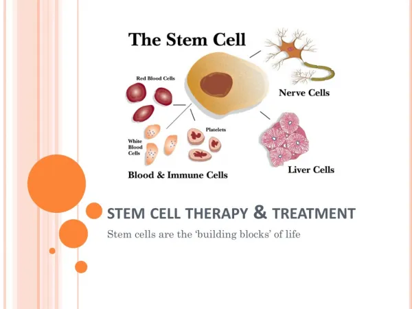
Advances in Andrology 5 and treatment efficacy of IC injection of autologous ADSC in patients with organic ED. In addition to the above-mentioned research and clinical trials, SC’s therapeutic potential has also inspired several business opportunities. By searching the Internet with “stem cell erectile,” one can find many “treatment centers” that offer autologous ADSC treatment for ED at prices of several thousand US dollars. While most of these businesses are located in developing countries such as the Philippines and Thailand, some indicate their presence in the USA. This is surprising because for-profit SC treatment of any disease is illegal in the USA, and criminal convictions have been handed down on cases that involved treating patients with SCs. Therefore, at issue are probable fraudulent advertise- ments. In any event, SC therapy for ED remains largely in the preclinical investigational stage, and this is what will be discussed in the following sections. a lower abdominal incision and, with the aid of a dissecting microscope, identifies the two branches of CN, which arise from the MPG and course proximally on both sides of the prostate and distally alongside the urethra toward the penis. Aninjuryisthenappliedatalocation2–5mmfromtheMPG. The types of injury can be crush, freezing, transection, or resection. Crush is commonly done by applying a hemostatic clamp to the CN so as to simulate accidental nerve contusion or traction that occurs during nerve-sparing RP. Freezing is commonly done by applying a cryoprobe to the CN so as to offer another way of preserving nerve structural continuity. Both transection (a simple cut) and resection (removal of a CN segment) can be considered as representing nonnerve sparing RP, but transection is more likely to allow nerve reconnection than resection is. Thus, among all types of experimental CN injuries, resection is the most severe. In the majority of SC for CN injury ED studies, crush is most frequently used (11/19, Table 4). While hundreds of studies have employed post-RP ED animal models, only 5 studies have worked with post-RT ED models [68, 69, 105, 113, 114]. These studies used similar procedures but with slightly different dosage, duration, and frequency of radiation. In the study by van der Wielen 8. Animal Models et al. [114], a 3mm thick lead shield with a 3cm × 4cm for 5 consecutive days. This protocol was initially adopted in our recently published study [105], but after getting a 50% death rate, we modified it by using a lead shied with a All but one preclinical SC-for-ED studies used rats as animal models; the singularexception used mice (Tables 2, 3, and 4). Aging animal models were used in 3 studies (Table 2), and they were rats between 20 and 30 months of age. Hyperlipi- demia rat model was used in one study (Table 2), and it was established by feeding with a commercially formulated high- fat diet (mainly 2% cholesterol and 10% lard) for 5 months. The high cost of this diet and the lengthy time requirement are hindering factors for this model’s widespread usage. TA injury animal models were used in two studies (Table 2), and they were established by two different procedures. In the earlierstudy,a0.5cmincisionwasmadethroughbothlateral sides of the rat’s TA, and a SIS graft seeded or not seeded with ADSCs was then interpositioned into the incision and sutured.Assuch,thisanimalmodelshouldprobablybecalled a TA reconstruction rather than a TA injury ED model. In window was placed on the rat to confine the radiation to the lower abdomen. Radiation was then delivered at 7.4Gy smaller window (2.5cm × 3cm) and delivering radiation at in the irradiated rats. Thus, some adjustments appear to be necessary for each research group to produce the optimal post-RT ED animal model. a smaller dosage (4Gy/day for 5 days). These modifications resulted in no death, yet achieving the goal of inducing ED the later study, TA injury was induced by injection of 50휇g farastreatmenttargetisconcerned,thisisanacuteTAinjury, not PD, animal model, because PD is characterized clinically as a chronic disease. Type1DM(T1DM)animalmodelswereusedin8studies (Table 3), and they were all established by intraperitoneal injection of streptozotocin (STZ). The ease of this DM- induction procedure is certainly the main reason why this animal model is frequently used in research despite the fact that only 5% of DM patients are type 1. On the other hand, only one study has used T2DM animal model—the Zucker of TGF-훽 into the TA, and one day later, ADSC was injected 9. Stem Cell Basics SCs can be classified according to their differentiation poten- tial into pluripotent and multipotent, where pluripotent sig- nifies the potential to differentiate into all cell types, whereas multipotent, some but not all cell types. SCs can also be classifiedaccordingtotheirsourceintoembryonic(ESC)and adult (ASC), where embryonic indicates the inner cell mass of a blastocyst, whereas adult, various tissues of a developed ordevelopingindividual.Intermsofdifferentiationpotential, ESCispluripotentwhereasASCmultipotent.However,many studies, including several in ED field, have stated that SCs isolated from adult tissues are pluripotent. This misunder- standing is a result of accepting in vitro differentiation data that were generated by using nonphysiological conditions and nonspecific cell markers (see below for further details). Furthermore, even if in vitro evidence is acceptable, there is no possibility to demonstrate ASC’s differentiation into “all” cell types. As such, it is prudent to define ASCs as multipotent. In any event, depending on their tissue origin or the tissue type they can differentiate into, ASCs can be further classified into hematopoietic, neural, epithelial, and into the same site as a treatment for the injured TA. Thus, as Diabetic Fatty (ZDF) rat. The high cost (>$300 per rat) of induced DM animal models are well accepted as clinically relevant. CN injury models were used in 19 SC-for-ED studies (Table 4), and their purposes were to simulate postradical prostatectomy (post-RP) or postradiation therapy (post-RT) ED. To simulate post-RP ED, the investigator performs this mutant rat is probably prohibitory for most researchers to consider its usage, especially when knowing that STZ-
6 Advances in Andrology Table 2: Published studies of stem cell therapy for noncavernous nerve injury, nondiabetic erectile dysfunction. Modification/supplementation∗ eNOS transduction None None None None None Publication year 2007 2008 2010 2010 2012 2013 First author Animal model Stem cell type Transplantation route Reference Bivalacqua Nolazco Abdel Aziz Huang Ma Castiglione Aging rat Aging rat Aging rat Allogeneic BMSC Mouse SkMSC Allogeneic BMSC IC IC IC IC [81] [82] [83] [84] [78] [79] Hyperlipidemia rat Autologous ADSC TA injury rat TA injury rat Autologous ADSC Human ADSC SIS graft Intratunical ∗For enhancing stem cell’s therapeutic efficacy. Table 3: Published studies of stem cell therapy for diabetic erectile dysfunction. Modification/supplementation∗ None None VEGF transfection None VEGF transduction None None None KCNMA1 transduction VEGF transduction Publication year 2010 2010 2011 2011 2012 2012 2012 2012 2013 2013 Patients/animal model T2DM patients ZDF rat STZ rat STZ rat STZ rat STZ rat STZ rat STZ mouse STZ rat STZ rat First author Stem cell type Transplantation route Reference Bahk Garcia Gou Qiu Qiu Sun Nishimatsu Ryu He Liu Allogeneic UCBSC Autologous ADSC Allogeneic EPC# Allogeneic BMSC Allogeneic BMSC Allogeneic BMSC Allogeneic ADSC Syngeneic SVF Allogeneic BMSC Human ADSC IC IC IC IC IC IC IC IC IC IC [80] [85] [86] [87] [88] [89] [90] [91] [92] [93] ∗For enhancing stem cell’s therapeutic efficacy. #Possibly BMMNC. mesenchymal. Because mesenchymal SCs (MSCs) are by far the most frequently used cell types in SC-for-ED research, some of their characteristics relevant to ED research are discussed below. MSCs have the potential to differentiate into mesenchy- mal tissues, such as bone, cartilage, and fat. They were first identified in the bone marrow but have now been shown to exist in virtually all postnatal tissues, including skeletal muscle and adipose tissue [115, 116]. MSCs have also been consistently localized to the vicinity of blood vessels, and, therefore, the term “perivascular” has been used to denote MSC’s tissue location [115, 116]. While “perivascular” can mean near but outside of blood vessels, our investigation of the tissue location of ADSCs indicated that they reside in the capillaries and in the adventitia of blood vessels [117]. Thus,inthatpaper’sDiscussionsectionweproposedtheterm “vascular stem cells (VSCs)” to denote ADSCs. Since then, severalotherresearchgroupsandamorerecentstudyofours have confirmed the adventitial localization of ADSCs [118– 122]. Thus, based on these ADSC studies and many other MSC studies, we have recently redefined MSCs as VSCs, and we also defined VSCs as “cells that reside within the blood vessel wall and can differentiate into all of the cell types that constitute a functional blood vessel” [123]. Based on this definition, VSCs can differentiate into pericytes, smooth muscle, and endothelial cells, which are then assembled into maturebloodvessels,duringangiogenesisorneovasculogen- esis within any particular tissue. Additionally, during normal tissue cycling and in times of tissue injury, VSCs can also differentiate into tissue-specific cells (e.g., adipocytes) so as to maintain tissue homeostasis and to replace lost cells. MSC’s multipotent differentiation potential is what ini- tially attracted interests to this field of research. However, up to this date, it remains controversial whether cellular differentiation is truly responsible for MSC’s therapeutic efficacy. First, it is important to know that virtually all claims of MSC’s differentiation potential are based on in vitro evidence. In fact, the International Society for Cellular Therapy (ISCT) recommended using chemical or immuno- histochemical (IHC) staining of cultured MSCs as a means to demonstrate their differentiation potential [124]. As such, while numerous MSC studies have fulfilled this in vitro criterion, relatively few have investigated whether MSCs do indeed differentiate into particular cell types after transplan- tation into a host. In the few studies that did conduct such investigations,theresultsareeitherutterlynegativeorkind-of positive [125]. Such difficulties in recognizing MSC’s in vivo differentiation potential are mainly due to the lack of specific MSC markers and the unreliability of current cell-tracking labels, as discussed in our recent review article [126]. The ISCT also recommended that MSCs be verified by flow cytometric analysis for expression of cell surface
Advances in Andrology 7 Table 4: Published studies of stem cell therapy for cavernous nerve injury erectile dysfunction. Modification/supplementation∗Transplantation route Reference BDNF transduction None None None p75LNGFR selection None None None BDNF transduction Oral sildenafil BDNF PLGA None None BDNF PLGA Oral udenafil NGF hydrogel None None None None Publication year 2004 2006 2009 2010 2010 2011 2011 2012 2012 2012 2012 2012 2012 First author CN injury method# Stem cell type Bochinski Kim Fall Albersen Kendirci Lin Woo Fandel Kim Kovanecz Piao Qiu Qiu Crush Transection 5-mm resection Crush Crush 5-mm resection Transection Crush Crush 5-mm resection Crush Radiation Crush Allogeneic ESC Allogeneic SkMSC Allogeneic BMMNC Autologous ADSC Allogeneic BMSC Autologous ADSC Allogeneic SkMSC Autologous ADSC Allogeneic BMSC Mouse SkMSC Human ADSC Allogeneic ADSC Autologous SVF IC or intra-MPG IC IC IC IC Nerve graft IC IC Intra-MPG IC CN scaffold IV IC [94] [95] [96] [97] [98] [99] [100] [101] [102] [103] [104] [105] [106] Jeong Crush Human ADSC CN scaffold 2013 [107] Kim You You Choi Ying Crush Human ADSC Human BMSC Human ADSC Human testis SC Autologous ADSC CN scaffold IC + Periprostatic IC + Periprostatic Periprostatic IC [108] [109] [110] [111] [112] 2013 2013 2013 2013 2013 Not described Stretch Crush Crush ∗For enhancing stem cell’s therapeutic efficacy. markers [124]. Specifically, ≥95% of the population must CD19, and HLA class II. However, all three recommended positive markers, CD105, CD73, and CD90, are expressed in a wide variety of cells while one of the recommended negativemarker,CD34,hasproventobehighlyusefulforthe identificationandisolationofseveralMSCtypes,particularly, ADSCs [127]. In fact, CD34+ bone marrow cells were used to generatetheStro-1monoclonalantibody[128,129],whichhas since been used in hundreds of studies for the identification and isolation of a wide variety of MSC types [130, 131]. As such, the ISCT-recommended MSC criteria are in need of a revision. The scarcity of evidence supporting cellular differentia- tion as a therapeutic mechanism has prompted considera- tions for alternative explanations, and presently the leading theory is that MSCs exert therapeutic effects through secre- tion of paracrine factors [125, 132–134]. Specifically, MSCs have been shown to secrete trophic and immunomodulatory factors that have the potential to (1) stimulate local tissue regeneration, (2) modulate local and systematic inflamma- tory responses, and (3) mobilize host cells, particularly bone marrow SCs (BMSCs), as repair cells for injured tissues. However, while gaining increasing acceptance, the paracrine theory raises an obvious question: Why not just use the paracrine factors as therapeutic agents? The answer may lie in the fact that MSCs are live cells and therefore capable #All performed in rats. express CD105, CD73, and CD90, and ≤2% of the population of responding to host tissue needs, and their paracrine repertoire is obviously much larger and more diverse than single factors. Nevertheless, just like cellular differentiation, paracrine action as a mechanism for MSC’s therapeutic effects remains largely unproven. Further details, especially those relevant to SC-for-ED studies, will be discussed in the “Therapeutic Mechanism” section. must not express CD45, CD34, CD14 or CD11b, CD79a or 10. Stem Cells Used in Published Erectile Dysfunction Studies 10.1.EmbryonicStemCell. ESChasbeenusedinonlyoneSC- for-ED study, and it is the first SC-for-ED study (published in 2004) [94]. The study was conducted in our laboratory and was a very challenging task mainly because of the difficulty in establishing rat ESC in culture [135–137]. In any event, while we finally succeeded in establishing the culture and demonstrated its therapeutic efficacy, ethical concernsaboutusingembryosasasourceoftherapeuticcells and the emergence of MSCs as substitutes led us to begin exploring ADSC as a more clinically feasible therapeutic agent. Subsequently, we published our first ADSC paper in 2006[138],andsincethenwehavefocusedentirelyonADSC as a promising therapeutic agent. 10.2. Bone Marrow Stem Cell. Because MSCs were first identified and isolated from bone marrow, many published
8 Advances in Andrology studies, including several SC-for-ED studies, have simply used the term “MSCs” to denote BMSCs. However, MSCs have now been isolated from many nonbone marrow tissues [115, 116]; therefore, “BMSCs” should be used as a more discriminative term for bone marrow-derived MSCs. In any event, BMSCs have been investigated in thousands of studies, including 9 SC-for-ED studies, with overall proven therapeutic efficacy. However, it should be noted that in preclinical studies that use small animals as cell donors, the isolation of BMSCs is commonly done after sacrificing these animals.Therefore,transplantationofBMSCsinsuchstudies is mostly allogeneic and sometimes xenogeneic. This would of course raise immunocompatibility issues, but such issues are rarely addressed. In any event, for clinical applications, which are likely to employ autologous transplantation, the preparation of BMSCs requires bone marrow aspiration and several weeks of cell culture/expansion [139]. monocytes contaminated with platelet microparticles [152], or they might be injured or senescent vascular endothelial cells that slough into the bloodstream [153]. Regardless, accumulating evidence has shown that different EPC subsets are in fact various hematopoietic lineage cells; thus, it has been suggested that the term EPC be retired [154, 155]. In the one and only EPC-for-ED study, the so-called EPCs were isolated from the bone marrow, not the expected circulating blood. Furthermore, in the study’s Methods sec- tion, the isolated bone marrow cells were called “mononu- clear cells,” and the cited studies also suggest that the cells were BMMNCs. One of these studies is titled “Endothelial progenitor cells: identity defined?” [156], and another study concludes: “the adult EPCs we find so far may be mononu- clear/macrophages.” [157] Thus, despite being called EPCs, the cells used in this ED study are likely BMMNCs. 10.5. Umbilical Cord Blood Stem Cell. Umbilical cord blood SC (UCBSC) has been used in only one SC-for-ED study, and it is a clinical study, the only published clinical study [80]. However, umbilical cord blood is known to contain both hematopoietic and mesenchymal SCs [158], and based on the following considerations, the UCBSC in this study is most likely MSC. First, the cells were provided by a company (also partaking in the study) that commercializes umbilical cord blood-derived MSCs. Second, one of the described 10.3. Bone Marrow Mononuclear Cell. A preclinical ED study employed BMMNCs to treat CN injury ED [96]. The cells were harvested from donor rats and injected into ED rats without characterization; therefore, their relevance to SC research cannot be determined with certainty. However, the authors hinted at such relevance by stating in the paper’s Introduction that “bone marrow is a well-established source of multipotent stem cells..., including mesenchymal stem BMMNCs can be reasonably expected to contain some SCs. Thus,despitelackingexperimentalevidenceforSCrelevance, thestudyisincludedfordiscussioninthisSC-focusedreview article. More importantly, this preclinical study apparently provided the scientific basis for the afore-mentioned French clinical trial (Identifier: NCT01089387) that is currently recruiting patients. characteristics of these cells was their being CD34−. Third, company was involved, the cells were clearly described as umbilical cord blood-derived MSCs [159]. cells, endothelial progenitor cells, and hematopoietic stem cells.”Furthermore,whileheterogeneousincellcomposition, MSC’s characteristics were addressed extensively in the report’sdiscussion.Forth,inarecentstudyinwhichthesame 10.6. Skeletal Muscle-Derived Stem Cell. Three ED studies have used skeletal muscle-derived stem cells (SkMSCs) as treatment cells. The first study isolated such cells from rats [95] while the two more recent studies from mice [82, 103]. These cells were all used to treat ED in rat models; therefore, the transplantation was xenogeneic in the two more recent studies.Inoneofthesetwostudies[82],thereasonforchoos- ing mice over rats as cell donor was “these (mouse) MDSCs are the only ones prepared by this method that have been extensivelycharacterizedasstemcells.”However,ratSkMSCs isolated by the same method (see below) were described in the ED study by Kim et al. [95] that was published 2 years earlier.And,severalurology-relatedpapersthatemployedrat SkMSCs were also published earlier, including two that were published 7 years earlier [160, 161]. In any event, the same group of researchers still used mouse SkMSCs to treat ED in rats in their more recent study [103], this time giving no reason. The cell isolation method used in all three SkMSC-for- ED studies is termed “preplating,” meaning the selection of SkMSCs through several rounds of cell seeding (plating) in plastic cell culture flasks. Only cells that do not adhere in one particular round are seeded in the next round, because early adhering cells (1st and 2nd rounds) have been shown to be more fibroblastic whereas late adhering cells (5th and 6th rounds) more myogenic [160, 161]. Thus, for 10.4. Endothelial Progenitor Cell. Endothelial progenitor cell (EPC)hasbeenusedorclaimedtohavebeenusedinoneSC- for-ED study [86]. However, EPC is concerned with several outstandingissuesthatareimportanttobothEDandnon-ED research;therefore,despitebeingusedinonlyoneSC-for-ED study, EPC will be discussed at a disproportional length. EPC was first reported as circulating blood cells that display endothelial-specific cell surface markers [140]. In recent years, both its relevance to ED and its identity as a SC type have been controversial. In regard to its ED relevance, several studies have shown that ED patients had a reduced number of circulating EPC [141–145], and another study even suggested that boosting EPC number through dietary supplementationcouldenhanceerectilefunction[146].How- ever, three studies from another research team have found the opposite; that is, ED patients had higher numbers of circulatingEPC[147–149].Theselatterstudiesthussuggested that the discrepancy might be due to differences in the immunophenotypes that were used to evaluate the identity of the isolated EPCs. However, the actual reason is more likely due to uncertainties about what EPC really is [150, 151]. For example, it has been shown that EPC might be
Advances in Andrology 9 clinical applications, which are likely to employ autologous transplantation, the preparation of a sufficient number of SkMSCsrequiresseveralweeksofcellseeding,culturing,and expansion [162]. any event, the authors stated that these cells might represent apromisingnewautologouscellsourceforclinicaluse.But,it remainstobeseenwhetherpatientswillbewillingtosacrifice portions of their testicles for the treatment of their ED. 10.7. Adipose-Derived Stem Cell. ADSC is now the most frequently used cell type in SC-for-ED research, with 18 studies versus 9 for the second-place BMSC. The principal reasons for such an increased interest in ADSC are (1) its overall proven similarities with BMSC, (2) its overall proven therapeutic efficacy across medical disciplines, (3) its abundant tissue source, (4) its ease of isolation, and (5) the availability of automated isolation devices. The first demonstration of adipose-derived stromal cells as SCs is generally credited to a study published in 2001 by Zuk et al. [163]. But, the fact is that one year earlier Halvorsen et al. [164] already showed that such cells could be induced to express adipocyte- and osteoblast-specific proteins. Nevertheless, Zuk’s study is far more thorough in the experimental approach and far more convincing in the outcome data; thus, it indeed deserves being commonly referred to as the first ADSC paper. Still, it should be pointed out that in 2001, there were 5 other ADSC publications [165– 169], and they too need to be acknowledged as pioneering studies in this field of research. The isolation of ADSC involves a centrifugation step that results in the separation of the stromal vascular fraction (SVF) from the rest of the adipose tissue homogenate. The SVF,whichappearsasapelletinthecentrifugetube,contains endothelial cells, smooth muscle cells, pericytes, fibroblasts, mast cells, and preadipocytes [170]. Culturing of SVF cells under standard conditions (i.e., in Dulbecco modified essen- tial medium) eventually (within 2-3 passages) results in the appearance of a relatively homogenous population of mesenchymal cells that are now commonly called ADSC (or ASC,whichisrecommendedbytheInternationalFatApplied Technology Society but can be confused with ASC for adult stem cells). Thus, the relationship between SVF cells and ADSCs is similar to that between BMNNCs and BMSCs, as discussed earlier. While cultured ADSCs have been used in most SC-for- ED studies, uncultured SVF cells have been increasingly tested as therapeutic agents. The reasons are mainly the emergence of machines that can rapidly process a human lipoaspirate into SVF cells and reports that such cells are as effectiveasADSCsinexperimentaltherapies.However,when considering cell dosage for transplantation, it is important to know that, from one gram of fat, the SVF pellet contains approximately 250,000 cells, but only 2% of these cells can adhere to plastic culture dish and be further propagated to become the eventual ADSC [171]. 11. Stem Cell Modification A few studies have tested SCs that were transduced or trans- fected with genes known to encode proteins that positively regulateerectilefunction[81,86,88,92–94,102].Transfection with plasmids results in cells that only transiently express the transfected gene while transduction requires using a viral vector and a lengthy process to select for positively transduced cells [172]. Therefore, the implementation of transfected or transduced SCs in clinical trials will require preclinical demonstration of highly significant benefits when compared to their untransfected counterparts. Whether this has been accomplished in ED research will be discussed in the “main therapeutic outcomes” section. 12. Stem Cell Labeling For the purpose of tracking SCs after their transplantation, various compounds or genes have been used as cell labels. However, every label has its shortcomings, as discussed in detailinourrecentreview[126].Briefly,theuseofLacZgene, which encodes bacterial 훽-gal, is hampered by endogenous by autofluorescence in mammalian tissues. DAPI, a DNA labeling dye, binds DNA noncovalently; therefore, it can leak from transplanted cells to host cells. DiI, a cell mem- brane labeling dye, binds to cell membrane noncovalently; therefore, it can also leak from transplanted cells to host cells. BrdU is a thymidine analog, and its detection requires harshtreatmentoftissuesamples.Therefore,cellularproteins can no longer be detected by IHC or immunofluorescence (IF) due to loss of antigenicity. EdU is a newer thymidine analogthatrequiresnospecial tissue treatmentfordetection; therefore, costaining by IHC or IF for cellular proteins is feasible. However, similar to the situation with BrdU, long- termdetectionoftransplantedcellsispossibleonlyifthecells are relatively quiescent, as the label gets diluted with each round of cell replication. expression of 훽-gal in mammalian tissues. The use of GFP gene,whichencodesagreenfluorescenceprotein,istroubled 13. Immunocompatibility In preclinical studies that employ rats or mice as disease models, transplantationof SCs is most commonlyallogeneic. This is due to the following: (1) allogeneic transplantation requires SC isolation from as few as one animal whereas autologous transplantation requires SC isolation from each test animal and (2) vital tissues such as bone marrow and skeletal muscle cannot be harvested in sufficient quantities for autologous SC preparation without seriously harming the animals. In the case with ADSC, despite its more abundant harvestabletissuesourcethanBMSCorSkMSC,increasingly more studies are conducting xenogeneic transplantation, 10.8. Testis-Derived Stem Cell. A recently published study demonstrated the isolation of SCs from human testis [111]. These cells were positive for CD34 and localized to the testicular stroma; therefore, they are most likely MSCs. However,whileitwouldseemmorelogictousethesecellsfor infertilitytreatment,thestudychoseCNinjuryEDinstead.In
10 Advances in Andrology nonetheless, and the donor species has been exclusively humans [173]. The reason is obviously the high availability of adipose tissues that are removed from millions of patients who elect to undergo liposuction or lipectomy. In any event, whether allogeneic or xenogeneic, studies that performed such transplantations usually do not talk about the immuno- compatibility issue [173]. Thus, it cannot be certain whether the investigators were unaware of or chose to ignore such issues. Theimmunocompatibilityissuecanactuallybeaddressed easily by citing certain published studies that focused on MSC’s immunogenicity. For example, we have recently pub- lished a review article on MSC’s immunocompatibility issues [173], and although the MSC in question is ADSC, the discussion is actually applicable to all MSCs. This review article itself or some of its referenced studies can be cited to explain why MSCs can be transplanted allogeneically or xenogeneically in immunocompetent recipients without the use of immunosuppressants. Briefly, MSCs have been ratherconsistentlyshowntopossessimmunosuppressiveand immunomodulatory properties, and there is also convincing evidence that MSCs do not express major histocompatibility complex-II and therefore elicit no immune reaction when transplanted allogeneically or xenogeneically. For concerns that animal studies may not translate into human out- comes, a recently published phase 1/2 clinical study [174] can be a helpful reference. In this study, 30 patients with ischemic cardiomyopathy were equally randomized into an allogeneic and an autologous groups for transendocardial BMSC injection. The resulting findings are (1) a satisfactory safety profile for both groups, (2) improvement in structural and functionalcardiac measures for both groups,and (3) low (3.7%) alloimmune reactions in patients receiving allogeneic MSCs. As intra-MPG injection is less clinically feasible than IC injection, it has not been used again until recently in which CN injury ED was the treatment target [102]. While positive outcomes were reported, no reason was given as to why choosing this more difficult injection method. In any event, the same group of researchers also tested other transplantation methods in three additional CN injury ED studies. Specifically, SCs were applied to the injured CNs and then covered with a scaffold material of either poly(lactic- co-glycolic acid) (PLGA) or hydrogel [104, 107, 108]. While positive outcomes were observed, it is presently unknown whether this scaffold approach is better than IC injection in terms of treatment efficacy. In three other CN injury- ED studies by two other research groups, SCs were injected periprostatically with [109, 110] or without [111] concomitant IC injection. Direct comparison showed that periprostatic and IC injections produced similar therapeutic effects, but concomitant periprostatic and IC injections did not produce better outcomes than either injection alone [110]. Autologous nerve graft has been tested for the repair of damaged CN [178, 179]. However, the harvest of autologous nerves causes donor site morbidity. Thus, in a recently published study we tested whether acellular matrix seeded with SCs can be used as a nerve graft for CN repair [99]. Specifically, based on consideration that large quantities of adipose tissue are routinely removed from plastic surgery patients, we proposed that such “medical wastes” could be processed into acellular matrix for the seeding of ADSCs. Grafting of the seeded matrix to repair resected CNs resulted in improved erectile function that slightly missed statistical significance (푃 < 0.07, due to large variations). Thus, we We have previously shown that both intraorgan (urinary bladderorurethra)andintravenous(IV)injectionsofADSCs improved urinary function in rat models of stress urinary incontinence and detrusor overactivity [180, 181]. In a more recent study we showed that IV injection of ADSC also improved erectile function in post-RT ED rats [105]. Thus, IV injection appears to be a feasible option for SC transplan- tation. TA reconstruction with grafts is often performed on PD patients with large plaques. However, the procedure is associated with postoperation complications, such as ED. In a recent study, TA reconstruction with ADSC-seeded SIS was found to have a lower rate of ED complication than using unseeded SIS [78]. In another recent study, ADSC was injected into the TA that was undergoing fibrotic believe that the approach is feasible but requires improve- ments in fabrication of the acellular matrix. 14. Stem Cell Transplantation IC injection of erectogenic agents is a well-established method to induce erection in ED patients; therefore, in the first SC-for-ED study we tested whether IC injection of SCs could treat CN injury ED in rats [94]. Meanwhile, because CNsoriginatefromMPG,wealsotestedintra-MPGinjection ofSCs.Astheresultsshowedthatbothtransplantationroutes were equally effective in treating CN injury ED, and due to thefactthatICinjectioniseasiertoperformthanintra-MPG injection, IC injection has become the preferred method for the transplantation of SC into CN-injury and other types of ED animal models (25 out of 35 studies). However, it should be pointed out that CN injury causes pathological changes in the MPG; therefore, for treating CN injury, IC injected SCs need to be able to migrate to the MPG and/or release soluble factors that can reach the MPG via blood flow. Such therapeutic mechanisms have received supports from our studies that showed (1) IC injected ADSCs can migrate to the MPG in response to CN injury [101, 175], (2) cultured ADSCs abundantly secrete CXCL5 cytokine, and (3) CXCL5 possesses both angiogenic and neurotrophic properties [176, 177]. changesduetoapriorTGF-훽injection[79].Thisintratunical injection. ADSC injection resulted in significantly less TA fibrosis and better erectile function when compared to intratunical saline 15. Functional Assessment Whether SC treatment improves erectile function is most commonly assessed at 4 weeks after treatment by mea- surement of intracavernous pressure (ICP) in response to
Advances in Andrology 11 electrostimulation of CN. Such stimulation mimics sexual stimulation and causes an increase of ICP that can approach systemic blood pressure. In fact, to account for variations in individual animal’s systemic blood pressure, ICP is often normalized to mean arterial pressure (MAP). Thus, after the animalisanesthetized, a 25G needle connectedto aheparin- filled PE-50 tubing is inserted into the aortic bifurcation, the carotid artery, or the femoral artery for the measurement of MAP,andanothersimilarlyconnected25Gneedleisinserted into the crura for the measurement of ICP. The MAP and ICP values are acquired via a pressure transducer connected to a data acquisition system. Electrostimulation is applied through a stainless steel bipolar hook placed on the CN approximately5mmdistaltotheMPG.Electricityistypically applied at a fixed frequency of 20Hz, pulse width of 0.2ms, anddurationof50seconds.Threedifferentamperagesettings of 0.5, 1, and 1.5mA are typically tested to obtain a range of erectile responses, and in between stimulations a rest period of 5–10min is allowed for the CN to recover. injury (19 studies) or DM (10 studies). So, the discussion of SC-for-ED studies’ main outcomes will be divided according to these two types of ED. 17.1. Diabetic ED. The first such studies were published in 2010, both dealing with T2DM, with one being clinical trial and the other preclinical. The clinical trial was carried out in KoreaandinvolvedtreatingeachofsevenT2DMpatients(57 to 87 years of age) with IC injection of 15 million allogeneic UCBSCs [80]. At one month after treatment, three patients regainedmorningerection,andat3months,threeadditional patients regained morning erection. However, despite having increased penile rigidity, these patients were still unable to achieve vaginal penetration unless taking sildenafil before coitus. Nevertheless, at 11-month follow-up, one patient was abletomaintainsufficienterectionforcoitus.Interestingly,all patientsexcepttheoldesthadreducedlevelsofbloodglucose and glycosylated hemoglobin, suggesting SC therapy might have antidiabetes effects, and supporting our observation that IC injection is a systemic application (see Stem Cell Transplantation). The preclinical study was conducted by our group [85] and it utilized the ZDF rats, which were found to develop T2DM within the first 10 weeks of age and to develop ED at 22 weeks of age. These T2DM-ED rats were randomized for treatment with IC injection of autologous ADSC or with saline. Three weeks later, the ADSC-treated rats were found to have significantly higher ICP/MAP values than the saline- treated rats. Histological examination of erectile tissue also found that ADSC-treated rats had lower levels of apoptosis and higher numbers of endothelia cells and nNOS-positive nerves. Apart from the two studies mentioned above, all SC-for- diabetic ED studies employed STZ-induced T1DM animal models. In one of two studies published in 2011, Gou et al. [86] reported using IC injection of VEGF-transfected allogeneicEPCtotreatdiabeticEDinrats.Whilethecellmay actually be BMMNC (see Endothelial Progenitor Cell), ICP values obtained at 3 weeks after treatment were 56.22, 20.46, and 20.24 for rats treated with VEGF-transfected cell, blank vector-transfected cell, and untransfected cell, respectively. However, it should be noted that there were no normal or untreated controls; therefore, it cannot be determined whethertheICPvalueof56.22indicatestreatmentefficacy,or whether the ICP values of 20.46 and 20.24 indicate treatment inefficacy. It should also be pointed out that the SCs were transfected with a VEGF-expressing plasmid; therefore, they cannot be expected to maintain VEGF expression. In the other 2011 study Qiu et al. [87] employed IC injec- tion of allogeneic BMSC, and ICP/MAP values obtained four weeks later were 0.8, 0.6, and 0.4 for normal (nondiabetic), BMSC-treated, and untreated rats, respectively. In 2012, the same team of researchers published a similar study, this time adding a group of rats treated with VEGF-transduced BMSC [88]. The resulting ICP/MAP values were 0.82, 0.59, 0.48, and 0.36 for normal (nondiabetic), VEGF-BMSC-treated, BMSC-treated, and untreated rats, respectively. Therefore, the two studies had similar ICP/MAP values for both 16. Histological Assessment The above-described functional assessment requires laparo- tomyfollowedbysacrificingtheanimal.Thesacrificedanimal is then subjected to the harvest of the penis, MPG, and any other tissues of interest. These tissues are then assessed histologically by IHC, IF, and/or chemical staining. In the penis, IHC or IF staining is routinely used to assess the three key structures that regulate penile erection, namely, CEC, CSMC, and CN. The most commonly used markers for CEC are CD31, eNOS, RECA-1, and vWF. CN is mostly commonlyevaluatedbyIHCorIFstainingfortheexpression ofnNOSinthedorsalnerves.ForCSMC,themostcommonly used method is IHC or IF staining for -smooth muscle actin. However, we have recently reported that staining with Alexafluor-conjugatedphalloidinwaseasierandquicker,and the resulting images were superior [18]. Masson’s trichrome staining has also been commonly used to assess CSMC although the resulting red stains actually represent not only CSMC but also CEC, fibroblasts, and any other cells. For PD-related studies, the assessment of TA is usually done by trichromestainingorbyIHCorIFstainingforcollagen-Iand elastin. For studies using CN injury models, the examination of MPG is usually accomplished by IHC or IF staining for S100, neurofilaments, or other neural markers. A few earlier SC-for-ED studies have attempted to iden- tify transplanted SCs through histological examination of penile tissues. However, molecules or agents that were used to prelabel SCs all have inherent shortcomings [126]. In addition, we have shown that IC transplanted SCs quickly disappearedfrompeniletissues[101,175].Assuch,fewrecent studies have reported cell tracking data. 17. Main Outcomes Up to November 2013 a total of 35 SC-for-ED studies have beenpublished,and29ofthemareconcernedwitheitherCN
12 Advances in Andrology the positive (nondiabetic) and negative (untreated diabetic) controls.However,theICP/MAPvaluefortheBMSC-treated group changed from 0.6 in 2011 to 0.48 in 2012. Thus, this discrepancy needs to be resolved before a conclusion can be made on whether VEGF transduction indeed improved therapeutic efficacy. Three additional studies were published in 2012. Nishimatsu et al. [90] demonstrated that the therapeutic effects of IC injected allogeneic ADSC’s were mediated by adrenomedullin. The study by Sun et al. [89] is unusual in that an emphasis was placed on CN nerve regeneration (as opposed to endothelial preservation or restoration) as the therapeutic mechanism for IC injected BMSC in a diabetic ED animal model. The study by Ryu et al. [91] is unique in that the animal model was murine and the treatment cell was syngeneic SVF. More importantly, this study tested SVF cells at three different dosages. While 10,000 cells had no effects, 100,000and200,000improvedICPto82%ofnormalcontrol. Such data is valuable in that they validated the importance of SC and identified a potential optimal dosage. Twostudieswerepublishedin2013.Heetal.[92]reported thatBMSCtransducedwithKCNMA1(apotassiumchannel) was better than untransfected BMSC. Liu et al. [93] showed that VEGF-transduced ADSC was better than untransfected ADSC. injection route (IC) were employed, and similar functional outcome (ICP value) obtained [100]. The other is unique in that SC was employed as seeded cells on an acellular matrix, which was then grafted to repair a 5mm gap in resected CNs[99].Thefunctionaloutcomeswerefavorablebutslightly missed statistical significance. Six CN injury studies were published in each of 2012 and 2013. Three of the 2012 studies are from our group. First, Fandel et al. [101] provided the first evidence that IC injected SCs (autologous ADSCs in this case) rapidly disappeared from the injection site and migrated to MPG in CN injury rats. Second, Qiu et al. [105] for the first time established a post-RT ED as target for SC therapy and also for the first time used IV injection for SC transplantation. Third, for the firsttimeweshowedthatICinjectionofSCs(autologousSVF cellsinthiscase)wasabletotreatbothacute(immediate)and chronic (4 weeks) CN injury-induced ED [106]. This study is alsothefirsttoexaminefunctionalandhistologicaloutcomes at3monthspost-SCinjection,insteadofthemorecommonly chosen one-month time point. The remaining three 2012 studies all investigated com- binatory SC therapy. Kovanecz et al. [103] showed that IC injection of SkMSC (xenogeneic mouse cells) alone or in combination with oral sildenafil was able to normalize erec- tilefunctioninCNinjuryrats,butthecombinationofSkMSC with sildenafil was not superior to SkMSC or sildenafil alone. Kim et al. [102] employed intra-MPG injection of BDNF- transduced allogeneic BMSCs; the results showed that both BMSC and BDNF-BMSC were efficacious, but the latter was better. Piao et al. [104] employed BDNF-immobilized PLGA membrane with and without ADSC (xenogeneic human cells);theresultsshowedthatthecombinatorytreatmentwas better than single-agent treatments. The same research group that conducted the above- mentioned BDNF-BMSC and BDNF-ADSC studies pub- lished two additional CN-injury studies in 2013. The one by Jeong et al. [107] is essentially the same as Piao et al. [104], except for adding a third treatment agent, udenafil, which is a newly developed PDE5I. The other by Kim et al. [108] usedNGFinsteadofBDNFandhydrogelinsteadofPLGA.In any event, all four studies conducted by this research group (2 each in 2012 and 2013) indicated that any tested agent, whether BMSC, ADSC, BDNF, NGF, hydrogel, PLGA, or udenafil,wasabletoprovideanadditionalleveloftherapeutic efficacy. In two separate studies [109, 110], You et al. tested human BMSC and human ADSC (thus, both were xenogeneic), respectively, for treatment of CN-injury ED in rats. These studies’ novelty was the hypothesis that a combination of IC and periprostatic injections might be better than IC or periprostatic injection alone. But, while the BMSC study obtained results that supported this hypothesis, the ADSC study did not. Choi et al. [111] recently reported the isolation of MSCs from human testes. While such cells would seem suited for treating infertility, they were tested in a CN injury rat ED model. In any event, the results showed that these cells were aseffectiveasBMSCsinpreservingerectilefunction,andthis led the authors to conclude that these cells might represent 17.2. CN-Injury ED. Thefirst such study transplantedBDNF- transducedESCintoaCNcrushinjuryratmodelbyeitherIC or intra-MPG injection [94]. Erectile function was assessed byICPmeasurementat3months,andtheresultingpeakICP values (in cmH2O) were 96.9 for sham-treated group, 30.5 IC group. While both treated groups were significantly better than the untreated group, there was no significant difference between the two treated groups. Histological analysis out- comes paralleled erectile functional outcomes. The second study transplanted allogeneic SkMSC into a CN transection injury rat model by IC injection [95]. Func- tional analyses at 2 and 4 weeks both showed significantly better ICP values in treated versus untreated groups. The thirdstudytransplantedallogeneicBMMNCintoa5mmCN resection injury rat model by IC injection [96]. Functional analysesat3and5weeksbothshowedsignificantlybetterICP values in treated versus untreated groups. TwoCNcrushinjurystudieswerepublishedin2010.One emphasized on comparing between allogeneic BMSC and a specific population of allogeneic BMSC that was selected for cell surface expression of p75 low affinity nerve growth factorreceptor(p75LNGFR)[98].Theotherwasinterestedin comparingbetweenautologousADSCandcelllysatethatwas prepared from autologous ADSC [97]. Functional and histo- logical analyses at 4 weeks showed that p75LNGFR-selected BMSC was significantly better than unselected BMSC, and ADSCandADSClysateswereequallyeffectiveintreatingCN crush injury ED. Two additional studies were published in 2011. One is similar to the abovementioned 2006 study [95], as acknowledged in its discussion, in that the same animal model (CN transection), same SC type (SkMSC), and same for untreated group, 55.1 for intra-MPG group, and 54.1 for
Advances in Andrology 13 a promising new autologous cell source for clinical use. However, it should be pointed out that in this study the SC was xenogeneic, not autologous, and if indeed future clinical application of this SC will be done autologously, patients mayhavedifficultydecidingwhetherexcisionoftheirtesticle tissue for SC isolation is acceptable. The last CN injury study [112] to be discussed here per- formed functional and histological assessments at 3 months after IC injection of ADSC. Thus, it reinforced our earlier observation [106] that a single-bolus IC injection of autolo- gous ADSC could preserve erectile function for a prolonged period. study can be considered as providing solid direct evidence for paracrine action as a mechanism for SC’s therapeutic effects. 19. Conclusion SC therapy for ED has been investigated in 34 preclinical studies and one clinical trial. In responding to clinical needs, 19 of these studies targeted CN injury ED and 10 diabetic ED. ADSC is the most commonly employed SC, likely due to its ease of isolation from an abundant tissue source. However, deviating from expected clinical practices, allogeneic and xenogeneic SCs have increasingly been used in these studies. The reasons are (1) autologous SCs must be isolated from each tested animal, and this usually means more than 30 SC preparations in a typical preclinical study, (2) only one SC preparation is needed for allogeneic or xenogeneic transplantation, and it can even be obtained through purchase, and (3) the abundance of lipoaspirates produced from plastic surgeries has encouraged the use of human ADSC as a convenient SC source. Regardless of the reason, when allogeneic or xenogeneic SCs are used, their immunocompatibility issue should be properly addressed. In more recent studies, SCs tend to be modified by transfection with angiogenic or neurotrophic genes, or their transplantation supplemented with such growth factors. While these modifications and supplementations were usu- ally reported to increase therapeutic efficacy, the degree of improvement has always been small. So, it remains to be seen whether the benefit can outweigh the risk when it comestoclinicalapplications.Regardless,SCtransplantation has mostly been done by IC injection, but more recent studies tend to try alternative routes, such as IV injection, periprostatic injection, and scaffolding. However, with few head-on comparisons with IC injection, it remains uncertain whether these alternatives are more advantageous. All 35 studies reported improved erectile function with SC transplantation in ED patients or animal models. But the improvement as determined by ICP measurement is usually around 70% when compared to non-ED controls. Thus, it remains to be seen whether this level of improve- ment is sufficient for ED patients to successfully perform intercourse. In some preclinical studies SC transplantation alsoresultedinimprovedmuscle,endothelium,and/ornerve in the erectile tissue, but few of these studies reported differentiation or engraftment of the transplanted SCs. Thus, authorsofthesestudiesoftenstatedthatparacrineactionwas likely responsible for SC’s therapeutic effects. However, while convenient,suchahypothesishasreceivedlittlesupportfrom actual investigations. Therefore, in terms of improving our understanding of how SCs exert their therapeutic effects, there needs to be a shift of research effort from the cur- rent preference for functional outcomes toward mechanism findings. For example, if indeed a particular growth factor is believed to be important, it should be investigated by means of specific inhibition or blockade of its action, or by using cell lines or animals that lack its expression. Such tasksareobviouslytechnicallychallenging,butifsuccessfully 18. Therapeutic Mechanism MSC’s therapeutic capacity has been demonstrated across all medical disciplines, but how they exert such effects remains poorly understood. As mentioned earlier in the “Stem Cell Basics” section, two seemingly opposing mechanisms, cellu- lar differentiation and paracrine action, have been consid- ered. For cellular differentiation as a therapeutic mechanism, directevidencehasbeenscarce,andthedifficultyinobtaining such evidence lies with the fact that cell-tracking labels are unreliable [126] and transplanted SCs rapidly disappear from the injection site [101, 175]. Thus, most SC-for-ED studieshavecometotheconclusionthatparacrineactionwas responsible for their SC’s therapeutic effects. However, direct evidence for paracrine action as a therapeutic mechanism is similarly scant, as summarized below. Kendirci et al. [98] found that rat BMSC abundantly secreted basic fibroblast growth factor in culture. Albersen et al. [97] showed that lysate preparation from ADSC was just as good as intact ADSC in repairing CN injury. And, Zhang et al. [176, 177] found that rat ADSC abundantly secreted CXCL5 in culture, and this cytokine had potent angiogenic andneurotrophicactivities.Thus,thesefourstudiesprovided indirect or suggestive evidence for paracrine action as a therapeutic mechanism. On the other hand, the study by Nishimatsu et al. [90] provided direct evidence by using siRNA to knock down the expression of adrenomedullin in ADSC and demonstrating that such ADSC had reduced ability to preserve erectile function in diabetic ED rats. However, it must be pointed out that in this study, ADSCs with or without adrenomedullin knockdown were cultured inamediumspecificallyformulatedforendothelialcells,and we have shown that ADSCs cultured in this medium sponta- neously differentiated into endothelial cells [182]. Therefore, while providing direct evidence for paracrine action as a therapeuticmechanism,thestudymighthaveunintentionally employed endothelially induced ADSC rather than regular (uninduced) ADSC. In another study by Ryu et al. [91] VEGF neutralizing antibody was injected subcutaneously immediately before IC injection of SVF cells in STZ-induced diabetic ED mice. Such treatment effectively abolished SVF cell-induced angiogenesis in the CC and improvement of erectile function. Thus, apart from the fact that SVF cell preparation is a mixture of ADSCs and non-SCs, this
14 Advances in Andrology executed, they can truly advance the field of SC-for-ED research. [3] G. Jackson, N. Boon, I. Eardley et al., “Erectile dysfunction and coronary artery disease prediction: evidence-based guidance and consensus,” International Journal of Clinical Practice, vol. 64, no. 7, pp. 848–857, 2010. [4] K. Chew, J. Finn, B. Stuckey et al., “Erectile dysfunction as a predictor for subsequent atherosclerotic cardiovascular events: findings from a linked-data study,” The Journal of Sexual Medicine, vol. 7, no. 1, pp. 192–202, 2010. [5] J. Dong, Y. Zhang, and L. Qin, “Erectile dysfunction and risk of cardiovascular disease: meta-analysis of prospective cohort studies,” Journal of the American College of Cardiology, vol. 58, no. 13, pp. 1378–1385, 2011. [6] G. Wagner, K. S. Fugl-Meyer, and A. R. Fugl-Meyer, “Impact of erectile dysfunction on quality of life: patient and partner perspectives,” International Journal of Impotence Research, vol. 12, supplement 4, pp. S144–S146, 2000. [7] J. J. S´ anchez-Cruz, A. Cabrera-Le´ on, A. Mart´ ın-Morales, A. Fern´ andez, R. Burgos, and J. Rejas, “Male erectile dysfunction andhealth-relatedqualityoflife,”EuropeanUrology,vol.44,no. 2, pp. 245–253, 2003. [8] T. Kushiro, A. Takahashi, F. Saito et al., “Erectile dysfunction and its influence on quality of life in patients with essential hypertension,” American Journal of Hypertension, vol. 18, no. 3, pp. 427–430, 2005. [9] M. S. Sand, W. Fisher, R. Rosen, J. Heiman, and I. Eardley, “Erectile dysfunction and constructs of masculinity and quality of life in the multinational Men’s Attitudes to Life Events and Sexuality (MALES) study,” The Journal of Sexual Medicine, vol. 5, no. 3, pp. 583–594, 2008. [10] G. V. Fernandes, R. R. dos Santos, W. Soares et al., “The impact of erectile dysfunction on the quality of life of men undergoing hemodialysis and its association with depression,” The Journal of Sexual Medicine, vol. 7, no. 12, pp. 4003–4010, 2010. [11] A. Avasthi, S. Grover, A. Bhansali et al., “Erectile dysfunction in diabetes mellitus contributes to poor quality of life,” Interna- tional Review of Psychiatry, vol. 23, no. 1, pp. 93–99, 2011. [12] I.A.Aytac ¸,J.B.McKinlay,andR.J.Krane,“Thelikelyworldwide increase in erectile dysfunction between 1995 and 2025 and some possible policy consequences,” BJU International, vol. 84, no. 1, pp. 50–56, 1999. [13] A. Melman and K. Davies, “Gene therapy for erectile dysfunc- tion: what is the future?” Current Urology Reports, vol. 11, no. 6, pp. 421–426, 2010. [14] A. R. McCullough, J. H. Barada, A. Fawzy, A. T. Guay, and D. Hatzichristou, “Achieving treatment optimization with silde- nafil citrate (Viagra) in patients with erectile dysfunction,” Urology, vol. 60, no. 2, pp. 28–38, 2002. [15] M. Kendirci and W. J. G. Hellstrom, “Current concepts in the management of erectile dysfunction in men with prostate cancer,” Clinical Prostate Cancer, vol. 3, no. 2, pp. 87–92, 2004. [16] V. Ph´ e and M. Rouprˆ et, “Erectile dysfunction and diabetes: a review of the current evidence-based medicine and a synthesis of the main available therapies,” Diabetes and Metabolism, vol. 38, no. 1, pp. 1–13, 2012. [17] T. F. Lue, “Erectile dysfunction,” The New England Journal of Medicine, vol. 342, no. 24, pp. 1802–1813, 2000. [18] G. Lin, X. Qiu, T. M. Fandel et al., “Improved penile histology byphalloidinstain:circularandlongitudinalcavernoussmooth muscles, dual-endothelium arteries, and erectile dysfunction- associated changes,” Urology, vol. 78, no. 4, pp. 970.e1–970.e8, 2011. Abbreviations ADSC: BDNF: BMNNC: BMSC: CC: CEC: CN: CSMC: DM: ED: eNOS: EPC: IC: ICP: IF: IHC: IV: MAP: MPG: MSC: NGF: NO: nNOS: p75LNGFR: p75 low affinity nerve growth factor receptor PD: Peyronie’s disease PDE5: Phosphodiesterase 5 PDE5I: Phosphodiesterase 5 inhibitor PKG: Protein kinase G PLGA: Poly(lactic-co-glycolic acid) RP: Radical prostatectomy RT: Radiation therapy SC: Stem cell SIS: Small intestine submucosa STZ: Streptozotocin SVF: Stromal vascular fraction T1DM: Type 1 diabetes mellitus T2DM: Type 2 diabetes mellitus TA: Tunica albuginea VSC: Vascular stem cell ZDF: Zucker Diabetic Fatty. Adipose-derived stem cell Brain-derived neurotrophic factor Bone marrow mononucleated cell Bone marrow stem cell Corpus cavernosum Cavernous endothelial cell Cavernous nerve Cavernous smooth muscle cell Diabetes mellitus Erectile dysfunction Endothelial nitric oxide synthase Endothelial progenitor cell Intracavernous Intracavernous pressure Immunofluorescence Immunohistochemistry Intravenous Mean arterial pressure Major pelvic ganglia Mesenchymal stem cell Nerve growth factor Nitric oxide Neuronal nitric oxide synthase Conflict of Interests The author declares no conflict of interests. References [1] N. C. Panel, “Impotence,” NIH Consens Statement, vol. 10, pp. 1–33, 1992. [2] B. A. Inman, J. L. St. Sauver, D. J. Jacobson et al., “A population- based, longitudinal study of erectile dysfunction and future coronary artery disease,” Mayo Clinic Proceedings, vol. 84, no. 2, pp. 109–113, 2009.
Advances in Andrology 15 [19] X. Qiu, T. M. Fandel, G. Lin et al., “Cavernous smooth muscle hyperplasia in a rat model of hyperlipidaemia- associated erectile dysfunction,” BJU International, vol. 108, no. 11, pp. 1866–1872, 2011. [20] A. Awad, B. Alsaid, T. Bessede, S. Droupy, and G. Benoˆ ıt, “Evolution in the concept of erection anatomy,” Surgical and Radiologic Anatomy, vol. 33, no. 4, pp. 301–312, 2011. [21] W.G.Dail,D.Trujillo,D.delaRosa,andG.Walton,“Autonomic innervation of reproductive organs: analysis of the neurons whose axons project in the main penile nerve in the pelvic plexus of the rat,” Anatomical Record, vol. 224, no. 1, pp. 94–101, 1989. [22] C. S. Lin, “Lue TF Cyclic nucleotide signaling in vascular and cavernous smooth muscle: aging-related changes,” in Advances in Cell Aging and Gerontology, E. Bittar, Ed., vol. 16, pp. 57–106, Elsevier, 2004. [23] C. Lin, G. Lin, and T. F. Lue, “Cyclic nucleotide signaling in cavernous smooth muscle,” The Journal of Sexual Medicine, vol. 2, no. 4, pp. 478–491, 2005. [24] C. Lin, Z. Xin, Z. Wang, G. Lin, and T. F. Lue, “Molecular Yin andYangoferectilefunctionanddysfunction,”AsianJournalof Andrology, vol. 10, no. 3, pp. 433–440, 2008. [25] K. J. Hurt, B. Musicki, M. A. Palese et al., “Akt-dependent phosphorylation of endothelial nitric-oxide synthase mediates penileerection,”ProceedingsoftheNationalAcademyofSciences of the United States of America, vol. 99, no. 6, pp. 4061–4066, 2002. [26] T. J. Bivalacqua, M. F. Usta, H. C. Champion, P. J. Kadowitz, and W. J. G. Hellstrom, “Endothelial dysfunction in erectile dysfunction: role of the endothelium in erectile physiology and disease,” Journal of Andrology, vol. 24, pp. S17–S37, 2003. [27] A. L. Burnett, “Novel nitric oxide signaling mechanisms reg- ulate the erectile response,” International Journal of Impotence Research, vol. 16, supplement 1, pp. S15–S19, 2004. [28] G. Corona, L. Petrone, E. Mannucci et al., “Difficulties in achieving vs maintaining erection: organic, psychogenic and relational determinants,” International Journal of Impotence Research, vol. 17, no. 3, pp. 252–258, 2005. [29] H. Wessells, T. H. Teal, K. Engel et al., “Fluid shear stress- induced nitric oxide production in human cavernosal endothe- lial cells: inhibition by hyperglycaemia,” BJU International, vol. 97, no. 5, pp. 1047–1052, 2006. [30] T. Michel and O. Feron, “Nitric oxide synthases: which, where, how,andwhy?”TheJournalofClinicalInvestigation,vol.100,no. 9, pp. 2146–2152, 1997. [31] C. Lin, A. Lau, E. Bakircioglu et al., “Analysis of neuronal nitric oxide synthase isoform expression and identification of human [36] A. A. Carvalheira, N. M. Pereira, J. Maroco et al., “Dropout in thetreatmentoferectiledysfunctionwithPDE5:astudyonpre- dictorsandaqualitativeanalysisofreasonsfordiscontinuation,” The Journal of Sexual Medicine, vol. 9, pp. 2361–2369, 2012. [37] National Diabetes Statistics, National Diabetes Information Clearinghouse, National Institute of Diabetes and Digestive and Kidney Diseases (NIDDK), National Institutes of Health, 2011, http://diabetes.niddk.nih.gov/dm/pubs/statistics/index.aspx. [38] G.Danaei,M.M.Finucane,Y.Luetal.,“National,regional,and global trends in fasting plasma glucose and diabetes prevalence since 1980: systematic analysis of health examination surveys and epidemiological studies with 370 country-years and 2⋅7 [39] IDF Diabetes Atlas, International Diabetes Federation, http://www.idf.org/diabetesatlas/5e/the-global-burden. [40] R. W. Lewis, K. S. Fugl-Meyer, G. Corona et al., “Defini- tions/epidemiology/risk factors for sexual dysfunction,” The Journal of Sexual Medicine, vol. 7, no. 4, pp. 1598–1607, 2010. [41] H. A. Feldman, I. Goldstein, D. G. Hatzichristou, R. J. Krane, and J. B. McKinlay, “Impotence and its medical and psychoso- cial correlates: results of the Massachusetts Male Aging study,” Journal of Urology, vol. 151, no. 1, pp. 54–61, 1994. [42] A. Ponholzer, C. Temml, K. Mock, M. Marszalek, R. Obermayr, and S. Madersbacher, “Prevalence and risk factors for erectile dysfunction in 2869 men using a validated questionnaire,” European Urology, vol. 47, no. 1, pp. 80–85, 2005. [43] V. Fonseca, A. Seftel, J. Denne, and P. Fredlund, “Impact of diabetes mellitus on the severity of erectile dysfunction and response to treatment: analysis of data from tadalafil clinical trials,” Diabetologia, vol. 47, no. 11, pp. 1914–1923, 2004. [44] D. F. Penson, D. M. Latini, D. P. Lubeck, K. L. Wallace, J. M. Henning, and T. F. Lue, “Do impotent men with diabetes have more severe erectile dysfunction and worse quality of life than the general population of impotent patients? Results from the ExploratoryComprehensiveEvaluationofErectileDysfunction (ExCEED) database,” Diabetes Care, vol. 26, no. 4, pp. 1093– 1099, 2003. [45] M.Albersen,G.Lin,T.M.Fandeletal.,“Functional,metabolic, and morphologic characteristics of a novel rat model of type 2 diabetes-associatederectiledysfunction,”Urology,vol.78,no.2, pp. 476.e1–476.e8, 2011. [46] M. R. Dashwood, A. Crump, X. Shi-Wen, and A. Loesch, “Identification of neuronal nitric oxide synthase (nNOS) in human penis: a potential role of reduced neuronally-derived nitric oxide in erectile dysfunction,” Current Pharmaceutical Biotechnology, vol. 12, no. 9, pp. 1316–1321, 2011. [47] F.Zhou,H.Xin,T.Liuetal.,“EffectsoficarisideIIonimproving erectile function in rats with streptozotocin-induced diabetes,” Journal of Andrology, vol. 33, pp. 832–844, 2012. [48] S. Cellek, N. A. Foxwell, and S. Moncada, “Two phases of nitrergic neuropathy in streptozotocin-induced diabetic rats,” Diabetes, vol. 52, no. 9, pp. 2353–2362, 2003. [49] X. Qiu, G. Lin, Z. Xin et al., “Effects of low-energy shockwave therapy on the erectile function and tissue of a diabetic rat model,” The Journal of Sexual Medicine, vol. 10, pp. 738–746, 2013. [50] C. Costa, R. Soares,ˆA. Castela et al., “Increased endothelial apoptotic cell density in human diabetic erectile tissue— comparison with clinical data,” The Journal of Sexual Medicine, vol. 6, no. 3, pp. 826–835, 2009. million participants,” The Lancet, vol. 378, no. 9785, pp. 31–40, 2011. nNOS-휇,” Biochemical and Biophysical Research Communica- [32] H. Ning, X. Qiu, L. Baine et al., “Effects of high glucose on human cavernous endothelial cells,” Urology, vol. 80, pp. 1162. e7–1162. e11, 2012. [33] C. Lin, “Phosphodiesterase type 5 regulation in the penile corpora cavernosa,” The Journal of Sexual Medicine, vol. 6, supplement 3, pp. 203–209, 2009. [34] C. Lin, G. Lin, Z. Xin, and T. F. Lue, “Expression, distribution andregulationofphosphodiesterase5,”CurrentPharmaceutical Design, vol. 12, no. 27, pp. 3439–3457, 2006. [35] R. Shamloul and H. Ghanem, “Erectile dysfunction,” The Lancet, vol. 381, pp. 153–165, 2013. tions, vol. 253, no. 2, pp. 388–394, 1998.
16 Advances in Andrology [51] H.Ning,G.Lin,T.F.Lueetal.,“Acoculturesystemofcavernous endothelial and smooth muscle cells,” International Journal of Impotence Research, vol. 25, pp. 63–68, 2013. [52] K. Park, K. S. Ryu, W. J. Li, S. W. Kim, and J. Paick, “Chronic treatment with a type 5 phosphodiesterase inhibitor suppresses apoptosis of corporal smooth muscle by potentiating Akt signalling in a rat model of diabetic erectile dysfunction,” European Urology, vol. 53, no. 6, pp. 1282–1288, 2008. [53] A. Jemal, R. Siegel, J. Xu, and E. Ward, “Cancer statistics,” CA Cancer Journal for Clinicians, vol. 60, no. 5, pp. 277–300, 2010. [54] J. Brandeis, C. L. Pashos, J. M. Henning et al., “A nationwide charge comparison of the principal treatments for early stage prostate carcinoma,” Cancer, vol. 89, pp. 1792–1799, 2000. [55] R. C. Dean and T. F. Lue, “Neuroregenerative strategies after radical prostatectomy,” Reviews in Urology, vol. 7, supplement 2, pp. S26–S32, 2005. [56] M. Kendirci, J. Bejma, and W. J. G. Hellstrom, “Update on erec- tiledysfunctioninprostatecancer patients,”CurrentOpinion in Urology, vol. 16, no. 3, pp. 186–195, 2006. [57] M. J. Resnick, T. Koyama, K. H. Fan et al., “Long-term func- tional outcomes after treatment for localized prostate cancer,” The New England Journal of Medicine, vol. 368, pp. 436–445, 2013. [58] V. Ficarra, G. Novara, T. E. Ahlering et al., “Systematic review andmeta-analysisofstudiesreportingpotencyratesafterrobot- assisted radical prostatectomy,” European Urology, vol. 62, pp. 418–430, 2012. [59] Y. Pardo, F. Guedea, F. Aguilo et al., “Quality-of-life impact of primary treatments for localized prostate cancer in patients without hormonal treatment,” Journal of Clinical Oncology, vol. 28, pp. 4687–4696, 2010. [60] M. Fode, D. A. Ohl, D. Ralph et al., “Penile rehabilitation after radical prostatectomy: what the evidence really says,” BJU International, vol. 112, no. 7, pp. 998–1008, 2013. [61] M. Albersen, M. Kendirci, F. van der Aa, W. J. G. Hellstrom, T. F. Lue, and J. L. Spees, “Multipotent stromal cell therapy for cavernous nerve injury-induced erectile dysfunction,” The Journal of Sexual Medicine, vol. 9, no. 2, pp. 385–403, 2012. [62] M. G. Ferrini, H. H. Davila, I. Kovanecz, S. P. Sanchez, N. F. Gonzalez-Cadavid, and J. Rajfer, “Vardenafil prevents fibrosis and loss of corporal smooth muscle that occurs after bilateral cavernosalnerveresectionintherat,”Urology,vol.68,no.2,pp. 429–435, 2006. [63] L. T. Klein, M. I. Miller, R. Buttyan et al., “Apoptosis in the rat penis after penile denervation,” Journal of Urology, vol. 158, no. 2, pp. 626–630, 1997. [64] J. J. Lysiak, S. Yang, A. P. Klausner, H. Son, J. B. Tuttle, and W. D. Steers, “Tadalafil increases Akt and extracellular signal- regulated kinase 1/2 activation, and prevents apoptotic cell death in the penis following denervation,” Journal of Urology, vol. 179, no. 2, pp. 779–785, 2008. [65] S. Leungwattanakij, T. J. Bivalacqua, M. F. Usta et al., “Cav- ernous neurotomy causes hypoxia and fibrosis in rat corpus cavernosum,” Journal of Andrology, vol. 24, no. 2, pp. 239–245, 2003. [66] W. Hu, L. Hu, J. Song et al., “Fibrosis of corpus cavernosum in animals following cavernous nerve ablation,” Asian Journal of Andrology, vol. 6, no. 2, pp. 111–116, 2004. [67] F. Iacono, R. Giannella, P. Somma, G. Manno, F. Fusco, and V. Mirone, “Histological alterations in cavernous tissue after radical prostatectomy,” Journal of Urology, vol. 173, no. 5, pp. 1673–1676, 2005. [68] S. Carrier, H. Hricak, S.-S. Lee et al., “Radiation-induced decrease in nitric oxide synthase—containing nerves in the rat penis,” Radiology, vol. 195, no. 1, pp. 95–99, 1995. [69] M. Kimura, H. Yan, Z. Rabbani et al., “Radiation-induced erec- tile dysfunction using prostate-confined modern radiotherapy in a rat model,” The Journal of Sexual Medicine, vol. 8, no. 8, pp. 2215–2226, 2011. [70] G. Garaffa, L. W. Trost, E. C. Serefoglu et al., “Understanding thecourseofPeyronie’sdisease,”InternationalJournalofClinical Practice, vol. 67, pp. 781–788, 2013. [71] T. J. Walsh, J. M. Hotaling, T. F. Lue et al., “How curved is too curved? The severity of penile deformity may predict sexual disability among men with Peyronie’s disease,” International Journal of Impotence Research, vol. 25, pp. 109–112, 2013. [72] J. A. Lopez and J. P. Jarow, “Penile vascular evaluation of men with Peyronie’s disease,” Journal of Urology, vol. 149, no. 1, pp. 53–55, 1993. [73] M. C ¸ulha, B. Alici, O. Acar, N. Mutlu, and A. G¨ okalp, “The relationship between diabetes mellitus, impotence and veno- occlusive dysfunction in Peyronie’s disease patients,” Urologia Internationalis, vol. 60, no. 2, pp. 101–104, 1998. [74] J. F. Stecker Jr. and C. J. Devine Jr., “Evaluation of erectile dysfunction in patients with Peyronie’s disease,” Journal of Urology, vol. 132, no. 4, pp. 680–681, 1984. [75] T. J. Bivalacqua, E. K. Diner, T. E. Novak et al., “A rat model of Peyronie’s disease associated with a decrease in erectile activity and an increase in inducible nitric oxide synthase protein expression,” Journal of Urology, vol. 163, no. 6, pp. 1992–1998, 2000. [76] D. Martinez, C. E. Ercole, T. S. Hakky et al., “Peyronie’s disease: still a surgical disease,” Advances in Urology, vol. 2012, Article ID 206284, 5 pages, 2012. [77] A. Kadioglu, O. Sanli, T. Akman et al., “Graft materials in Peyronie’sdiseasesurgery:acomprehensivereview,”TheJournal of Sexual Medicine, vol. 4, pp. 581–595, 2007. [78] L. Ma, Y. Yang, S. C. Sikka et al., “Adipose tissue-derived stem cell-seeded small intestinal submucosa for tunica albug- inea grafting and reconstruction,” Proceedings of the National Academy of Sciences of the United States of America, vol. 109, no. 6, pp. 2090–2095, 2012. [79] F. Castiglione, P. Hedlund, F. van der Aa et al., “Intratunical injection of human adipose tissue-derived stem cells prevents fibrosis and is associated with improved erectile function in a rat model of Peyronie’s disease,” European Urology, vol. 63, pp. 551–560, 2013. [80] J. Y. Bahk, J. H. Jung, H. Han, S. K. Min, and Y. S. Lee, “Treatment of diabetic impotence with umbilical cord blood stem cell intracavernosal transplant: preliminary report of 7 cases,” Experimental and Clinical Transplantation, vol. 8, no. 2, pp. 150–160, 2010. [81] T. J. Bivalacqua, W. Deng, M. Kendirci et al., “Mesenchymal stem cells alone or ex vivo gene modified with endothelial nitric oxide synthase reverse age-associated erectile dysfunc- tion,” American Journal of Physiology—Heart and Circulatory Physiology, vol. 292, no. 3, pp. H1278–H1290, 2007. [82] G. Nolazco, I. Kovanecz, D. Vernet et al., “Effect of muscle- derived stem cells on the restoration of corpora cavernosa smooth muscle and erectile function in the aged rat,” BJU International, vol. 101, no. 9, pp. 1156–1164, 2008. [83] M. T. Abdel Aziz, S. El-Haggar, T. Mostafa et al., “Effect of mesenchymal stem cell penile transplantation on erectile
Advances in Andrology 17 signaling of aged rats,” Andrologia, vol. 42, no. 3, pp. 187–192, 2010. [84] Y. Huang, H. Ning, A. W. Shindel et al., “The effect of intracavernousinjectionofadiposetissue-derivedstemcellson hyperlipidemia-associated erectile dysfunction in a rat model,” TheJournalofSexualMedicine,vol.7,no.4,pp.1391–1400,2010. [85] M. M. Garcia, T. M. Fandel, G. Lin et al., “Treatment of erectile dysfunction in the obese Type 2 diabetic ZDF rat with adipose tissue-derivedstemcells,” The Journal of Sexual Medicine,vol.7, no. 1, pp. 89–98, 2010. [86] X. Gou, W. He, M. Xiao et al., “Transplantation of endothelial progenitor cells transfected with VEGF165 to restore erectile function in diabetic rats,” Asian Journal of Andrology, vol. 13, no. 2, pp. 332–338, 2011. [87] X.Qiu,H.Lin,Y.Wangetal.,“Intracavernoustransplantationof bone marrow-derived mesenchymal stem cells restores erectile function of streptozocin-induced diabetic rats,” The Journal of Sexual Medicine, vol. 8, no. 2, pp. 427–436, 2011. [88] X.Qiu,C.Sun,W.Yuetal.,“Combinedstrategyofmesenchymal stem cell injection with vascular endothelial growth factor gene therapy for the treatment of diabetes-associated erectile dysfunction,”JournalofAndrology,vol.33,no.1,pp.37–44,2012. [89] C.Sun,H.Lin,W.Yuetal.,“Neurotrophiceffectofbonemarrow mesenchymal stem cells for erectile dysfunction in diabetic rats,” International Journal of Andrology, vol. 35, pp. 601–607, 2012. [90] H. Nishimatsu, E. Suzuki, S. Kumano et al., “Adrenomedullin mediates adipose tissue-derived stem cell-induced restoration of erectile function in diabetic rats,” The Journal of Sexual Medicine, vol. 9, no. 2, pp. 482–493, 2012. [91] J. K. Ryu, M. Tumurbaatar, H. R. Jin et al., “Intracavernous delivery of freshly isolated stromal vascular fraction rescues erectile function by enhancing endothelial regeneration in the streptozotocin-induced diabetic mouse,” The Journal of Sexual Medicine, vol. 9, pp. 3051–3065, 2012. [92] Y.He,W.He,G.Qinetal.,“TransplantationKCNMA1modified bone marrow-mesenchymal stem cell therapy for diabetes mellitus-induced erectile dysfunction,” Andrologia, 2013. [93] G. Liu, X. Sun, J. Bian et al., “Correction of diabetic erectile dysfunction with adipose derived stem cells modified with the vascular endothelial growth factor gene in a rodent diabetic model,” PLoS ONE, vol. 8, Article ID e72790, 2013. [94] D. Bochinski, G. T. Lin, L. Nunes et al., “The effect of neural embryonic stem cell therapy in a rat model of cavernosal nerve injury,” BJU International, vol. 94, no. 6, pp. 904–909, 2004. [95] Y. Kim, F. de Miguel, I. Usiene et al., “Injection of skeletal muscle-derived cells into the penis improves erectile function,” International Journal of Impotence Research, vol. 18, no. 4, pp. 329–334, 2006. [96] P. A. Fall, M. Izikki, L. Tu et al., “Apoptosis and effects of intracavernous bone marrow cell injection in a rat model of postprostatectomyerectiledysfunction,”EuropeanUrology,vol. 56, pp. 716–725, 2009. [97] M. Albersen, T. M. Fandel, G. Lin et al., “Injections of adipose tissue-derived stem cells and stem cell lysate improve recovery of erectile function in a rat model of cavernous nerve injury,” TheJournalofSexualMedicine,vol.7,no.10,pp.3331–3340,2010. [98] M. Kendirci, L. Trost, B. Bakondi, M. J. Whitney, W. J. G. Hell- strom, and J. L. Spees, “Transplantation of nonhematopoietic adult bone marrow stem/progenitor cells isolated by p75 nerve growth factor receptor into the penis rescues erectile function inaratmodelofcavernousnerveinjury,”JournalofUrology,vol. 184, no. 4, pp. 1560–1566, 2010. [99] G. Lin, M. Albersen, A. M. Harraz et al., “Cavernous nerve repair with allogenic adipose matrix and autologous adipose- derived stem cells,” Urology, vol. 77, no. 6, pp. 1509.e1–1509.e8, 2011. [100] J. C. Woo, W. J. Bae, S. J. Kim et al., “Transplantation of muscle- derived stem cells into the corpus avernosum restores erectile function in a rat model of cavernous nerve injury,” Korean Journal of Urology, vol. 52, no. 5, pp. 359–363, 2011. [101] T. M. Fandel, M. Albersen, G. Lin et al., “Recruitment of intracavernously injected adipose-derived stem cells to the major pelvic ganglion improves erectile function in a rat model of cavernous nerve injury,” European Urology, vol. 61, no. 1, pp. 201–210, 2012. [102] S. J. Kim, S. W. Choi, K. J. Hur et al., “Synergistic effect of mesenchymal stem cells infected with recombinant adenovirus expressing human BDNF on erectile function in a rat model of cavernous nerve injury,” Korean Journal of Urology, vol. 53, pp. 726–732, 2012. [103] I. Kovanecz, S. Rivera, G. Nolazco et al., “Separate or com- binedtreatmentswithdailysildenafil,molsidomine,ormuscle- derived stemcells prevent erectile dysfunctioninaratmodelof cavernosal nerve damage,” The Journal of Sexual Medicine, vol. 9, pp. 2814–2826, 2012. [104] S. Piao, I. G. Kim, J. Y. Lee et al., “Therapeutic effect of adipose- derivedstemcellsandBDNF-immobilizedPLGAmembranein a rat model of cavernous nerve injury,” The Journal of Sexual Medicine, vol. 9, pp. 1968–1979, 2012. [105] X. Qiu, J. Villalta, L. Ferretti et al., “Effects of intravenous injection of adipose-derived stem cells in a rat model of radiation therapy-induced erectile dysfunction,” The Journal of Sexual Medicine, vol. 9, pp. 1834–1841, 2012. [106] X. Qiu, T. M. Fandel, L. Ferretti et al., “Both immediate and delayedintracavernousinjectionofautologousadipose-derived stromalvascularfractionenhancesrecoveryoferectilefunction inaratmodelofcavernousnerveinjury,”EuropeanUrology,vol. 62, pp. 720–727, 2012. [107] H. H. Jeong, S. Piao, J. N. Ha et al., “Combined therapeutic effect of udenafil and adipose-derived stem cell (ADSC)/brain- derivedneurotrophicfactor(BDNF)-membranesysteminarat model of cavernous nerve injury,” Urology, vol. 81, pp. 1108. e7– 1108. e14, 2013. [108] I. G. Kim, S. Piao, J. Y. Lee et al., “Effect of an adipose-derived stem cell and nerve growth factor-incorporated hydrogel on recovery of erectile function in a rat model of cavernous nerve injury,” Tissue Engineering A, vol. 19, pp. 14–23, 2013. [109] D. You, M. J. Jang, J. Lee et al., “Periprostatic implantation of human bone marrow-derived mesenchymal stem cells poten- tiates recovery of erectile function by intracavernosal injection in a rat model of cavernous nerve injury,” Urology, vol. 81, pp. 104–110, 2013. [110] D. You, M. J. Jang, J. Lee et al., “Comparative analysis of periprostatic implantation and intracavernosal injection of human adipose tissue-derived stem cells for erectile function recoveryinaratmodelofcavernousnerveinjury,”Prostate,vol. 73, pp. 278–286, 2013. [111] W. Y. Choi, H. G. Jeon, Y. Chung et al., “Isolation and char- acterization of novel, highly proliferative human CD34/CD73- double-positive testis-derived stem cells for cell therapy,” Stem Cells and Development, vol. 22, pp. 2158–2173, 2013.
18 Advances in Andrology [112] C. Ying, M. Yang, X. Zheng et al., “Effects of intracavernous injection of adipose-derived stem cells on cavernous nerve regeneration in a rat model,” Cellular and Molecular Neurobi- ology, vol. 33, pp. 233–240, 2013. [113] B. F. Koontz, H. Yan, M. Kimura, Z. Vujaskovic, C. Donatucci, and F. Yin, “Feasibility study of an intensity-modulated radia- tion model for the study of erectile dysfunction,” The Journal of Sexual Medicine, vol. 8, no. 2, pp. 411–418, 2011. [114] G. J. van der Wielen, M. Vermeij, B. W. D. de Jong et al., “Changes in the penile arteries of the rat after fractionated irradiation of the prostate: a pilot study,” The Journal of Sexual Medicine, vol. 6, no. 7, pp. 1908–1913, 2009. [115] L. da Silva Meirelles, P. C. Chagastelles, and N. B. Nardi, “Mesenchymalstemcellsresideinvirtuallyallpost-natalorgans and tissues,” Journal of Cell Science, vol. 119, no. 11, pp. 2204– 2213, 2006. [116] M. Crisan, S. Yap, L. Casteilla et al., “A perivascular origin for mesenchymal stem cells in multiple human organs,” Cell Stem Cell, vol. 3, no. 3, pp. 301–313, 2008. [117] G.Lin,M.Garcia,H.Ningetal.,“Definingstemandprogenitor cells within adipose tissue,” Stem Cells and Development, vol. 17, no. 6, pp. 1053–1063, 2008. [118] J. Braun, A. Kurtz, N. Barutcu et al., “Concerted regulation of CD34 and CD105 accompanies mesenchymal stromal cell derivation from human adventitial stromal cell,” Stem Cells and Development, vol. 22, pp. 815–827, 2013. [119] M. Corselli, C. W. Chen, B. Sun et al., “The tunica adventitia of human arteries and veins as a source of mesenchymal stem cells,” Stem Cells and Development, vol. 21, pp. 1299–1308, 2012. [120] G. Lin, Z. Xin, H. Zhang et al., “Identification of active and quiescent adipose vascular stromal cells,” Cytotherapy, vol. 14, no. 2, pp. 240–246, 2012. [121] M. Maumus, J.-A. Peyrafitte, R. D’Angelo et al., “Native human adiposestromalcells:localization,morphologyandphenotype,” InternationalJournalofObesity,vol.35,no.9,pp.1141–1153,2011. [122] L. Zimmerlin, V. S. Donnenberg, M. E. Pfeifer et al., “Stromal vascular progenitors in adult human adipose tissue,” Cytometry A, vol. 77, no. 1, pp. 22–30, 2010. [123] C.S.LinandT.F.Lue,“Definingvascularstemcells,”StemCells and Development, vol. 22, pp. 1018–1026, 2013. [124] M. Dominici, K. Le Blanc, I. Mueller et al., “Minimal crite- ria for defining multipotent mesenchymal stromal cells. The International Society for Cellular Therapy position statement,” Cytotherapy, vol. 8, no. 4, pp. 315–317, 2006. [125] C. S. Lin and T. F. Lue, “Adipose-derived stem cells: therapy throughparacrineactions,”inStemCellsandCancerStemCells, M. A. Hayat, Ed., vol. 4, pp. 203–216, Springer, New York, NY, USA, 2012. [126] C.S.Lin,Z.C.Xin,J.Daietal.,“Commonlyusedmesenchymal stem cell markers and tracking labels: limitations and chal- lenges,” Histology and Histopathology, vol. 28, pp. 1109–1116, 2013. [127] C.S.Lin,H.Ning,G.Linetal.,“IsCD34trulyanegativemarker for mesenchymal stromal cells?” Cytotherapy, vol. 14, pp. 1159– 1163, 2012. [128] P. J. Simmons and B. Torok-Storb, “Identification of stromal cell precursors in human bone marrow by a novel monoclonal antibody, STRO-1,” Blood, vol. 78, no. 1, pp. 55–62, 1991. [129] P.J.SimmonsandB.Torok-Storb,“CD34expressionbystromal precursorsinnormalhumanadultbonemarrow,”Blood,vol.78, no. 11, pp. 2848–2853, 1991. [130] C. M. Kolf, E. Cho, and R. S. Tuan, “Mesenchymal stromal cells. Biology of adult mesenchymal stem cells: regulation of niche, self-renewal and differentiation,” Arthritis Research and Therapy, vol. 9, no. 1, pp. 204–213, 2007. [131] G. Lin, G. Liu, L. Banie et al., “Tissue distribution of mesenchy- mal stem cell marker stro-1,” Stem Cells and Development, vol. 20, no. 10, pp. 1747–1752, 2011. [132] A.I.CaplanandD.Correa,“TheMSC:aninjurydrugstore,”Cell Stem Cell, vol. 9, no. 1, pp. 11–15, 2011. [133] D. J. Prockop and J. Y. Oh, “Medical therapies with adult stem/progenitor cells (MSCs): a backward journey from dra- maticresultsinvivotothecellularandmolecularexplanations,” Journal of Cellular Biochemistry, vol. 113, no. 5, pp. 1460–1469, 2012. [134] M. B. Murphy, K. Moncivais, and A. I. Caplan, “Mesenchymal stem cells: environmentally responsive therapeutics for regen- erative medicine,” Experimental & Molecular Medicine, vol. 45, article e54, 2013. [135] D. Brenin, J. Look, M. Bader, N. H¨ ubner, G. Levan, and P. Iannaccone, “Rat embryonic stem cells: a progress report,” Transplantation Proceedings, vol. 29, no. 3, pp. 1761–1765, 1997. [136] M.KawamataandT.Ochiya,“Establishmentofembryonicstem cells from rat blastocysts,” Methods in Molecular Biology, vol. 597, pp. 169–177, 2010. [137] M.Schulze,H.Ungefroren,M.Bader,andF.F¨ andrich,“Deriva- tion, maintenance, and characterization of rat embryonic stem cells in vitro,” Methods in Molecular Biology, vol. 329, pp. 45–58, 2006. [138] H. Ning, G. Lin, T. F. Lue, and C. Lin, “Neuron-like differentia- tionofadiposetissue-derivedstromalcellsandvascularsmooth musclecells,”Differentiation,vol.74,no.9-10,pp.510–518,2006. [139] A. B. Mathiasen, E. Jorgensen, A. A. Qayyum et al., “Rationale and design of the first randomized, double-blind, placebo- controlled trial of intramyocardial injection of autologous bone-marrow derived Mesenchymal Stromal Cells in chronic ischemic Heart Failure (MSC-HF Trial),” American Heart Jour- nal, vol. 164, pp. 285–291, 2012. [140] T. Asahara and J. M. Isner, “Endothelial progenitor cells for vascular regeneration,” Journal of Hematotherapy and Stem Cell Research, vol. 11, no. 2, pp. 171–178, 2002. [141] C. Foresta, N. Caretta, A. Lana, A. Cabrelle, G. Pal` u, and A. Ferlin, “Circulating endothelial progenitor cells in subjects with erectile dysfunction,” International Journal of Impotence Research, vol. 17, no. 3, pp. 288–290, 2005. [142] C. Foresta, N. Caretta, A. Lana et al., “Relationship between vascular damage degrees and endothelial progenitor cells in patients with erectile dysfunction: effect of vardenafil admin- istration and PDE5 expression in the bone marrow,” European Urology, vol. 51, no. 5, pp. 1411–1419, 2007. [143] C. Foresta, A. Ferlin, L. de Toni et al., “Circulating endothelial progenitorcellsandendothelialfunctionafterchronicTadalafil treatment in subjects with erectile dysfunction,” International Journal of Impotence Research, vol. 18, no. 5, pp. 484–488, 2006. [144] M. Baumh¨ akel, N. Werner, M. B¨ ohm, and G. Nickenig, “Cir- culating endothelial progenitor cells correlate with erectile function in patients with coronary heart disease,” European Heart Journal, vol. 27, no. 18, pp. 2184–2188, 2006. [145] K. Esposito, M. Ciotola, M. I. Maiorino et al., “Circulating CD34+KDR+ endothelial progenitor cells correlate with erec- tile function and endothelial function in overweight men,” The Journal of Sexual Medicine, vol. 6, no. 1, pp. 107–114, 2009.
Advances in Andrology 19 [146] T. E. Ichim, Z. Zhong, N. A. Mikirova et al., “Circulating endothelialprogenitorcellsanderectiledysfunction:possibility of nutritional intervention?” Panminerva Medica, vol. 52, no. 2, pp. 75–80, 2010. [147] S. la Vignera, R. Condorelli, E. Vicari, R. D’Agata, and A. E. Calogero, “Circulating endothelial progenitor cells and endothelial microparticles in patients with arterial erectile dysfunction and metabolic syndrome,” Journal of Andrology, vol. 33, no. 2, pp. 202–209, 2012. [148] S. la Vignera, R. Condorelli, E. Vicari, R. D’Agata, and A. Calogero, “Original immunophenotype of blood endothelial progenitor cells and microparticles in patients with isolated arterial erectile dysfunction and late onset hypogonadism: effects of androgen replacement therapy,” Aging Male, vol. 14, no. 3, pp. 183–189, 2011. [149] S. la Vignera, R. A. Condorelli, E. Vicari, R. D’Agata, and A. E. Calogero, “New immunophenotype of blood endothelial progenitor cells and endothelial microparticles in patients with arterial erectile dysfunction and late-onset hypogonadism,” Journal of Andrology, vol. 32, no. 5, pp. 509–517, 2011. [150] M. C. Yoder and D. A. Ingram, “Endothelial progenitor cell: ongoing controversy for defining these cells and their role in neoangiogenesis in the murine system,” Current Opinion in Hematology, vol. 16, no. 4, pp. 269–273, 2009. [151] A.MohammadandA.Shuaib,“Endothelialprogenitorcellsand vasculardisease:aretheyforreal?”Neurology,vol.75,no.23,pp. 2050–2051, 2010. [152] M. Prokopi, G. Pula, U. Mayr et al., “Proteomic analysis reveals presenceofplateletmicroparticlesinendothelialprogenitorcell cultures,” Blood, vol. 114, no. 3, pp. 723–732, 2009. [153] M. C. Yoder, “Is endothelium the origin of endothelial progen- itor cells?” Arteriosclerosis, Thrombosis, and Vascular Biology, vol. 30, pp. 1094–1103, 2010. [154] M. R. Richardson and M. C. Yoder, “Endothelial progenitor cells: quo vadis?” Journal of Molecular and Cellular Cardiology, vol. 50, no. 2, pp. 266–272, 2011. [155] M. C. Yoder, “Endothelial progenitor cell: a blood cell by many other names may serve similar functions,” Journal of Molecular Medicine, vol. 91, pp. 285–295, 2013. [156] F. Timmermans, J. Plum, M. C. Y¨ oder, D. A. Ingram, B. Vandekerckhove, and J. Case, “Endothelial progenitor cells: identity defined?” Journal of Cellular and Molecular Medicine, vol. 13, no. 1, pp. 87–102, 2009. [157] J. Z. Shi, H. Zhang, M. Hou et al., “Is it possible to obtain “true endothelialprogenitorcells” byinvitrocultureofbonemarrow mononuclear cells?” Stem Cells and Development, vol. 16, no. 4, pp. 683–690, 2007. [158] J.L.Goldberg,M.J.Laughlin,andV.J.Pompili,“Umbilicalcord blood stem cells: implications for cardiovascular regenerative medicine,”JournalofMolecularandCellularCardiology,vol.42, no. 5, pp. 912–920, 2007. [159] J.H.Park,I.Hwang,S.H.Hwangetal.,“Humanumbilicalcord blood-derived mesenchymal stem cells prevent diabetic renal injurythroughparacrineaction,”DiabetesResearchandClinical Practice, vol. 98, pp. 465–473, 2012. [160] T. Yokoyama, R. Pruchnic, J. Y. Lee et al., “Autologous primary muscle-derived cells transfer into the lower urinary tract,” Tissue Engineering, vol. 7, no. 4, pp. 395–404, 2001. [161] T. Yokoyama, N. Yoshimura, R. Dhir et al., “Persistence and survival of autologous muscle derived cells versus bovine collagen as potential treatment of stress urinary incontinence,” Journal of Urology, vol. 165, no. 1, pp. 271–276, 2001. [162] A. Furuta, L. K. Carr, N. Yoshimura et al., “Advances in the understandingofsressurinaryincontinenceandthepromiseof stem-cell therapy,” Reviews in Urology, vol. 9, pp. 106–112, 2007. [163] P. A. Zuk, M. Zhu, H. Mizuno et al., “Multilineage cells from human adipose tissue: implications for cell-based therapies,” Tissue Engineering, vol. 7, no. 2, pp. 211–228, 2001. [164] Y. C. Halvorsen, W. O. Wilkison, and J. M. Gimble, “Adipose- derivedstromalcells—theirutilityandpetentialinboneforma- tion,”InternationalJournalofObesity,vol.24,supplement4,pp. S41–S44, 2000. [165] S.Gronthos,D.M.Franklin,H.A.Leddyetal.,“Surfaceprotein characterizationofhumanadiposetissue-derivedstromalcells,” Journal of Cellular Physiology, vol. 189, pp. 54–63, 2001. [166] Y. C. Halvorsen, A. Bond, A. Sen et al., “Thiazolidinediones and glucocorticoids synergistically induce differentiation of human adipose tissue stromal cells: biochemical, cellular, and molecular analysis,” Metabolism, vol. 50, no. 4, pp. 407–413, 2001. [167] Y.-D.C.Halvorsen,D.Franklin,A.L.Bondetal.,“Extracellular matrixmineralizationandosteoblastgeneexpressionbyhuman adipose tissue-derived stromal cells,” Tissue Engineering, vol. 7, no. 6, pp. 729–741, 2001. [168] A. Sen, Y. R. Lea-Currie, D. Sujkowska et al., “Adipogenic potential of human adipose derived stromal cells from multiple donors is heterogeneous,” Journal of Cellular Biochemistry, vol. 81, pp. 312–319, 2001. [169] P.Wu,K.Sato,S.Yukawa,Y.Hikasa,andK.Kagota,“Differenti- ation of stromal-vascular cells isolated from canine adipose tis- sues in primary culture,” Journal of Veterinary Medical Science, vol. 63, no. 1, pp. 17–23, 2001. [170] P. Pettersson, M. Cigolini, and L. Sjostrom, “Cells in human adipose tissue developing into adipocytes,” Acta Medica Scan- dinavica, vol. 215, no. 5, pp. 447–451, 1984. [171] B. M. Strem, K. C. Hicok, M. Zhu et al., “Multipotential differ- entiation of adipose tissue-derived stem cells,” Keio Journal of Medicine, vol. 54, no. 3, pp. 132–141, 2005. [172] T.K.KimandJ.H.Eberwine,“Mammaliancelltransfection:the present and the future,” Analytical and Bioanalytical Chemistry, vol. 397, no. 8, pp. 3173–3178, 2010. [173] C. S. Lin, G. Lin, and T. F. Lue, “Allogeneic and xenogeneic transplantation of adipose-derived stem cells in immunocom- petentrecipientswithoutimmunosuppressants,”StemCellsand Development, vol. 21, pp. 2770–2778, 2012. [174] J. M. Hare, J. E. Fishman, G. Gerstenblith et al., “Comparison ofallogeneicvsautologousbonemarrow-derivedmesenchymal stem cells delivered by transendocardial injection in patients with ischemic cardiomyopathy: the POSEIDON randomized trial,” Journal of the American Medical Association, vol. 308, pp. 2369–2379, 2012. [175] G. Lin, X. Qiu, T. Fandel et al., “Tracking intracavernously injected adipose-derived stem cells to bone marrow,” Interna- tional Journal of Impotence Research, vol. 23, no. 6, pp. 268–275, 2011. [176] H. Zhang, H. Ning, L. Banie et al., “Adipose tissue-derived stem cells secrete CXCL5 cytokine with chemoattractant and angiogenic properties,” Biochemical and Biophysical Research Communications, vol. 402, no. 3, pp. 560–564, 2010. [177] H. Zhang, R. Yang, Z. Wang, G. Lin, T. F. Lue, and C. Lin, “Adiposetissue-derivedstemcellssecreteCXCL5cytokinewith neurotrophic effects on cavernous nerve regeneration,” The Journal of Sexual Medicine, vol. 8, no. 2, pp. 437–446, 2011.
20 Advances in Andrology [178] E.D.Kim,R.Nath,K.M.Slawin, D.Kadmon,B.J.Miles,andP. T. Scardino, “Bilateral nerve grafting during radical retropubic prostatectomy: extended follow-up,” Urology, vol. 58, no. 6, pp. 983–987, 2001. [179] B. A. Nelson, S. S. Chang, M. S. Cookson, and J. A. Smith Jr., “Morbidity and efficacy of genitofemoral nerve grafts with radical retropubic prostatectomy,” Urology, vol. 67, no. 4, pp. 789–792, 2006. [180] G. Lin, G. Wang, L. Banie et al., “Treatment of stress urinary incontinence with adipose tissue-derived stem cells,” Cytother- apy, vol. 12, no. 1, pp. 88–95, 2010. [181] Y. Huang, A. W. Shindel, H. Ning et al., “Adipose derived stem cellsamelioratehyperlipidemiaassociateddetrusoroveractivity inaratmodel,”JournalofUrology,vol.183,no.3,pp.1232–1240, 2010. [182] H.Ning,G.Liu,G.Lin,R.Yang,T.F.Lue,andC.Lin,“Fibroblast growth factor 2 promotes endothelial differentiation of adipose tissue-derived stem cell,” The Journal of Sexual Medicine, vol. 6, no. 4, pp. 967–979, 2009.
MEDIATORS INFLAMMATION of The Scientific World Journal Hindawi Publishing Corporation http://www.hindawi.com Gastroenterology Research and Practice Journal of Disease Markers Diabetes Research Hindawi Publishing Corporation http://www.hindawi.com Hindawi Publishing Corporation http://www.hindawi.com Hindawi Publishing Corporation http://www.hindawi.com Hindawi Publishing Corporation http://www.hindawi.com Volume 2014 Volume 2014 Volume 2014 Volume 2014 Volume 2014 International Journal of Journal of Endocrinology Immunology Research Hindawi Publishing Corporation http://www.hindawi.com Hindawi Publishing Corporation http://www.hindawi.com Volume 2014 Volume 2014 Submit your manuscripts at http://www.hindawi.com BioMed Research International PPAR Research Hindawi Publishing Corporation http://www.hindawi.com Hindawi Publishing Corporation http://www.hindawi.com Volume 2014 Volume 2014 Journal of Obesity Evidence-Based Complementary and Alternative Medicine Stem Cells International Journal of Journal of Ophthalmology Oncology Hindawi Publishing Corporation http://www.hindawi.com Hindawi Publishing Corporation http://www.hindawi.com Hindawi Publishing Corporation http://www.hindawi.com Hindawi Publishing Corporation http://www.hindawi.com Hindawi Publishing Corporation http://www.hindawi.com Volume 2014 Volume 2014 Volume 2014 Volume 2014 Volume 2014 Parkinson’s Disease Computational and Mathematical Methods in Medicine Research and Treatment AIDS Behavioural Neurology Oxidative Medicine and Cellular Longevity Hindawi Publishing Corporation http://www.hindawi.com Hindawi Publishing Corporation http://www.hindawi.com Hindawi Publishing Corporation http://www.hindawi.com Hindawi Publishing Corporation http://www.hindawi.com Hindawi Publishing Corporation http://www.hindawi.com Volume 2014 Volume 2014 Volume 2014 Volume 2014 Volume 2014