Download
1 / 104
1.04k likes | 1.06k Views
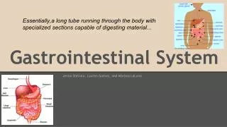
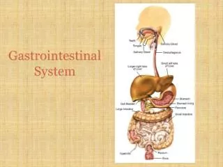
Gastrointestinal System. Chapter 23. GI: Overview: Organ systems. Gastrointestinal (GI) tract [Alimentary canal] a continuous muscular digestive tube Digests : breaks food into smaller fragments Absorbs : digested material is moved through mucosa into the blood Eliminates :

E N D
Gastrointestinal System Chapter 23
GI: Overview: Organ systems • Gastrointestinal (GI) tract [Alimentary canal] a continuous muscular digestive tube • Digests: • breaks food into smaller fragments • Absorbs: • digested material is moved through mucosa into the blood • Eliminates: • unabsorbed & secreted wastes.
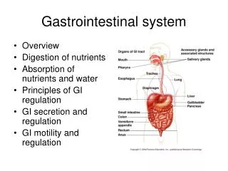
Organ systems • Includes: • Mouth, pharynx & esophagus • Stomach • Small intestine • Large intestine • Accessory digestive organs: teeth, tongue, gall bladder, salivary glands, liver & pancreas Figure 23.1
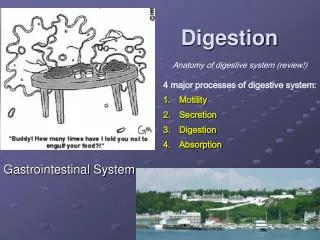
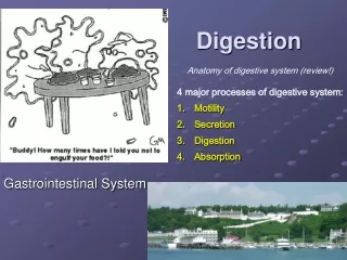
Processes • Ingestion • Propulsion • Mechanical digestion • Chemical digestion • Absorption • Defecation
Processes • Ingestion: obtaining food • Propulsion: moves food along the GI tract by peristalsis (wave-like muscular contraction) • Mechanical digestion : • chewing & mixing with saliva • mixing in stomach • segmentation (local constriction in intestine to mix food & digestive juices)
Processes • Chemical digestion: breaks down food to molecular fragments (monomers) (Hydrolysis). • Begins in the mouth with saliva & continues into the small intestine. • Absorption: movement of nutrients across the mucosal membrane into blood/lymph • Defecation: eliminates unused/indigestible & secreted substances from the body
Functional Considerations : • Substances in the GI tract lumen are outside of the body. • Multiple sensors & receptors line the GI tract to monitor contents & respond to conditions. • Controls: intrinsic (local control) & extrinsic (CNS)
Peritoneum : serous membrane • Visceral peritoneum: covers the external surfaces of most digestive organs • Parietal Peritoneum: lines the body wall • Peritoneal Space: potential space containing fluid that separates the visceral & parietal peritoneum Figure 23.5a
Peritoneum • Mesentery: double layer of peritoneum fused together that extends to the organs from the posterior body wall. • Provides support for the organs • Provides support for vessels & nerves supplying the organs Figure 23.5a
Peritoneum • Retroperitoneal organs • Organs that adhere to the posterior abdominal wall & lose their peritoneum by resorption • Parts of the large & small intestine & most of the pancreas; (also kidneys) Figure 23.5b
GI blood supply • Blood supply: about 25% of cardiac output • Arterial: Abdominal aorta g celiac trunk • Celiac trunk g Hepatic, splenic & gastric branches which serve the liver, spleen & stomach • Celiac trunk g superior & inferior mesenteric branches serve small & large intestine
Histology • GI tract wall has 4 layers: • Mucosa • Submucosa • Muscularis Externa • Serosa or Adventitia
Histology of the Alimentary Canal Figure 23.6
Histology • Mucosa: The epithelial membrane that lines the GI tract from mouth g anus. • Secretes mucous, digestive enzymes & hormones • Absorbs nutrients • Protects from disease & from the GI contents
Histology • Mucosa; 3 layers: • Epidermis • Lamina propria (loose ct : contain capillaries & some elements of MALT) • Muscularis mucosa
Histology • Submucosa: moderately dense CT with blood, nerve, lymph vessels & lymphoid follicles; rich in elastic fibers • Muscularis externa: smooth muscle • Responsible for peristalsis & segmentation • Circular layer • Longitudinal layer • Sphincters: in some areasthe circular layer thickens; act as valves
Histology • Serosa of intraperitoneal organs = visceral peritoneum • Esophagus has an outer covering of fibrous connective tissue = adventitia • Retroperitoneal organs: visceral serosa on the surface facing the peritoneal cavity & adventitia on the surface facing the body wall.
Nerves • Intrinsic: (Local): Short reflex • Submucosal nerve plexus:regulates glands & mucosal muscle • Myenteric plexus: controls GI wall & GI motility • Extrinsic: (CNS): Long reflex • Parasympathetic NS: enhances gut motility & secretion • SNS: inhibits gut motility & secretion
Nerves Figure 23.4 • Intrinsic: (Local): Short reflex • Submucosal nerve plexus: regulates glands & mucosal muscle • Myenteric plexus: controls GI wall & GI motility • Extrinsic: (CNS): Long reflex • Parasympathetic NS: enhances gut motility & secretion • SNS: inhibits gut motility & secretion
Functional Anatomy: Mouth • Mouth: lips, palate, & tongue • Mouth cavity = Buccal cavity
Functional Anatomy: Mouth • Lips: extend from inferior margin of the nose to the superior margin of the chin. Red area = red margin, is poorly keratinized & lacks sweat or sebaceous glands. • Palate: • Hard palate: rigid surface against which food is forced in chewing • Soft palate: muscular structure that rises & blocks off the nasopharynx during swallowing
Functional Anatomy: Mouth • Tongue: muscular tentacle composed of interlaced muscle fibers that grips & repositions food, mixes food with saliva & compresses food to form a food bolus, prior to swallowing.
Functional Anatomy: Mouth • Filiform papillae: rough surface • Fungiform papillae: house taste buds • Circumvallate papillae: house taste buds, • Foliate papillae: posterolateral; taste buds
Functional Anatomy: Mouth • Salivary Glands: intrinsic & extrinsic • Intrinsic glands: scattered throughout the buccal cavity mucosa • Extrinsic glands: supply most of the saliva; outside buccal cavity & supply secretions via ducts: • Parotid • Submandibular • Sublingual
Functional Anatomy: Mouth • Composition of saliva: • 97-99.5% H2O • Electrolytes: • pH 6.75-7.0 • Amylase: (digestive enzyme) • Proteins: mucin, lysozyme, & IgA • Protection from microbes by saliva: • IgA: immunglobulins in secretions • Lysozyme: bacteriostatic (inhibits bacterial growth) • Cyanide • Defensins: local antibiotic activity & when activated promote chemotaxis by WBCs • Normal flora: convert salivary components to nitrates then to NO. NO is toxic & bacteriocidal
Functional Anatomy: Mouth • Control of Salivation: • Continuous baseline secretory activity • With food ingestion, salivation increases dramatically • Parasympathetic NS: chemoreceptors & pressoreceptors stimulate salivatory nuclei to increase salivation
Functional Anatomy: Mouth, Pharynx • Teeth: • Primary: 2I 1C 2M x 2 = 20 2I 1C 2M • Permanent: 2I 1C 2PM 3M x 2 = 32 2I 1C 2PM 3M • Structures • Crown: exposed above gingiva (gum) • Root: anchored by periodontal ligament to the bone by a fibrous joint (gomphosis) Figure 23.07 Figure 23.11
Functional Anatomy: Throat & Esophagus • Pharynx: oropharynx & laryngopharynx; muscular wall propels food to the esophagus • Esophagus: • Muscular 25cm tube from laryngopharynx to stomach • Passes through the diaphragm at the esophageal hiatus • Gastroesophageal (cardiac) sphincter: A physiologic sphincter that helps keep esophagus closed when empty
Functional Anatomy: Esophagus • Esophagus (continued) • Wall has all 4 GI tract tunics: • Epithelial layer changes at the junction with the stomach from stratified squamous epithelium to simple columnar epithelium • Esophageal mucous glands lubricate food bolus • Muscularis externa • Superior 1/3 of muscularis externa is skeletal muscle • Middle 1/3 is mixed skeletal & smooth • Lower 1/3 is smooth muscle • Adventitia: external covering
Digestive Processes: Mouth, Pharynx & Esophagus • Ingestion • Mechanical digestion: chewing • Chemical digestion: mixing food with saliva • Propulsion: swallowing & initiating peristalsis
Functional Anatomy: Stomach • Cardiac region: narrow, receives food bolus • Fundus: bulge that extends supero-laterally to the cardia, reaches the diaphragm • Body: mid-portion • Pyloric antrum : funnel shaped portion narrows to form the; • Pyloric canal • Pylorous • Pyloric sphincter • small intestine • Rugae • longitudinal mucosalfolds • volume about 4L Figure 23.14a
Microscopic Anatomy : Stomach • Stomach: has the 4 tunics of the GI tract. • Epithelium: Simple columnar epithelium (goblet cells-mucous); • Muscularis externa has an additional oblique layer of muscle (allows another dimension of contraction).
MicroscopicAnatomy : Stomach • Gastric glands secrete gastric juices Figure 23.15
Microscopic Anatomy : Stomach • Mucous neck cells: in the duct portion Figure 23.15
Microscopic Anatomy : Stomach • Gastric glands secrete gastric juices • Mucous neck cells: in the duct portion • Parietal cells: mid portion secrete HCl & intrinsic factor for B12 absorption Figure 23.15
Microscopic Anatomy : Stomach • Gastric glands secrete gastric juices • Mucous neck cells: in the duct portion • Parietal cells: mid portion of glands secrete HCl & intrinsic factor • Chief cells: base of gland; secretes pepsinogen a precursor molecule to pepsin (an enzyme that digests protein) Figure 23.15
Microscopic Anatomy : Stomach • Enteroendocrine cells: secrete multiple hormonal products; • Gastrin, histamine, endorphins, serotonin, cholecystokinin, & somatostatin, which influence several digestive system organs Figure 23.15
Microscopic Anatomy : Stomach • Mucosal barrier: protects the stomach from its own secretions • Viscous mucous overlies a thick coating of HCO3- rich mucous • Tight junctions between epithelial cellPM of glandular cells are impermeable to HCl • Epithelium is replaced every 3-6 days
Digestive Processes (Stomach) • Acts as a holding vessel for ingested food • Participates in mechanical & chemical digestion • Propulsion: Delivers its product (chyme) to the small intestine
Digestive Processes (Stomach) • Protein digestion: HCl denatures protein • HCl activates pepsinogen to pepsin • Pepsin breaks peptide bonds of proteins • Rennin: an enzyme that breaks down casein (milk protein) secreted in infants • Intrinsic factor: required for Vit. B12 absorption (needed to mature RBC); • Absence of B12 results in pernicious anemia
Regulation of gastric secretion (3 phases) • Cephalic Phase • Gastric Phase • Intestinal Phase
Cephalic phase: Stimulation • Cephalic phase: CNS response to presentation of food; enhances gastric gland secretion • Loss of appetite; satiety / depression Cephalic phase: Inhibition
Gastric phase: Stimulation • Gastric phase: food entering stomach; • Stretch • Change in pH (increase) • Peptides • All cause increased gastric gland secretion
Gastric phase: Stimulation • Stretch: reflex arc causes increased Acetylcholine release which then causes increased gastric gland secretions • Increased pH / polypeptides / caffeine • All enhance Gastrin secretion by enteroendocrine Gcells
Gastric phase: Stimulation • 3 chemicals: cause enhanced HCl secretion through 2nd messenger systems • Gastrin • Acetylcholine • Histamine
Gastric phase: Inhibition • pH <2.0 inhibits Gastrin secretion • SNS inhibits Gastrin (G cell) activity
Intestinal phase: Stimulation • Excitatory: As chyme enters the duodenum the mucosa secretes entericgastrin which stimulates secretion by gastric glands
Intestinal phase: Inhibition • Inhibitory: As more chyme fills the small intestine, the enterogastricreflex is triggered • Inhibits CNS stimulation • Inhibits local reflexes • Controls gastric emptying • Activates sympathetic fibers that tighten the pyloric sphincter
Regulation of Gastric Activity Figure 23.16