Download
1 / 82
820 likes | 944 Views
INFLAMMATORY BOWEL DISEASE. BY Dr. Nwalozie J.C. Outline. Introduction Classification Epidemiology Aetiopathogenesis Pathology Clinical presentation -G.I. Complications -Extraintestinal manifestations Differential diagnoses Investigations/Work- up Treatment Prognosis

E N D
INFLAMMATORY BOWEL DISEASE BY Dr. Nwalozie J.C.
Outline • Introduction • Classification • Epidemiology • Aetiopathogenesis • Pathology • Clinical presentation -G.I. Complications -Extraintestinal manifestations • Differential diagnoses • Investigations/Work- up • Treatment • Prognosis • Summary
INTRODUCTION • Inflammatory bowel disease(IBD) is a chronic idiopathic , immune- mediated gastrointestinal condition that arises from a dysregulated immune response to host intestinal microflora. • There is a genetic predisposition. • They pursue a protracted relapsing and remitting course(usually extending over years) & give rise to extraintestinal manifestations.
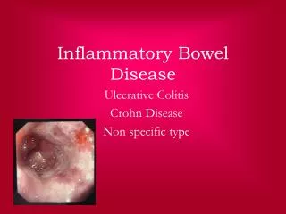
Classification • 2 major types(Typical IBD): Ulcerative colitis(UC)/Colitis ulcerosa & Crohn’s disease(CD)/ Crohn syndrome/Regional enteritis. • Atypical IBD: Lymphocytic colitis Collagenous colitis Ischaemic colitis Diversion colitis Indeterminate colitis Behcet’s disease
EPIDEMIOLOGY • Incidence increasing:0.5-24.5/100,000 person-years(UC). 0.1-16/100,000 person-years(CD). • Overall prevalence:396/100,000 person-years UC:80-150/100,000 person-years CD:25-100/100,000 person-years • The incidence of IBD varies within different geographic areas • Highest incidence occurs in Europe, United Kingdom and North America. • Jewish > non-Jewish white > African American >Hispanic>Asian
The peak age of onset is between 15-30yrs. A second peak occurs between the ages of 60-80yrs • M:F for UC is 1:1 and for CD is 1.1-1.8 :1 • More common in developed countries, colder climates, urban areas, higher socio-economic classes. • Appendectomy & cigarette smoking are protective in UC but are assoc. with increased risk for CD. • OCP use assoc. with increased risk of CD.
A retrospective study was done by Nwosu M.N et al between January 2007 and June 2010 at 3 teaching hospitals in Southern Nigeria. -Diagnosis was made from clinical features, colonoscopic and histopathologic findings. -12 patients presented with IBD: 8(66.7%) were males and 4(33.3%) were females. Age ranges from 18 to 80yrs with a median of 25.5yrs.
AETIOPATHOGENESIS • The aetiology of IBD is unknown.
Genetic susceptibility • A familial disease(5-10%) • Risk is increased(10%) among 1st degree relatives,5% if a parent has IBD & 36% if both parents have it. • Association with genetic syndromes( Turner, Hermansky-Pudlak, WAS,CGD,IPEX etc) • There is increased concordance of the disease in monozygotic twins in comparison to dizygotic twins • 163 susceptibility loci(as at 2012) • Genes associated : NOD 2, ATG16LI, IRGM, JAK2, STAT 3(innate immunity & autophagy), XBP1, ORMDL3, OCTN(ER stress & metabolism), IL23R, IL12B, IL10,PTPN 2(adaptive immunity), MST1, CCR6, TNFAIP3( devt. & resolution of inflammation)
Others: ECM 1,IL-26(12q15),1p36. HLA DRB0103,-0701,MICA 010,HLA B44,HLA B27 have modifying effect. • Presence of gene overlap. • Genetic influences increases the risk of 1 form while decreasing the risk for another.
ENVIRONMENTAL FACTORS • Nutrition- Many food and food components have been suggested to play a role in the aetiopathogenesis of IBD eg high sugar or fat intake.E3N prospective study: high animal protein • Smoking • Appendectomy • NSAIDS • Psychological factors • Hygiene
The Intestinal microbiota • Dysbiosis • Bacterial antigens • Specific bacterial pathogens • Defective chemical barrier/intestinal defensins • Impaired mucosal barrier function • Butyrate
HOST IMMUNE RESPONSE • Normally the mucosal immune system is unreactive to luminal content due to oral(mucosal) tolerance • This tolerance is induced by multiple mechanisms – deletion or anergy of antigen reactive T cells, induction of CD4 T cell type that suppress inflammation (those expressing the fox p3 transcription factor) • In IBD, this suppression is altered leading to uncontrolled inflammation
In IBD there is inappropriate innate immune sensing and reactivity to commensal bacteria triggering the inflammation pathway • This pathway entails activation of CD4 T cell that secrete inflammatory cytokines which recruits other inflammatory cells • These cytokines are also normally produced in response to infection but are turned off at appropriate time to limit tissue damage. But in IBD, their activity are not regulated resulting in the disease condition.
PATHOLOGY • Ulcerative colitis- there are macroscopic and microscopic features Macroscopic Features • It is a mucosal disease that usually involves the rectum and extends proximally to involve all or part of the colon • There are no skip areas • The mucosa is erythematous and has fine granular surface resembling sandpaper.
In severe disease the mucosa is hemorrhagic, edematous and ulcerated. • In long standing disease, inflammatory polyps as a result of epithelial regeneration. • In remission mucosa may appear normal but in patients with many years of disease it appears atrophic. it can be narrowed and shortened. Microscopic Features • Findings are limited to mucosal and submucosa • The crypt architecture of the colon is distorted. They can be bifid and reduced in number
There can also be cryptitis and cryptal abscess • There is plasma cells and multiple basal lymphoid aggregates • Mucosal vascular congestion with edema and focal hemorrhage • There is inflammatory cell infiltrate of plasma cells, neutrophil, lymphocytes and macrophages • If ileum is involved there is villous atrophy, neutrophil and mononuclear infiltration of lamina propria
Crohn’s disease Macroscopic features • It can affect any part of the G I tract from mouth to the anus • 30-40%(small bowel disease), 40-55%(terminal ileum and ascending colon), 15-25%(large bowel alone) • Affectation is segmental with skip areas in the midst of diseased intestine • It is a transmural process • In mild disease there is aphthous or superficial ulceration • Perirectal fistulas, fissures, abscess, and anal stenosis occurs in 1/3rd of patients
Rarely it can involve liver and pancreas. • Cobblestone appearance is characteristic of Crohn's dx and is seen on endoscopy and barium study Microscopic features • There are aphthoid ulcerations and focal crypt abscesses with loose aggregate of macrophages which form non Caseating granulomas in all layers of the bowel. • There is submucosal or subserosal lymphoid aggregate, occurring away from areas of ulceration with gross and microscopic skip areas
CLINICAL PRESENTATION • ULCERATIVE COLITIS • Diarrhea (post prandial/nocturnal) • Rectal bleeding • Tenesmus and passage of mucus • Cramps abdominal pain • Few constitutional symptoms- fever, malaise, anorexia On examination Abdomen- slightly distended and tender DRE-tender anal canal, blood on examining finger
CROHN’S DISEASE • About 15% of patients have constitutional symtoms • Site of the disease determines clinical features ILEOCOLITIS • Right lower abdominal pain • Bloody diarrhoea • Weight loss • Right iliac fossa mass • Bowel obstruction`
Jejunoilieitis • Steatorrhea and malabsorption • Nutritional deficiencies • Diarrhea Colitis and perianal disease • Constitutional symptoms • Haematochezia • Diarrhoea • Fecal incontinence, anorectal fistula and perirectal abscess Gastro duodenal disease • Nausea, vomiting, epigastric pain, features of GOO
G.I.COMPLICATIONS Ulcerative colitis- Haemorrhage • Toxic megacolon • Perforation • Stricture • Colonic Ca Crohn’s disease- Adhesions • Fistula formation- enterocutaneous, colovaginal • Perforation • Intra abdominal and pelvic abscess • Intestinal obstruction • Colonic Ca
Extra Intestinal Manifestations • Up to one-third of IBD patients have extra intestinal disease manifestation DERMATOLOGIC • Erythema nodosum- found in 15% of CD patient and 10% of UC patient • Digital clubbing • Pyoderma gangrenosum • Pyoderma vegetans • Sweet syndrome- neutrophilic dermatosis • Psoriasis- shared immunologic basis • Perianal skin tags
Rheumatologic • Arthropathy • Arthralgia • Inflammatory back pain • Ankylosing spondylitis • Sacroilitis Ocular • Occurs in 1-10% of patients • Conjunctivitis • Anterior uveitis/iritis • Episcleritis
Hepatobiliary • Fatty liver & cirrhosis • Cholelithiasis • Primary sclerosing cholangitis Urologic • Calculi formation • Ureteral obstruction • Ileo-vesical fistula Metabolic bone disease • Osteopenia • Osteonecrosis
Others include • Hypercoagulable states • Vasculitides • Endocarditis • Interstitial lung disease • Amyloidosis • Pancreatitis
Differential Diagnosis Infectious diseases • Bacterial- Campylobacter colitis, Salmonellosis, Shigellosis, Clostridium difficile, E.coli (enterohaemorrhagic,enteroinvasive) • Viral- Cytomegalovirus, Herpes simplex • Protozoal- Isospora belli, Entamoeba histolytica • Parasitic- Trichuris Trichura, Strongyloides • Fungal- Histoplasmosis, Candidiasis
Non infectious • Diverticulitis • Ischaemic bowel disease • Radiation colitis • Appendicitis • Neoplasia- Ca ileum, familial polyposis • Drugs- NSAIDS, OCP, Cocaine
Investigations • Full blood count: anaemia, leukocytosis • Acute phase reactants: ESR, CRP • Markers of intestinal inflammation- Fecal lactoferrin, Fecal calprotectin • Reduced albumin levels • Stool culture- to exclude superimposed enteric infection
Endoscopy • Sigmoidoscopy- used to view the rectum and sigmoid colon. Biospies can be taken for histology • Colonoscopy- shows active inflammation with ulcers and pseudopolyps. Complicating carcinoma can also be seen. Biopsies can be taken to determine disease extent. It should not be done in severe attacks for fear of perforation. • Capsule endoscopy
Barium studies • Barium enema- it is used in colonic disease. In ulcerative colitis the colon is shortened,there are pseudopolyps and loss of haustrations. In crohn’s colitis there are strictures and ulcers with skip lesions. • Barium meal and follow through- used to detect ulcers and strictures in the upper gastrointestinal tracts
OTHER IMAGING: • Abdominal ultrasound and CT scan- shows thickened bowel wall and mesentery as well as intra abdominal and paraintestinal abscess • Radionuclide scan- used to localize extraintestinal abscess • Plain abdominal x-ray- shows dilated bowel and multiple air fluid level in intestinal obstruction.
Serological markers • Can be used 2 differentiate btw CD and UC and help in predicting the course of the disease Perinuclear antineutrophil cytoplasmic antibodies(pANCA) – this is found in 60-70% of UC patients and 5-10% of CD patients, 5-15% of 1st degree relatives of UC patients are pANCA +ve. 2-3% of the general population are pANCA +ve. They are often associated with pancolitis, early surgery, primary sclerosing cholangitis
Anti Saccharomyces cerevisiae antibodies (ASCA)- found 60-70% of CD patients and 10-15% of UC patients.5% of non IBD patients are +ve. • Outer membrane porin C(OMPC)- about 55% of CD patients are +ve. Most are likely to have intestinal obstruction. • pANCA +ve with ASCA –ve results showed 57% sensitivity and 97% specificity for UC • pANCA –ve with ASCA+ve showed 49% sensitivity and 97% specificity for CD
Anti flagellin(anti CBir1)- identified in 55% of CD patients. It is associated with small bowel disease and is usually fibrostenosing type.
TREATMENT • The management of IBD involves the physician, surgeons, radiologist and dieticians. Medical Management(Stepwise approach) • Aminosalicylates(5ASA)- sulfasalazine, melsalazine, olsalazine, balsalazide • Used in the treatment of both CD and UC. • Sulfasalazine consist of sulfapyridine linked to 5ASA via an azo bond
Intestinal bacteria breaks the azo bond releasing sulfapyridine which is absorbed and excreted in urine.5ASA stays in the lumen in contact with the mucosa an eventually excreted in feaces. • Mechanism of action: modulates cytokine release from the mucosa thereby regulating inflammation • Side effects: abd. discomfort attributed to salicylate effect on the GI tract, folate deficiency due to folate competition with sulfasalazine for absorption. Others include skin eruptions, bone marrow suppression.
Other 5ASA preparations- much of the side effects are related to the sulfa portion. So some preparations have the sulfa portion replaced. • They include: -Olsalazine(diperitum )- consist of 2 5ASA moieties joined by an azo bond, requires bacteria degradation in the colon -Ascol- controlled release tablet form of 5ASA encapsulated by acrylic resin that dissolves at PH higher than 6.0
Pentasa- controlled release formulation of 5ASA encapsulated in ethylcellulose microgranules • Balasalazide- 5ASA preperation containing azo bond. 5ASA moiety is release in the colon by cleavage of azo bond by colonic bacteria. • They induce and maintain remission in UC. They have limited role in inducing remission in CD, but no clear role in maintaining remission.
Glucocorticoids • Used in moderate to severe disease of UC and CD • Both oral and parenteral formulations are used • Used in those who are intolerant or unresponsive to aminosalicylates • Oral prednisolone 40-60mg dly, methylprednisolone 40-60mg daily and hydrocortisone 300mg dly • Budesonide is a controlled ileal release steroid with fewer side effects • Dosage: 9mg dly then taper • Side effects: fluid retention, fat redistribution, hypergylcaemia, osteoporosis, myopathy etc
Antibiotics • Those used are metronidazole and ciprofloxacin • Metronidazole is used in active inflammation, fistulous and perianal CD • Dosage is 15-20mg/kg/day in 3 divided doses • Side effects: nausea, metallic taste, disulfiram like reaction, peripheral neuropathy • Ciprofloxacin beneficial in inflammatory, perianal and fistulous CD but has been associated with achilles tendinitis and rupture.