Download
1 / 46
460 likes | 548 Views
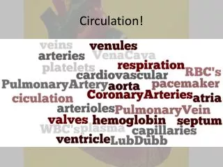
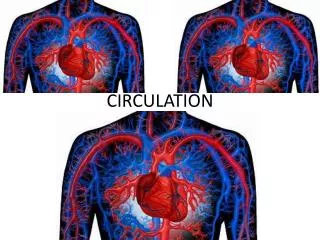
Circulation. Chapter 38. The First ECG. Waller recorded the electrical signals from his bulldog’s heart Signals were transmitted through skin and saltwater to monitoring device. Figure 38.1 Page 665 . Circulatory System.

E N D
Circulation Chapter 38
The First ECG • Waller recorded the electrical signals from his bulldog’s heart • Signals were transmitted through skin and saltwater to monitoring device Figure 38.1 Page 665
Circulatory System • Accepts oxygen, nutrients, and other substances from the respiratory and digestive systems and delivers them to cells • Accepts carbon dioxide and wastes from cells and delivers them to respiratory and urinary systems for disposal
Functional Connections food, water intake oxygen intake DIGESTIVE SYSTEM RESPIRATORY SYSTEM elimination of carbon dioxide nutrients, water, salts carbon dioxide oxygen CIRCULATORY SYSTEM URINARY SYSTEM water, solutes elimination of food residues rapid transport to and from all living cells elimination of excess water, salts, wastes Figure 38.2 Page 665
Circulatory Systems Open System Closed System pump pump spaces or cavities in body tissues heart aorta Figure 38.3 Page 666
Velocity of Flow Varies • Volume of blood flowing through vessels always has to equal heart’s output • Flow velocity is highest in large-diameter transport vessels • Flow velocity is slowest in capillary beds; blood spreads out into many vessels with greater total cross-sectional area
Flow Analogy river in river out lake 1 2 3 1 2 3 1 2 3 Figure 38.3 Page 666
Vertebrate Systems • Fish • Two-chambered heart pumps blood through one circuit • Amphibians • Heart pumps blood through two partially separate circuits • Birds and mammals • Four-chambered heart pumps blood through two entirely separate circuits
Functions of Blood • Transports oxygen and nutrients to cells • Carries carbon dioxide and wastes away from cells • Helps stabilize internal pH • Carries infection-fighting cells • Helps equalize temperature
Components of Blood Blood 6-8% of body weight Plasma portion (50-60% of total volume) • Water • Proteins • Ions, sugars, lipids, amino acids, hormones, vitamins, dissolved gases Cellular portion (40-50% of total volume) • White blood cells Neutrophils Lymphocytes Monocytes Eosinophils Basophils • Red blood cells • Platelets Figure 38.5 Page 668
Erythrocytes (Red Cells) • Most numerous cells in the blood • Transport oxygen and carbon dioxide • Colored red by oxygen-binding pigment (hemoglobin) • Have no nucleus when mature
Leukocytes (White Cells) • Function in housekeeping and defense • Cell types Basophils Eosinophils B lymphocytes Neutrophils T lymphocytes Macrophages NK cells Dendritic cells Mast cells
Platelets • Membrane-bound cell fragments • Derived from megakaryocytes, which arise from stem cells • Release substances that initiate blood clotting
Blood Cell Development • Stem cells in bone marrow are unspecialized cells that retain the capacity to divide • Some daughter cells of stem cells differentiate to form blood cells • Body must continually replace blood cells
Blood Disorders • Anemias - Too few white cells, or deformed ones • Polycythemias - Too many red cells • Leukemias - Cancer suppresses white cell formation • Mononucleosis - Too many monocytes and lymphocytes
Blood Type in Transfusions • Require that donor and recipient have same blood type • If bloods of incompatible types are mixed, recipient’s immune system will attack and destroy donor cells • This is an agglutination reaction
ABO Blood Type • Type A red cells have one type of marker at surface • Type B red cells have a different type of marker • Type AB cells have both markers • Type O cells have neither marker
Blood Type Compatibility Blood type of donor O A B AB O A Blood type of recipient B AB Figure 38.8 Page 670
Rh Blood Type • Based on presence or absence of Rh marker on red cells • Can cause problems during pregnancy • If mother is Rh negative, • has previously carried Rh positive child, • is carrying fetus that is Rh positive • Mother’s antibodies can attack fetal cells
Human Heart Is a Double Pump • Partition separates heart into left and right sides • Each pumps blood through a different circuit
Pulmonary Circuit right pulmonary artery left pulmonary artery Short loop that oxygenates blood capillary bed of left lung capillary bed of right lung pulmonary trunk (to systemic circuit) (from systemic circuit) pulmonary veins heart lungs Figure 38.10 Page 672
Systemic Circuit capillary beds of head and upper extremities aorta (to pulmonary circuit) (from pulmonary circuit) Longer loop that carries blood to and from body tissues heart capillary beds of other organs in thoracic cavity capillary bed of liver capillary beds of intestines Figure 38.10 Page 672
Hepatic Portal System • Carries blood from capillaries in digestive organs to capillaries in the liver • Allows liver to detoxify substances from digestive tract before they are carried to the body
Major Vessels carotid arteries jugular veins ascending aorta superior vena cava pulmonary arteries pulmonary veins coronary arteries hepatic portal vein brachial artery renal artery renal vein inferior vena cava abdominal aorta iliac arteries iliac veins femoral artery femoral vein Figure 38.11 Page 673
Figure 38.12 Page 674 Location of the Heart right lung left lung rib cage pericardium diaphragm Figure 38.12 Page 674
Four Chambers • Each side has two chambers • Upper atrium • Lower ventricle • Valves between atria and ventricles Figure 38.12 Page 674
arch of aorta superior vena cava Heart Anatomy trunk of pulmonary arteries left semilunar valve right semilunar valve left pulmonary veins right pulmonary veins left atrium right atrium left AV valve right AV valve right ventricle left ventricle endothelium and connective tissue inferior vena cava inner layer of pericardium septum heart’s apex Figure 38.12Page 674 myocardium
Cardiac Cycle Diastole (mid to late). Ventricles fill, atria contract. Ventricular systole (atria are still in diastole). Ventricles eject. Diastole (early). Both chambers relax. Figure 38.13 Page 675
Conduction and Contraction SA node • SA node in right atrium is pacemaker • Electrical signals cause contraction of atria • Signal flows to AV node and down septum to ventricles Figure 38.14 Page 675
Blood Vessels • Arteries: main transporters of oxygenated blood • Arterioles: diameter is adjusted to regulate blood flow • Capillaries: diffusion occurs across thin walls Figure 38.15 Page 676
Blood Pressure • Highest in arteries, lowest in veins • Systolic pressure is peak pressure (ventricular contraction) • Diastolic pressure is the lowest • Greatest pressure drop is in arterioles Figure 38.16 Page 676
Controlling Blood Pressure • Cardiac output adjusted by controls over rate and strength of heartbeat • Total resistance is controlled by vasoconstriction of arterioles • Baroreceptor response is main short-term control of blood pressure
Diffusion Zone • Capillary beds are the site of exchange between blood and interstitial fluid • Capillary is a single sheet of epithelial cells • Flow is slow; allows gasses to diffuse across membranes of blood cells and across endothelium
Bulk Flow in Capillary Bed blood to venule outward-directed bulk flow inward-directed osmotic movement blood from arteriole cells of tissue Figure 38.18 Page 678
Pressures in Capillary Bed ARTERIOLE END OF CAPILLARY BED VENULE OF CAPILLARY BED Outward-Directed Pressure: Outward-Directed Pressure: Hydrostatic pressure of blood in capillary: Osmotic force due to plasma proteins: Hydrostatic pressure of blood in capillary: Osmotic force due to plasma proteins: 35 mm Hg 28 mm Hg 15 mm Hg 28 mm Hg Inward-Directed Pressure: Inward-Directed Pressure: Hydrostatic pressure of interstitial fluid: Osmotic force due to interstitial proteins: Hydrostatic pressure of interstitial fluid: Osmotic force due to interstitial proteins: 0 23 mm Hg 0 3 mm Hg Net Ultrafiltration Pressure: Net Reabsorption Pressure: (35 - 0) - (28 - 3) = 10 mm Hg (15 - 0) - (28 - 3) = -10 mm Hg ULTRAFILTRATION FAVORED REABSORPTION FAVORED
Net Bulk Flow • Normally, ultrafiltration only slightly exceeds reabsorption • Fluid enters interstitial fluid and is eventually returned to blood by way of the lymphatic system • High blood pressure causes excessive ultrafiltration and results in edema
The Venous System • Blood flows from capillaries into venules, then on to veins • Veins are large-diameter vessels with some smooth muscle in wall • Valves in some veins prevent blood from flowing backward Figure 38.15 Page 676
Risk Factors for Cardiovascular Disease • Smoking • Gender (maleness) • Genetic factors • Old age • High cholesterol • Obesity • Lack of exercise • Diabetes mellitus
Hypertension • Blood pressure above 140/90 • Tends to be genetic • May also be influenced by diet • Contributes to atherosclerosis • “Silent killer” - few outward signs
Atherosclerosis • Arteries thicken, lose elasticity, and fill up with cholesterol and lipids • High LDL increases risk
Arrhythmias • Bradycardia - slow heart rate • Tachycardia - 100+ beats/minute • Atrial fibrillation - irregular heartbeat • Ventricular fibrillation - uncontrolled contraction of ventricles; quickly fatal
Hemostasis • Blood vessel spasm, platelet plug formation, blood coagulation • Clotting mechanism • Prothrombin is converted to thrombin • Fibrinogen is converted to fibrin • Fibrin forms net that entangles cells and platelets
Lymphatic System • The circulatory system is leaky • Some fluid is forced out of the smallest vessels and into the interstitial fluid • Vessels of the lymphatic system pick up this fluid, filter it, and return it to the circulatory system
Lymph Vascular System • Fluid enters lymph capillaries • Capillaries merge into lymph vessels • Lymph vessels converge into ducts that funnel fluid into veins in the lower neck Figure 38.24 Page 683
Lymph Nodes • Located at intervals along lymph vessels • Act as a filter for lymph • Contain lymphocytes that can recognize a foreign invader Figure 38.24 Page 683
Lymphoid Organs • Central to the body’s defense • Tonsils • Spleen • Thymus gland Figure 38.24 Page 683