Download
1 / 1
10 likes | 110 Views
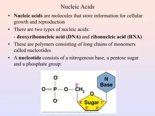
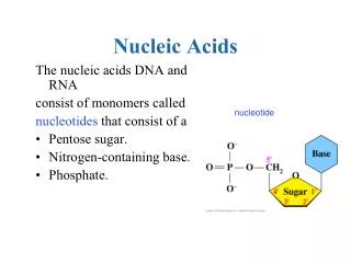
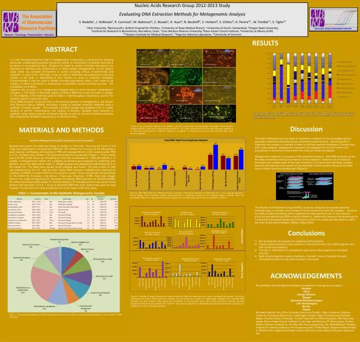
Nucleic Acids Research Group 2012-2013 Study Evaluating DNA Extraction Methods for Metagenomic Analysis V. Nadella 1 , J. Holbrook 2 , R. Carmical 3 , M. Robinson 4 , C. Rosato 5 , H. Auer 6 , N. Beckloff 7 , Z. Herbert 8 , S. Chittur 9 , A. Perera 10 , W. Trimble 11 , S. Tighe 12

E N D
Nucleic Acids Research Group 2012-2013 Study Evaluating DNA Extraction Methods for Metagenomic Analysis V. Nadella1, J. Holbrook2, R. Carmical3, M. Robinson4, C. Rosato5, H. Auer6, N. Beckloff7, Z. Herbert8, S. Chittur9, A. Perera10 , W. Trimble11, S. Tighe12 1Ohio University, 2Nemours/A.I. DuPont Hospital for Children, 3University of Texas Medical Branch, 4 University of Zurich, Switzerland, 5Oregon State University, 6Institute for Research in Biomedicine, Barcellona, Spain, 7Case Western Reserve University, 8Dana Farber Cancer Institute, 9University at Albany-SUNY, 10 Stowers Institute for Medical Research, 11Argonne National Laboratory, 12University of Vermont. A B C Bacillus cereus Enterococcus faecalisKlebsiellaterrigena D E F Bacillus megaterium Micrococcus luteus Pseudomonas aeruginosa G H I RESULTS Rhodospirillum rubra Sporosarcina ureae Streptomyces griseus ABSTRACT J K L After Multi-enzyme digestion Bacterial Cocktail Staphylococcus epidermidis It is well recognized that the field of metagenomics is becoming a critical tool for studying previously unobtainable population dynamics at both an identification of species level and a functional or transcriptional level. Because the power to resolve microbial information is so important for identifying the components in a mixed sample, metagenomics can be used to study nearly any possible environment or system including clinical, environmental, and industrial, to name a few. Clinically, it may be used to determine sub-populations colonizing regions of the body or determining a rare infection to assist in treatment strategies. Environmentally it may be used to identify microbial populations within a soil, water or air sample, or within a bioreactor to characterize a population- based functional process. The possibilities are endless. However, the accuracy of a metagenomics dataset relies on three important "gatekeepers" including 1) The ability to effectively extract all DNA or RNA from every cell within a sample, 2) The reliability of the methods used for deep or high-throughput sequencing, and 3) The software used to analyze the data. Since DNA extraction is the first step in the technical process of metagenomics, the Nucleic Acid Research Group (NARG) conducted a study to evaluate extraction methods using a synthetic microbial sample. The synthetic microbial sample was prepared from 11 known bacteria at specific concentrations and ranging in diversity. Samples were extracted in duplicate using various popular kit based methods as well as several homebrew protocols then analyzed by NextGen sequencing on an IlluminaHiSeq. Figure 5: Sequence coverage of different bacterial strains from different DNA extraction procedures. X-axis represents the various DNA extraction procedures and Y-axis has the percentage of sequence corresponding to different bacterial strains. Discussion Figure 2: (A-J) Bacterial cultures were diluted 1:100 and enumerated microscopically using Sybr Green/Acridine orange with the C-chip micro hemocytometer at 650 X . (K) Final cocktail was 0.25 OD at 600 nM as per NanoDrop at 1mm and enumerated at 1.08E+08 cells/80 uL of sample. (L) Staining of bacterial cells and DNA after multi enzyme digestion. MATERIALS AND METHODS The field of Metagenomics has been an important contributor to the knowledge base for population geneticists studying natural and environmental systems. Discerning which organisms are present in a sample of water or soil has revealed challenges in sample prep and in data analysis. Metagenomic research has expanded into the clinic where sub populations in microenvironments within the body are being investigated. Metagenomic research is increasing in the published literature. With DNA extraction as the first step toward discovering the presence of microorganism. However not all extraction techniques are created equal for the lysis of bacteria. It is well known that Gram negative bacterial cell walls are much easier to lyse then that of Gram positives because the latter have a thicker and more durable wall (Figure 6). This Nucleic Acid Research Group (NARG) study was designed to evaluate extraction methods using a synthetic mixed sample of known bacteria at know cell numbers. Goals for the study included identifying which organisms are detected from each of the extraction protocols and determining DNA extraction efficiency. Additionally, because the technology for running high throughput sequences continues to evolve, the assay was developed to utilize the most recent instrumentation (MiSeq, HiSeq2500). Synthetic Metagenomic Sample Components and Preparation Bacteria were grown the stationary phase (2 weeks) on TSA solid. One loop full (2mm) of cell mass was suspended in nuclease free PBS with 30% Ethanol for 72 hours (to fix) followed by a wash step by centrifuging and washing in PBS and resuspended in 0.02% sodium azide/ PBS to 5 mL. Samples were diluted 1:100 and enumerated microscopically (Figure 1). Final cocktail was 0.25 OD at 600 nM as per NanoDrop at 1mm and enumerated at 1.08E+08 cells/80 uL of sample. A metagenomics master mix containing all bacteria were prepared by combining each bacteria as per the table below (Table 1). Shipping tubes were prepared by distributing 80 uL of the master mix. The tubes were quickly mixed, pelleted, and frozen. The total number of cells was determined as 9.0 x10⁷ cells per tube. Seven DNA extraction methods were employed by members of NARG to extract DNA from the bacteria cocktail. These included the Omega Biotek kit, the MoBio Kit, Enzymes + hot phenol + Fast prep, Enzymes+ CTAB+ Fast prep, QiagenGentra Pure Yeast and Bacterial kit, Epicenter Soil Master DNA extraction kit, Modified MolBio Kit method and Sigma RED extract kit. IlluminaNexterra XT standard protocol was used to build libraries with barcodes. 0.79 to 1.44 ng of extracted DNA from each method was used as input material. The libraries were then pooled and run on two lanes of IlluminaHiseq. Table 1: Components of the Synthetic Metagenomic Sample Figure 3: Total DNA yield from different extraction methods. Theoretical estimation of DNA yield from 1.1E+8 cells per sample is around 430ng. Each extraction method was performed in duplicates except for the Prepman-Qiagen method. Note the quantitation data for the Sigma Extract-N-Amp Tissue kit is not vaild due to the chemical composition of the kit. Figure 6: Cell wall compositions of Gram positive and Gram negative bacteria. Gram Negative Gram Positive Conclusions • Not all extraction techniques are created equal for bacteria • Column based extraction may contribute to reduced recovery do to DNA fragment size and column inconsistency • The use of PEG 6000 in a precipitation step may be advantageous to increased recovery • Multi-enzyme digestion seem to facilitate a “broader”range of bacteria that gets extracted but does not help total recovery in this study ACKNOWLEDGEMENTS We gratefully acknowledge the following companies for their generous support Illumina Zymo Omega Biotech Qiagen Epicenter Biotechnologies Life Technologies Mo Bio Sigma We thank Rachel Yoho (Ohio University Genomics Facility), Marcy Kuentzel (UAlbany Center for Functional Genomics), Lydia Zeglin (Oregon State University) and Mehmet Balkan (Portland State University) for their help with the DNA extractions. We thank Amy Janiak (Dana-Farber Cancer Institute) for her help with Nextera XT library preps, Kendra Walton (Stowers institute) for her help with Hiseq sequencing, Jim Vallandingham (Stowers institute) for primary analysis of the sequencing data, Folker Meyer (Argonne National labs) for Bioinformatic support and Aimee Keithly (Illumina) for providing the Illumina sequence kits. Figure 4: Number of reads obtained from IlluminaHiseq for different organisms from various extraction procedures. The X-axis represents the various DNA extraction methods and the Y-Axis the number of usable reads obtained from duplicate DNA extraction for each method. Each panel has information on the bacterial strain, GC content and Gram staining. Genome Alignments were done using Bowtie V3—best-M1. The genome sequence for Klebsiellaterrigenaand Sporosarciniaureaeis not available to do the comparable analysis. Figure 1: Percentage distribution of different bacterial strains in the cocktail of sample that was shipped to various labs for DNA extraction.