Download
1 / 25
260 likes | 281 Views
Unit 6 Nucleic Acid Extraction Methods. Terry Kotrla, MS, MT(ASCP)BB Fall 2007. Purpose. To release nucleic acid from the cell for use in other procedures Must be free from contamination with protein, carbohydrate, lipids or other nucleic acids. Used pure nucleic acids for testing.

E N D
Unit 6 Nucleic Acid Extraction Methods Terry Kotrla, MS, MT(ASCP)BB Fall 2007
Purpose • To release nucleic acid from the cell for use in other procedures • Must be free from contamination with protein, carbohydrate, lipids or other nucleic acids. • Used pure nucleic acids for testing.
Isolation • Routinely isolated from human, fungal, bacterial and viral sources. • Pretreat to make nucleated cells available, • whole blood • Tissue samples • Microorganisms • Need sufficient sample for adequate yield.
Organic Isolation • Must purify DNA by removing contaminants. • Accomplished by using combination of high salt, low pH and an organic mixture of phenol and chloroform. • To avoid RNA contamination add RNAse, enzyme that degrades RNA.
Phenol/Chloroform • Biphasic emulsion forms • Hydrophobic layer on bottom has cell debris. • Hydrophilic layer on top has dissolved DNA • Remove top layer, add cold ethanol, DNA precipitates out.
Inorganic Isolation Methods • Also called “salting out”. • Uses low pH and high salt condition to selectively precipitate proteins. • DNA is left in solution (picture on far left). • Precipitate out DNA with isoproproanol (middle and right side pictures). • I know this is not scientific but the precipitated out DNA is usually referred to as “snotty”.
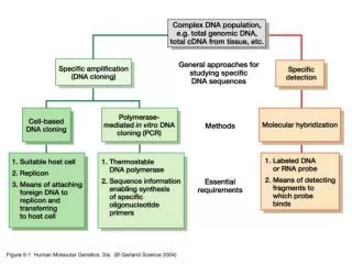
Solid Phase Isolation • More rapid and effective • Use solid matrix to bind the DNA. • Wash away contaminants. • Elute DNA from column • Textbook page 70, Figure 4.3
Solid Phase Isolation • The diagram below explains the attractive properties of solid phase for DNA and RNA.
Crude Lysis • Used for: • Screening large numbers of samples • Isolation of DNA in limited amounts • Isolation from challenging samples, ie, paraffin embedded tissue • Usually not done in clinical laboratory. • Textbook page 71, Figure 4-4
Isolation of Mitochondrial DNA • Mitochondrial DNA is passed from generation to generation along the maternal lineage. • Centrifugation to separate out • Lyse • Precipitate with cold ethanol.
Isolation of RNA • Requires STRICT precautions to avoid sample degradation. • RNA especially labile.
RNAses • RNases are naturally occurring enzymes that degrade RNA • Common laboratory contaminant (from bacterial and human sources) • Also released from cellular compartments during isolation of RNA from biological samples • Can be difficult to inactivate
RNAses • RNAses are enzymes which are small proteins that can renature and become active. • MUST be eliminated or inactivated BEFORE isolation. • CRITICAL to have a separate RNAse free area of lab.
Protecting Against RNAse • Wear gloves at all times • Use RNase-free tubes and pipet tips • Use dedicated, RNase-free, chemicals • Pre-treat materials with extended heat (180 C for several hours), wash with DEPC-treated water, NaOH or H2O2 • Supplement reactions with RNase inhibitors
Total RNA • 80-90% of total RNA is ribosomal RNA. • 2.5-5% is messenger RNA
Organic RNA Extraction • Lyse/homogenize cells • Add phenol:chloroform:isoamyl alcohol to lysed sample, and centrifuge • Organic phase separates from aqueous phase • Organic solvents on bottom • Aqueous phase on top (contains total RNA) • Cellular debris and genomic DNA appears as a “film” of debris at the interface of the two solutions • Remove RNA solution to a clean tube; precipitate RNA and wash with ethanol, then resuspend RNA in water
Affinity Purification of RNA • Lyse cells, and spin to remove large particulates/cell debris • Apply lysate (containing nucleic acids and cellular contaminants) to column with glass membrane • Wash with alcohol to remove contaminants; nucleic acids stick to glass membrane while contaminants wash through. Treat with DNase enzyme to remove contaminating DNA. • Apply water to the column; purified RNA washes off the glass and is collected
Isolation of PolyA (messenger) RNA • Only 2.5-5% • mRNA molecules have a tail of A’s at the 3’ end (polyA tail) • Oligo(dT) probes can be used to purify mRNA from other RNAs • mRNA can be eluted from oligo(dT) matrix using water or low-salt buffer • Textbook page 75, Figure 4.7
Electrophoresis • Analyze DNA and RNA for quality by electrophoresis. • Fluorescent dyes • Ethidium bromide • SybreGreen • Appearance depends on type of DNA isolated. • Will be covered in more detail in unit 7.
Staining with Ethidium Bromide and Sybr Green • DNA is electrophoresed and stained. • Left – Ethidium Bromide Right – Sybr Green • The lane in the left side of the picture on the left is a “ladder” which has fragments of known base pair (bp) sizes. This allows determination of the size of isolated fragments.
Spectrophotometry • Sample absorbances are determined on the spectrophotometer at 260nm and 280nm • nucleic acid (DNA, RNA, nucleotides) absorb light at 260nm • protein absorbs light at 280 nm 230nm: guanidine • A260/A280 ratio is a measure of DNA purity • The absorbance wavelength is directly proportional to the concentration of nucleic acid.
Determining Concentration • Formula Concentration = 260 Reading * Absorbance unit * Dilution factor • One optical density or absorbance unit at 260 nm is equal to • 50 ug/mL for DNA • 40 ug/mL for RNA. • Textbook page 77 has sample calculations.
Determining Purity • The OD at 260nm should be 1.6-2.00 times more than the absorbance at 280nm. • Divide the OD at 260nm by the OD at 280nm to get the ratio. • If the 260nm/280nm ratio is less than 1.6 for DNA, 2.0-2.3 for RNA this indicates contamination, usually with protein. • DNA -If the OD ratio is higher than 2.0 it may be contaminated with RNA. • Ratio of the readings : O.D.260/O.D.280 is a measure of purity. • Pure preparations of DNA and RNA have O.D 260/280 of 1.8 and 2.0 respectively.
Fluorometry • Fluorometry utilizes fluorescent dyes which specifically bind DNA or RNA. • It requires a negative control (to set the zero point on the fluorometer) and a standard of known concentration. • The fluorometer shines light on the sample (excitation) and then measures level of fluorescent light being emitted to the side (at a 90º angle) of the excitation light beam. • The fluorescent dyes are relatively specific to nucleic acids as opposed to protein and other cellular components. • The fluorescence of the dyes increases when they bind nucleic acids.
Fluorometry • Fluorometry is about 1,000x more sensitive than spectrophotometric absorbance (i.e. measurement of A260) and less susceptible to protein and RNA contamination. • However it also does not give a crude measurement of purity (like an A260/A280 ratio) nor does it assure that the DNA or RNA is not degraded (e.g. like size determination by gel electrophoesis). • Do not use glass (spectrophotmetry) cuvettes in a fluorometer because the frosted glass on the side of the cuvette interferes with detection of fluorescent light.