Download
1 / 35
410 likes | 499 Views
Cell Biology. CELL STRUCTURE. Levels of Organization. The levels of life are organized hierarchically: …from lowest to highest: 1. ATOMS eg. carbon, oxygen atoms 2. MOLECULES eg. glucose, proteins, neutral fats, cholesterol 3. ORGANELLES (structures that make up cells)

E N D
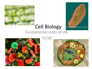
Cell Biology CELL STRUCTURE
Levels of Organization • The levels of life are organized hierarchically: …from lowest to highest: 1. ATOMS eg. carbon, oxygen atoms 2. MOLECULES eg. glucose, proteins, neutral fats, cholesterol 3. ORGANELLES (structures that make up cells) eg. nucleus, mitochondria, endoplasmic reticula 4. CELLS eg. nerve cells, smooth muscle cells 5. TISSUES (comprised of similar cells) eg. nerve tissue, muscle tissue, epithelial tissue, connective tissue
Nerve tissue – sense stimuli and transmit signals throughout animal. Muscle tissue – movement, support; three types: i. Skeletal (Striated) – voluntary bodily movements; ii. Smooth (Visceral) – involuntary contractions (blood vessels, intestine, bladder, etc); iii. Cardiac (Heart) – pumping of heart. Epithelial tissue – Protects: covers outside of body, lines organs and cavities within body. Connective tissue – binds and supports other three tissue types; six types: i. Loose Connective tissue – most widespread; attaches epithelia to other tissues and holds organs in place strong and elastic in nature; ii. Dense Connective tissue – tendons (muscle-to-bone) and ligaments (bone-to-bone); iii. Bone – mineralized (Ca2+ & Mg2+) for support and protection; iv. Cartilage – flexible support in certain areas (nose, ears, trachea, vertebrae); v. Blood – ‘connects’ different parts of body by transporting materials; vi. Fat (Adipose) tissue – pads and insulates body. 6. ORGANS (comprised of various tissue-types) eg. heart, brain, liver. 7. ORGAN SYSTEMS (comprised of various organs) eg. Nervous System, Digestive System
8. ORGANISM (comprised of organ systems) eg. human beings, cats, wildebeest 9. POPULATIONS/COMMUNITIES Definition: CELL – the structural and functional unit of life. It is the smallest structure capable of performing all of the functions necessary for life. FUNCTIONS NECESSARY FOR LIFE: 1. Reproduce ON OWN! (excludes viruses which require cells to repr.) 2. Grow 3. Respire (Metabolize) – Cellular Respiration (mitochondria) glucose (C6H12O6) + O2 CO2 + H2O + ATP energy 4. Contain genetic material (RNA and DNA) 5. Respond to stimuli
Macromolecules Making Up Cells • Carbohydrates (energy, identification) • Proteins (energy, digestion, structure, hormones etc) • Lipids (incl. fats, steroids) – (energy, insulation, hormones, cushioning) • Nucleic Acids (RNA/DNA) – (blueprint for proteins) • All of these molecules are made up of atoms (elements); the main elements comprising living organisms are: C, H, N, O. • Organic = C and H.
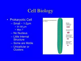
Classifying Cells CELLS PROKARYOTIC EUKARYOTIC -- smaller, less organized -- lack a true nucleus -- lack membrane-bound organelles -- single circular chromosome -- bacteria -- have a true nucleus with a cell membrane -- possess membrane-bound organelles -- 10X size of prok. cells -- focus of Biology 12
“Eu” = True “karyon” = nucleus -- p. 51 Figure 3.3 -- possess a cell wall -- have chloroplasts -- large, contractile vacuole -- p. 50 Figure 3.2 -- main focus of Biology 12 -- lacking the three structures mentioned to the left
Examples of Plant/Animal Cells The three differences…
Cell Structures/Organelles • All cells have a CELL MEMBRANE that acts as a “gatekeeper,” allowing only certain molecules into/out of the cell. • The cell membrane (mb) is therefore described as being SELECTIVELY PERMEABLE. • The cell mb separates the cytoplasm (the inner contents of the cell) from the extracellular fluid (ECF).
The cell mb also compartmentalizes the cell into various organelles which are highly specialized structures with specific functions. • Organelles are to the cell as organs are to our body. • The cell mb is a very fluid, flowing structure but it does work to maintain the cell’s shape (with help from the cytoskeleton).
Structure of the Cell Membrane • The cell mb is made up of a phospholipid bilayer along with embedded proteins. • Carbohydrates can be found associated with the cell mb as well, either attached to the proteins or to the phospholipids themselves. Cholesterol!
A closer look at the phospholipid molecules… • Organelles – small, membrane-bound structures found within the cytoplasm of the cell.
Eukaryotic Cell Organelles A recurring theme in Biology 12 is the relationship between structure and function, both at the cellular and organ system levels. CELL SIZE • As cells mature (post-mitosis), they grow. • However, for an organism to grow, its cells must divide. • Metabolic restrictions impose limits on the max. size of a cell. • As a cell grows, its volume and its surface area increase, but they do so at different rates. In fact, V grows faster than SA. • This is because V is a cubic function (V(sphere) = 4/3πr3), whereas SA is a quadratic function (SA(sphere) = 4πr2).
The SA of a cell governs its ability to import/export materials. • The V of a cell(the part of the cell housing the organelles) imposes the demandsupon the cell’s SA. • Thus, if the demands are increasing faster than the import/export abilities, the cell may eventually reach a point where it cannot sustain living; it will either have to divide or die. • therefore, as a cell grows, its surface area-to-volume ratio (SA:V)decreases.
furthermore, a cell’s nucleus has a limit as to what size of a volume that it can service. • Once a cell divides, its SA:V increases to a level that is conducive to ‘healthy living’. Organelles, which are structures within the cell that are compartmentalized by membranes similar in structure to the cell membrane, are able to sustain many different local environments that facilitate specific metabolic functions that might be incompatible with each other in an ‘open’ (prokaryotic-like) cytoplasm. AND NOW…THE ORGANELLES THEMSELVES!!!
VESICLES (fig. 3.2 p.50; 3.5 p.53; 3.6 p.54) VACUOLES (fig. 3.2 p.50)
ROUGH ENDOPLASMIC RETICULUM (ROUGH ER) (fig. 3.2 p.50; 3.5 p.53)
MITOCHONDRIA (fig. 3.2 p.50; 3.9 p.57) PEROXISOMES (fig. 3.2 p.50; 3.7 p.55)
CYTOSKELETON (fig. 3.2 p.50; 3.10 p.59) CELL WALL (fig. 3.3 p.51)
Relationship Between Organelles with respect to Importing/Exporting Importing: • Cell membrane indents to form a vesicle that is sent to the target if it is a hormone, enzyme, or neurotransmitter. • If the vesicle holds a macromolecule that is to be digested, the vesicle travels to a lysosome. • If the vesicle holds a macromolecule that is to be stored, the vesicle travels to a vacuole.
Exporting: • Proteins created at ribosomes on the Rough ER are funneled into the ER’s lumen where they are modified/packaged into a transition vesicle and sent to the cis-face of the Golgi Body. • Further processing/modifying occurs in the Golgi before the proteins are packaged into another vesicle (secretory vesicle) at the trans-face. • Secretory vesicle travels to the cell membrane and releases contents into the extracellular fluid (ECF) • Proteins created at ‘free’ ribosomes travel through the Golgi similar to Rough-ER-produced proteins, except that instead of being placed into a secretory vesicle, they are placed into a second transition vesicle bound for a lysosome (for digestion), a vacuole (for storage), or anywhere else in the cell where it is in demand. • See handout and fig. 3.6 p.54
Studying the Concepts Questions #1-9 p.65 • Testing Yourself Questions #1-7, 10, 11. • Thinking Scientifically Question #2 p.66 • Understanding the Terms Questions a-e.