Download
1 / 49
490 likes | 499 Views
LE 2-2. Sodium. Chlorine. Sodium chloride. Table 2-1. LE 2-3. Nitrogen deficiency. Iodine deficiency. Review of Chemistry. Helium 2 He. Hydrogen 1 H. 2 He 4.00. Atomic number. Atomic mass. Element symbol. First shell. Electron-shell diagram. LE 2-8. Lithium 3 Li.

E N D
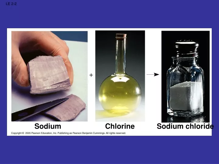
LE 2-2 Sodium Chlorine Sodium chloride
LE 2-3 Nitrogen deficiency Iodine deficiency
Helium 2He Hydrogen 1H 2 He 4.00 Atomic number Atomic mass Element symbol First shell Electron-shell diagram LE 2-8 Lithium 3Li Beryllium 4Be Boron 5B Carbon 6C Nitrogen 7N Oxygen 8O Fluorine 9F Neon 10Ne Second shell Sodium 11Na Magnesium 12Mg Aluminum 12Al Silicon 14Si Phosphorus 15P Sulfur 16S Chlorine 17Cl Argon 18Ar Third shell
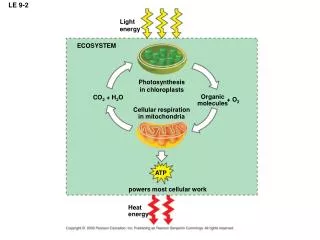
Cloud of negative charge (2 electrons) Electrons Nucleus LE 2-4
A ball bouncing down a flight of stairs provides an analogy for energy levels of electrons. LE 2-7a
Third energy level (shell) Energy absorbed LE 2-7b Second energy level (shell) First energy level (shell) Energy lost Atomic nucleus
Hydrogen atoms (2 H) LE 2-10 Hydrogen molecule (H2)
Ionic Bond Example • Take NaCl as example • Sodium: loses one electron • Chlorine: Gains one electron
Covalent Bonds • No gain or loss of electrons • Only shared electrons • Electrons can be shared equally or unequally
Name (molecular formula) Structural formula Space- filling model Electron- shell diagram Hydrogen (H2) Oxygen (O2) Covalently Bonded Molecules Water (H2O) Methane (CH4)
Nonpolar Covalent Bonds • Share e- equally • The non-polar compounds can NOT be dissolved in water
Polar Covalent Bonds • Share e- unequally • The polar compounds can EASILY be dissolved in water
Hydrogen Bonds • Water (H2O or H–O–H) is a polar molecule • H’s become slightly +, O slightly – • When polar molecules are dissolved in water • The H’s of water molecules are attracted to the negative parts of the solute molecules • Weak bond, easily broken
Water Molecule Animation
pH Scale • pH scale used to indicate acidity and alkalinity of a solution. • Values range from 0-14 • 0 to <7 = Acidic • 7 = Neutral • >7 to 14 = Basic (or alkaline) • pH = -log10 [H+]
pH Scale 0 1 Battery acid 2 Digestive (stomach) juice, lemon juice Vinegar, beer, wine, cola 3 Increasingly Acidic [H+] > [OH–] 4 Tomato juice Black coffee 5 Rainwater Urine 6 Neutral [H+] = [OH–] Pure water 7 Human blood 8 Seawater 9 10 Increasingly Basic [H+] < [OH–] Milk of magnesia 11 Household ammonia 12 Household bleach 13 Oven cleaner 14
Buffers in Biology • Human blood normally 7.4 • Many foods and metabolic processes add or subtract H+ orOH- ions • Reducing blood pH to 7.0 results in acidosis • Increasing blood pH to 7.8 results in alkalosis • Both life threatening situations • Bicarbonate ion (-HCO3) and proteins in the blood buffers pH to 7.4
Biological Molecules • Macromolecules • Carbohydrates • Lipids • Proteins • Nucleic Acids • Functional Groups and Isomers
Short polymer Unlinked monomer Dehydration removes a water molecule, forming a new bond Longer polymer Dehydration reaction in the synthesis of a polymer Hydrolysis adds a water molecule, breaking a bond Hydrolysis of a polymer
Macromolecules • large size • Consist of many repeating units • polymer • Repeating units are called monomers • Category • Example • Subunit(s) • Lipids • Fat • Glycerol & fatty acids • Carbohydrates • Polysaccharide • Monosaccharide • Proteins • Polypeptide • Amino acid • Nucleic Acids • DNA, RNA • Nucleotide
a Glucose b Glucose a and b glucose ring structures
Glucose in medicine • Blood glucose levels: • Fasting normal serum 60-115 mg/dl • Hyper-glycemia >140 mg/dl • Hypo-glycemia: man < 50 mg/dl; woman < 40 • Panic < 40 mg/dl or > 500 mg/dl • Extracellular levels are closely regulated, which is called glucose homeostasis • Factors controlling blood glucose levels: intake, storage and utilization. • Type I and type II diabetes: insulin deficiency and receptor deficiency.
a Glucose b Glucose a and b glucose ring structures
Chloroplast Starch 1 µm Amylose Amylopectin Starch: a plant polysaccharide
Glycogen granules Mitochondria 0.5 µm Glycogen Glycogen: an animal polysaccharide
Cellulose microfibrils in a plant cell wall Cell walls Microfibril 0.5 µm Plant cells Cellulose molecules b Glucose monomer
The structure of chitin. Chitin is used to make a strong and flexible surgical thread that decomposes after the wound or incision heals. Chitin forms the exoskeleton of arthropods. This cicada is molting, shedding its old exoskeleton and emerging in adult form.
CH2—R1 CH—R2 CH2—R3 Types of Lipids:Triglycerides R = R =
Fatty acid (palmitic acid) Dehydration reaction in the synthesis of a fat Ester linkage Fat molecule (triacylglycerol)
Stearic acid Saturated fat and fatty acid. Oleic acid cis double bond causes bending Unsaturated fat and fatty acid. Video
Protein: Levels of Structure • Primary: • the sequence of amino acids • A string of beads of 20 different colors with direction • Secondary: a-helix or b-sheets • Tertiary: Further folding • Quaternary: Subunits
The spiral strands (capture strands) are elastic, stretching in response to wind, rain, and the touch of insects. Abdominal glands of the spider secrete silk fibers that form the web. The radiating strands, made of dry silk fibers, maintain the shape of the web. Spider silk: a structural protein Containing b pleated sheets
Hydrophobic interactions and van der Waals interactions Polypeptide backbone Hydrogen bond Disulfide bridge Ionic bond
5¢ end Nucleoside Nitrogenous base Phosphate group Pentose sugar Nucleotide 3¢ end Polynucleotide, or nucleic acid
Nitrogenous bases Pyrimidines Uracil (in RNA) U Cytosine C Thymine (in DNA) T Purines Guanine G Adenine A Pentose sugars Deoxyribose (in DNA) Ribose (in RNA) Nucleoside components