Download
1 / 35

E N D
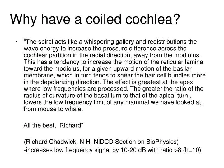
Why have a coiled cochlea? • “The spiral acts like a whispering gallery and redistributions the wave energy to increase the pressure difference across the cochlear partition in the radial direction, away from the modiolus. This has a tendency to increase the motion of the reticular lamina toward the modiolus, for a given upward motion of the basilar membrane, which in turn tends to shear the hair cell bundles more in the depolarizing direction. The effect is greatest at the apex where low frequencies are processed. The greater the ratio of the radius of curvature of the basal turn to that of the apical turn , lowers the low frequency limit of any mammal we have looked at, from mouse to whale. All the best, Richard” (Richard Chadwick, NIH, NIDCD Section on BioPhysics) -increases low frequency signal by 10-20 dB with ratio >8 (h=10)
Mid-basal turn guinea pig O.Corti • Tunnel of Corti efferent fibers OHC bodies 20 u
Tho stiff, longer stereocilia are more flexible. While not proven, longer stereocilia and a wider basilar membrane may aid in low frequency detection.
von Bekesy’s experiments (1950s) • Using cadavers (human and animal) • Temporal bone was rapidly dissected • In saline rubber replaced oval/round windows • Mechanical vibrator replaced stirrup • Cochlear wall opened, exposing Reissner’s mbr • Silver particles on mbr visualized with scope / strobe • Two types of experiments: • Opened length of cochlea, plotted waveforms and envelopes for sounds of different frequencies • Opened cochlea at certain points, measured vibrations at those points with different frequencies
Exposed cochlear partition; stimulation with one frequency FREQUENCY Low High BASILAR MEMBRANE DISPLACEMENT B a s e A p e x PLACE
FREQUENCY High Low BASILAR MEMBRANE DISPLACEMENT B a s e A p e x PLACE
Apex Wider basilar mbr Low frequency
From von Bekesy, 1960 Distance from stapes 24 mm 50 100 200 300 500 Each curve represents the reponses von Bekesy observed when he studied the reponses of one place along the cochlea to stimulation with a range of frequencies with loud tones.
von Bekesy’s findings • Insights: • Sound vibration gives traveling wave with gradual rise and rapid fall off • Cochlea has frequency-specific place code • Limitations: • Used non-physiological amplitude • Needed 130 dB to see Ag+ particles move • Measurements made on cadavers • Normal responses require live animal • The active process • Nobel prize in Medicine, 1961
B a s e A p e x PLACE FREQUENCY LINEAR Low High Bekesy Stimulus BASILAR MEMBRANE DISPLACEMENT INTENSITY
Cochlear Amplifier B a s e A p e x PLACE FREQUENCY NONLINEAR Low High BASILAR MEMBRANE DISPLACEMENT INTENSITY
Displacement as a function of frequency Parameter: sound intensity Threshold as a function Of sound intensity From Sellick, ea 1982
Cochlear Amplifier B a s e A p e x PLACE FREQUENCY NONLINEAR Low High BASILAR MEMBRANE DISPLACEMENT INTENSITY Threshold level
Characteristics of the Active Process • Is frequency specific, following place code of the cochlea • All cellular machinery aligned at frequency • Requires metabolic energy • Not present in dead cochlea • Depends on the endolymphatic cochlear battery • Furosemide decreases [K+], stops process
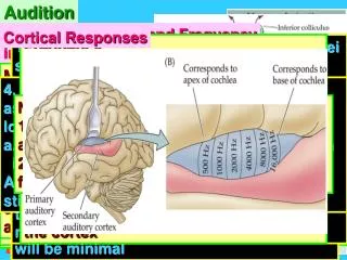
Compare: Principal Receptor Sends frequency-specific Information to brain based on the vibratory pattern of the basilar membrane Receptor/Effector (cochlear amplifier) Provides frequency-specific energy to the basilar membrane. Compressive nonlinearity - enhance responses to low level stimuli. Powered by endocochlear potential.
Efferent Afferent Cochlear nerve axons How can we measure the energy of the cochlear amplifier?
Primary Tones 50 dB SPL 0 0.9 1.0 1.1 2f1-f2 f1 f2 Distortion Product Otoacoustic Emissions (DPOAEs) S. Blatrix & R. Pujol. www.the-cochlea.info Emission generated by OHCs through an active process
The outer hair cell motor protein: Prestin - isolated 2002. One member of a large class of sugar transporters. Co-opted to be a motor protein. Localized to basolateral membrane. Voltage sensitive. From Adler, ea. Hearing Res. 184:27, 2003 Receptor/Effector (cochlear amplifier) Provides frequency-specific energy to the basilar membrane. Compressive nonlinearity - enhance responses to low level stimuli. Powered by endocochlear potential.
Cloned prestin gives motility to tsa cells • Transfected cell sucked into pipet • Response to 200Hz stim by 2 prestin cells (top); control • Reduced by salicylate (comp at anion site) • Responses to 200Hz and 1kHz commands • Fourier transform of responses in d.
Prestin KO mice Electromotility is lost in -/- mice ABRs, DPOAEs are lost in -/- mice From Liberman, etal. Nature 419:300, 2002
Summary - Cochlea • Traveling waves distribute frequency specific excitation along cochlear epithelia • Outer hair cells amplify displacement of basilar membrane - the cochlear amplifier! • Inner hair cells transform frequency specific mechanical activity into electrical activity - auditory encoding based on a place code! • Molecular components are being defined largely through study of deafness genes