Download
1 / 129
1.3k likes | 1.44k Views
Proper structural fold of protein molecule is essential to execute its precise functional mission. Md Abu Reza, PhD. Associate Professor Dept of Genetic Eng & Biotech University of Rajshahi. Bioinformatics Workshop-1. Date : 24 th March, 2012 Venue : Dept of Statistics, RU.

E N D
Proper structural fold of protein molecule is essential to execute its precise functional mission Md Abu Reza, PhD Associate Professor Dept of Genetic Eng & Biotech University of Rajshahi Bioinformatics Workshop-1 Date : 24th March, 2012 Venue : Dept of Statistics, RU Higher Education Quality Enhance Project
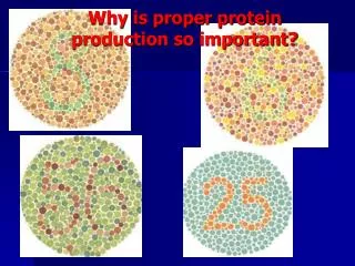
Protein – The Master Molecule • Proteins control all biological systems in a cell • They either act in constituting structure or perform distinct biological function in any physiological system • Many proteins perform their functions independently, the vast majority of proteins interact with others for proper biological activity • To perform the function effectively a proper structure is essential. Without proper structure a protein is useless or even cause malfunction in system • Conformation and functional-group chemistry controls function • Made up of 20 different types of amino-acid monomers • Proteins define what an organism is, what it looks like, how it behaves, etc. (responsible for most phenotype)
What are Proteins ? • Proteinsare biochemical compounds consisting of one or morepolypeptidestypically folded into a globular or fibrous form in a biologically functional way. • A polypeptide is a single linear polymer chain ofamino acidsbonded together bypeptide bonds • 20 natural amino acids join in different permutation and combinations in different lengths • Once linked in the protein chain, an individual amino acid is called aresidue,and the linked series of carbon, nitrogen, and oxygen atoms are known as the main chain orprotein backbone
Amino Acids Lysine with the carbon atoms in the side-chain labeled Amino Terminal Carboxy Terminal
How peptide bonds are formed ? • Here amino acids are both Alanine in which the R group is a single hydrogen. • The carboxyl acid end on the first amino acid is orientated to the amino group of the second amino acid. • The -OH group and -H are removed to form water (condensation reaction). • The bond forms between the terminal carbon on the first amino acid and the nitrogen on the second amino acid. • The backbone of the molecule has the sequence N-C-C-N-C-C • Polypeptides maintain this sequence no matter how long the chain. • The R groups project from the backbone. • As the amino acids are added in translation the polypeptide folds up into it specific shape.
Stereochemistry The CORN Law H H View in 3D
Amino Acid Properties The 20 amino acids can be divided into several groups based on their properties. Important factors are charge, hydrophilicity or hydrophobicity, size, and functional groups water-soluble proteins tend to have their hydrophobic residues (Leu, Ile, Val, Phe, and Trp) buried in the middle of the protein, whereas hydrophilic side-chains are exposed to the aqueous solvent. Livingstone & Barton, CABIOS, 9, 745-756, 1993
Group I: Nonpolar amino acids Group I amino acids are alanine, valine, leucine, isoleucine, proline, phenylalanine, methionine, and tryptophan. The R groups of these amino acids have either aliphatic or aromatic groups. This makes them hydrophobic (“water fearing”). In aqueous solutions, globular proteins will fold into a three-dimensional shape to bury these hydrophobic side chains in the protein interior.
Group II: Polar, uncharged amino acids Group II amino acids are glycine, serine, cysteine, threonine, tyrosine, asparagine, and glutamine. The side chains in this group possess a spectrum of functional groups. However, most have at least one atom (nitrogen, oxygen, or sulfur) with electron pairs available for hydrogen bonding to water and other molecules. Polar aa are hydrophilic.
Group III: Acidic amino acids The two amino acids in this group are aspartic acid and glutamic acid. Each has a carboxylic acid on its side chain that gives it acidic (proton-donating) properties. In an aqueous solution at physiological pH, all three functional groups on these amino acids will ionize, thus giving an overall charge of −1. In the ionic forms, the amino acids are called aspartate and glutamate. .
Group IV: Basic amino acids The three amino acids in this group are arginine, histidine, and lysine. Each side chain is basic (i.e., can accept a proton). Lysine and arginine both exist with an overall charge of +1 at physiological pH. The guanidino group in arginine’s side chain is the most basic of all R groups (a fact reflected in its pKa value of 12.5). As mentioned above for aspartate and glutamate, the side chains of arginine and lysine also form ionic bonds. The chemical structures of Group IV amino acids are
Why Proteins Need Structure ! Functions • Diverse functions related to structure • Structural components of cells • Motor proteins • Enzymes • Antibodies • Hormones • Hemoglobin/myoglobin • Transport proteins in blood
Protein structure - bonding • Interactions (forces) governing protein structure • Covalent Interaction • Peptide bond • Disulfide bond • Non Covalent interaction • Hydrogen bond • Ionic bond (Electrostatic interactions) • Salt bridge • Van-der-Waals interactions • Hydrophobic force
Disulfide bond • Covalent bond between sulfur atoms on two cysteine amino acids • Very strong Intereaction From: Elliott, WH. Elliott, DC. (1997) Biochemistry and Molecular Biology. Oxford: Oxford University Press. p32
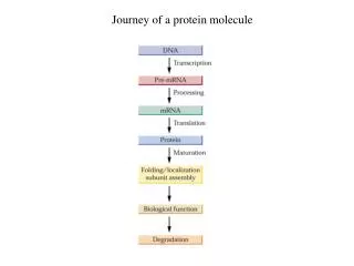
Hierarchical nature of protein structure Primary structure (Amino acid sequence) ↓ Secondary structure (α-helix, β-sheet ) ↓ Tertiary structure (Three-dimensional structure formed by assembly of secondary structures) ↓ Quaternary structure (Structure formed by more than one polypeptide chains)
Primary protein structure Primary structure of insulin • Linear sequence of amino acids forms primary structure • Sequence essential for proper physiological function Bettelheim & March (1990) Introduction to Organic & Biochemistry (International Edition) Philadelphia: Saunders College Publishing, p299
Secondary structure = local folding of residues into regular patterns
Secondary protein structure • Peptide chains fold into secondary structures: • - helix • - pleated sheet • Random coil
Peptide Bonds are Planar • For a pair of amino acids linked by a peptide bond , six atoms lie in the same plane: the carbon atom and CO group of the first amino acid and the NH group and carbon atom of the second amino acid • The C-N distance in a peptide bond is typically 1.32Å • Two configurations are possible for a planar peptide bond. In the trans configuration, the 2 carbon atoms are on opposite sides of the peptide bond. In the cis confi guration, these groups are on the same side of the peptide bond. Almost all peptide bonds are trans
Torsion Angle • In contrast with the peptide bond, the bonds between the amino group and the carbon atom and between the carbon atom and the carbonyl group are pure single bonds. The two adjacent rigid peptide units may rotate about these bonds, taking on various orientations • This freedom of rotation about two bonds of each amino acid allows proteins to fold in many different ways. The rotations about these bonds can be specified by torsion angles • The angle of rotation about the bond between the nitrogen and the carbon atoms is called phi ( ) • The angle of rotation about the bond between the carbon and the carbonyl carbon atoms is called psi ( ) • A clockwise rotation about either bond as viewed from the nitrogen atom toward the carbon atom or from the carbonyl group toward the carbon atom corresponds to a positive value • The and angles determine the path of the polypeptide chain
Ramachandran plot -- showsf and yangles for secondary structures A measure of the rotation of a f and ybond usually lie between - 180 and + 180
f and yangles for secondary structures Secondary structure conformation Residue Conformational Preference
Alpha Helix • In the a-helix, the carbonyl oxygen of residue “i” forms a hydrogen bond with the amide of residue “i+4”. • Although each hydrogen bond is relatively weak in isolation, the sum of the hydrogen bonds in a helix makes it quite stable. • The propensity of a peptide for forming an a-helix also depends on its sequence.
- helix • Shape maintained by hydrogen bonds between C=O and N-H groups in backbone • R groups directed outward from coil From: Elliott, WH. Elliott, DC. (1997) Biochemistry and Molecular Biology. Oxford: Oxford University Press. p28
α-Helix • A loop of 13 atoms is formed between the hydrogen bond. • 3.6 amino acids per turn of helix. • Helices observed in proteins can range from four to over forty residues long, but a typical helix contains about ten amino acids (about three turns). • α-Helix is also called 3.613 helix, compared to π-helix 4.416 and 310 helix. • Proline is the α-breaker.
Propensities for forming α-helical structure Different amino-acid sequences have different propensities for forming α-helical structure. Methionine, alanine, leucine, uncharged glutamate, and lysine ("MALEK" in the amino-acid 1-letter codes) all have especially high helix-forming propensities, whereas proline and glycine have poor helix-forming propensities. Proline either breaks or kinks a helix, both because it cannot donate an amide hydrogen bond (having no amide hydrogen), and also because its side-chain interferes sterically with the backbone of the preceding turn - inside a helix, this forces a bend of about 30° in the helix axis
α-helical coiled coil proteins: Form superhelix Found in myosin, tropomyosin (muscle), fibrin (blood clots), keratin (hair) Examples of α-Helical Proteins: Hair Also fingernails and wool are α-helical proteins; silk is β
β-sheet (-pleated sheet)- • A polypeptide chain, called a β-strand, in a β-sheet is almost fully extended rather than being tightly coiled as in the -helix • The distance between adjacent amino acids along a strand is approximately 3.5Å, in contrast to a distance of 1. 5Å along an helix • sheet is formed by linking two or more strands lying next to one another through hydrogen bonds • All residues in Beta sheet have nearly the same and angle • Hydrogen bonds can only formed between adjacent polypeptide chains. • R groups are directed above and below backbone
Parallel or Anti-parallel -Sheet • The adjacent polypeptide chains in a -sheet can be either parallel or anti-parallel (having the same or opposite amino-to-carboxyl orientations, respectively). H bonds between 2 same aa H bonds between different aa
Examples of β-sheet Proteins: Fatty acid binding protein -> β barrels structure Antibodies more β sheets OmpX: E. coli porin 44
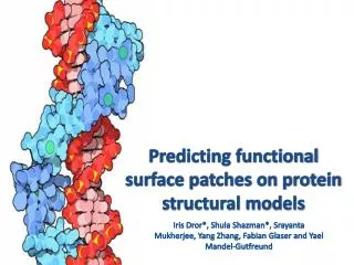
Tertiary Structure: 3D structure of a polypeptide chain Quaternary Structure: Polypeptide chains assemble into multisubunit structures Tetramer of hemoglobin Cell-surface receptor CD4
QUATERNARY STRUCTURE Deoxyhaemoglobin
B-Turns and Loops • -turns allow the protein backbone to make abrupt turns. • Again, the propensity of a peptide for forming b-turns depends on its sequence. • In this reverse turns, the CO group of residue i of a polypeptide is hydrogen bonded to the NH group of residue i + 3 • In other cases, more elaborate structures are responsible for chain reversals. These structures are called loops or sometimes loops (omega loops) to suggest their overall shape
Random coil • Not really random structure, just non-repeating • ‘Random’ coil has fixed structure within a given protein • Commonly called ‘connecting loop region’ • Structure determined by bonding of side chains (i.e. not necessarily hydrogen bonds) From: Elliott, WH. Elliott, DC. (1997) Biochemistry and Molecular Biology. Oxford: Oxford University Press. p27