Download
1 / 27
290 likes | 478 Views
Vesicles, Inclusions, Cytoskeleton. It’s best to view this in Slide Show mode. This module will take approximately 60 minutes to complete. After completing this exercise, you should be able to: Describe the function of each of the following cellular organelles / structures: Endosomes

E N D
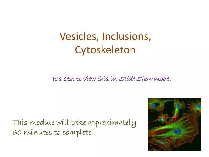
Vesicles, Inclusions, Cytoskeleton It’s best to view this in Slide Show mode. This module will take approximately 60 minutes to complete.
After completing this exercise, you should be able to: • Describe the function of each of the following cellular organelles / structures: • Endosomes • Lysosomes • Secretory granules • Peroxisomes • Lipid droplets • Inclusions • Cytoskeletal elements • Microtubules • Microfilaments • Intermediate filaments • Centrioles This module includes electron micrographs for your viewing pleasure. However, you do NOT need to be able to identify any of these structures in electron micrographs for the histology practical exam.
Endosomes Endosomes are vesicles that pinch off of the plasma membrane. Therefore, the lipid bilayer of endosomes contains plasma membrane proteins, and the lumen contains extracellular components. There are three types of endocytosis: Pinocytosisis nonspecific intake of small molecules. Receptor-mediated endocytosis is specific uptake using receptors. Phagocytosis is the uptake of large molecules and organisms. Figure A is a cartoon showing the process of pinocytosis: the processes of endocytosis and phagocytosis are similar. In figure B, pinocytotic vesicles are shown as clear vesicles just underneath the plasma membrane. Blood vessel lumen
Lysosomes Lysosomesare membrane-bound organelles involved in intracellular digestion. Lysosomal proteins, mostly enzymes, are produced by the rER and processed in the Golgi apparatus. Lysosomes that bud from the trans-Golgi are small, and typically have a dense stain in electron micrographs (red arrows). The right image shows a neutrophil (a white blood cell), with small, purple lysosomes (blue arrows) barely visible with the light microscope. Blood vessel lumen
Lysosomes Lysosomes released from the Golgi-apparatus ultimately fuse with a structure to be degraded, such as an endosome, phagosome, or a senile organelle. Although the size and appearance of lysosomes at this stage varies widely, most have dark clumps within them, presumably a reflection of digested material. Here, Ly, lysosomes, autophagosomes, and * all represent lysosomes. Ly Ly
Lysosomes There are a wide variety of genetic disorders that disrupt the function of lysosomes. As one would expect, disrupting digestive function leads to accumulation of undigested material within the cell (see cartoon), which inhibits normal cellular function. This has given rise to the term lysosomal-storage diseases. nucleus Ly One classic example of a lysosomal storage disease is Tay-Sach’s disease. A is a low-magnificationlight micrograph showing normal neurons (blue arrows) and a cell that has accumulated gangliosides. B is a high-magnification electron micrograph showing part of a cell; note the two large lysosomes (outlined) Ly
Secretory granules Like lysosomes, secretory granules are products of the rER / Golgi network. Their contents vary, depending on the specific function of the cell. Typically, when seen in electron micrographs, they are clustered near the plasma membrane on the side of the cell from which they will be released. This image shows most of one cell, and a portion of one other (bottom left). The lumen is the duct into which these cells secrete the contents of their secretory granules (SG). Note the extensive rER; the Golgi is not as apparent. Recall that secretory vesicles will be present in cells that use regulated secretion – secretion in response to a signal. Cells that secrete constitutively (constant secretion), will have few or no visible secretory granules. SG SG lumen
Secretory granules We have already looked at these cells from the pancreas that demonstrated cytoplasmic basophilia. Recall that they are organized into clusters of cells called acini (one acinus is outlined). The lumen into which these cells secrete their product is at the center of each acinus (green X). Note that the portion of the cell adjacent to the acinus is highly eosinophilic; this is due to the numerous secretory granules contained in these cells. X These pancreatic cells (and other epithelia) are polar. Their apical side is facing the lumen; the side of the cell with the secretory granules. Their basal side is the side nearest the connective tissue; the side with cytoplasmic basophilia.
Secretory granules Video of pancreas showing secretory granules – SL108 • Link to SL 108 • Be able to identify: • Secretory granules
Peroxisomes Peroxisomes (aka microbodies) contain oxidative enzymes, and are involved in detoxification and oxidation of long-chain fatty acids. They are, therefore, prominent in liver cells. In all species except humans, they can be identified by a single colloid density in electron micrographs. Because they are vesicles with a size and appearance similar to lysosomes, one would suspect that peroxisomes are generated in the same manner as lysosomes (i.e. from ER/Golgi). However, peroxisomal proteins are synthesized in the cytosol and imported into the peroxisome. Presumably, the initial structural vesicle that will become a peroxisome arises as a bud from the smooth endoplasmic reticulum.
Lipid Droplets Lipid droplets (L) are droplets of lipid within the cytoplasm (no kidding). Although they tend to be round, they will accommodate their shape to other components in the cell. They can be of various size, although typically they are fairly large, larger than mitochondria. Their density varies with staining in electron micrographs. Although most cells have one or a few lipid droplets, they are prominent in two cell types: Steroid secreting cells that use the lipids as substrates for hormone production. Adipose cells, which are storing energy.
Slide Preparation artifacts Video of H&E cytoplasm – SL52 The upper image and video link (from the original light microscopy homework assignment) were taken from the adrenal cortex. Here we add a detailed micrograph below to show that the pale cytoplasm is due to numerous lipid droplets that are washed away during tissue preparation (blue arrows). The larger spaces labeled lumen are blood vessels. These cells use lipids as substrates for hormone (steroid) production. FYI, for now, other cells that secrete steroid hormones include the Leydig cells of the testis and interstitial cells of the ovary. However, one principle you should understand now: Cells that produce steroid hormones have abundant lipid droplets; cells that secrete peptide or protein hormones (e.g. insulin) have abundant RER and Golgi. lumen lumen
Glycogen Glycogen is a complex carbohydrate most likely present in all cells, but abundant in the liver and skeletal muscle. In electron micrographs, it appears as clusters of dense round particles (Gl), and is often associated with smooth endoplasmic reticulum, which has at least one of the enzymes for glycogen metabolism on its membrane. Kinda looks like clusters of ribosomes. Each individual glycogen particle (red arrow) is approximately the same size and shape as a ribosome. However, glycogen will cluster as seen here, ribosomes do not.
Slide Preparation artifacts Again, another image and video from the microscopy homework assignment. Here, the liver cells contain abundant glycogen, which is the body’s preferred method for storage of carbohydrates. Upon tissue preparation, the glycogen washes away, leaving numerous pale regions within the liver cells. Video of H&E cytoplasm – SL102
Pigments Pigments are cellular deposits. The one you are most familiar with is melanin, which is deposited in skin cells (brown in outlined region), and provides the epithelial cells of the epidermis protection against ultraviolet radiation. This is an H&E stained slide, but the pigment is not stained by this method; it is visible naturally (which is the definition of a pigment).
Pigments Other pigments you will encounter underly potential pathology. For example, aging cells, particularly liver and heart, often exhibit yellowish-brown lipofuscin granules near the nucleus (blue arrow), which are electron-dense in electron micrographs (red arrow). These granules are not harmful, but commonly accumulate within the cell in response to oxidative stress, and, therefore, are suggestive of underlying pathology.
Pigments Another pigment that underlies pathology is hemosiderin. Small amounts of hemosiderin exist normally in many cell types involved in red blood cell breakdown, including macrophages, spleen, and liver. Hemosiderin accumulates within cells as a yellow-brown pigment (left image), and can be specifically visualized using Prussian blue (right image).
Pigments Excess hemosiderin can occur either as a localized or systemic process. Localized hemosiderin accumulation occurs during bruising, when hemorrhaging results in accumulation of hemosiderin within macrophages. The different states of iron during hemoglobin breakdown are reflected in the colors that appear during bruise healing. Systemic hemosiderin accumulation occurs during iron overload. This can occur in situations that include excess iron in the diet or excess hemolysis, among others. In these cases, the hemosiderin accumulates largely in the liver, bone marrow, spleen, and lymph nodes, although it also increases in other organs such as the pancreas and kidneys.
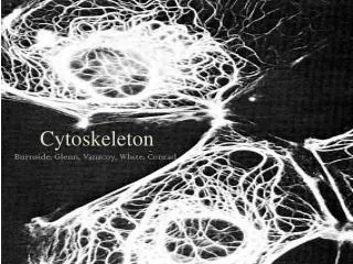
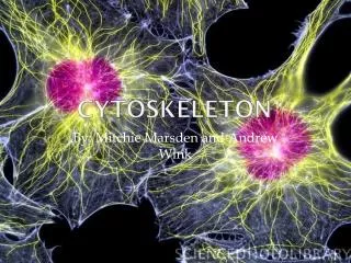
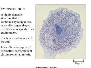
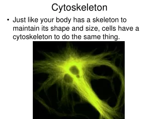
The Cytoskeleton The cytoskeleton is composed thread-like proteins of the cell that are involved in activities such as cell structure and intracellular trafficking. There are three major components of the cytoskeleton: Microtubules – hollow tubes, ~25nm in diameter Microfilaments – solid tubes, 5nm in diameter Intermediate filaments – solid tubes, 10nm in diameter This cultured cell was treated with a solution that dissolved the lipid portion of the plasma membrane. This allowed the soluble cytosolic elements to be washed away, leaving the cytoskeleton behind, which mirrors the shape of the cell. nucleus
The Cytoskeleton Because microtubules are hollow, they appear as parallel double lines in longitudinal sections (left EM), and as rings in cross section (right EM). The major function of microtubules include organelle movement with the cell, movement of cilia and flagella, and movement of chromosomes during mitosis.
The Cytoskeleton As noted on the previous slide and the first laboratory, microtubules are the major component of the mitotic spindle, which is involved in movement of chromosomes during mitosis and meiosis. Agents that disrupt microtubule dynamics, either by preventing microtubule formation (vinca alkaloids or colchicine) or inhibiting microtubule disassembly (Taxol) interrupt mitosis. These drugs target dividing cells, and are effective chemotherapies to treat cancers. Typically, you do not see any cytoskeletal elements in light micrographs. However, the concentration of microtubules in mitotic spindles is high enough that they are readily seen.
The Cytoskeleton Even in high concentrations, microfilaments can only be seen in cells through specialized staining techniques. In this image, the actin filaments are visualized using phallacidin conjugated with a green fluorescent dye (red = mitochondria, blue = DNA). Microfilaments are involved in cell motility and shape, and are major components of microvilli.
The Cytoskeleton The function of intermediate filaments are unclear, though it is likely that, at least for the keratins in skin, they are involved in resistance to shear force at the level of the tissue. In this image, vimentin (an intermediate filament) is red, and actin is green.
The Cytoskeleton To underscore the role of intermediate filaments in tissue integrity, mice were created with a deletion in a keratin intermediate filament protein. These mice were prone to skin blisters, similar to a human condition known as epidermolysisbullosa simplex. Electron microscopic analysis of skin from these mice demonstrated that the blistering occurred due to disruption of the basal aspect of the epidermal cells.
Centrioles Centrioles are paired organelles that are composed of microtubules arranged in 9 triplets. In this image, the centriole on the left is cut longitudinally, while the one on the right is cut in cross section. On the right, you can see 8 of the 9 triplets, the last triplet (at 9 o’clock) is not visible, presumably due to the angle of sectioning.
Centrioles In interphase cells, the centrioles (red) are located in the center of the cell, and are part of the centrosome, a complex of proteins from which radiate the microtubule network (green). The centrosome organizes the microtubule network, thus it is also called the microtubule-organizing center (MTOC). As mentioned before, microtubules are involved in intracellular trafficking of vesicles. In this regard, the centrosome is indeed the “center” of the cell, located adjacent to the Golgi apparatus. Newly processed proteins are packaged in the trans Golgi, and move from the center of the cell outward along these microtubule tracks to various destinations. Incoming vesicles from endocytosis also move toward the centrosome along the same microtubule tracks, some eventually reaching the Golgi apparatus for processing.
Centrioles During mitosis, centrioles are duplicated, and each pair migrates to opposite poles of the cell (arrows) where they play a role in organization of the mitotic spindle.