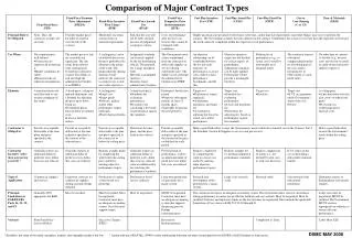
Download
1 / 36
360 likes | 600 Views
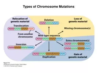
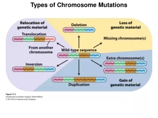
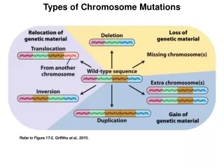
Major types of mutations. Major types of mutations. Major types of mutations. Example for conditional mutation is temperature-sensitive Mutation : Usually the restrictive temperature is high (29°indrosophila) and the phenotype is mutant above it. The permissive

E N D
Example for conditional mutation is temperature-sensitive Mutation: Usually the restrictive temperature is high (29°indrosophila) and the phenotype is mutant above it. The permissive temperature is low (18° in drosophila) and the phenotype is wildtype under it. • loss-of-function is also called knock out or null mutation. • A commontype of frameshift mutation is a single-base addition or deletion. CUG CUG CUG Leu Leu Leu CUG CAU GCU G Leu His Ala
Example (PAH)phenylalanine hydroxylase CGG = codon on mRNA for Argenine (AA) UGG = codon on mRNA for Tryptophan (AA) wildtypemutant DNA: 5’ ……CGG…… .…..TGG……3’ DNA(T): 3’……GCC…… .…..ACC……5’ mRNA: 5’……CGG…… ……UGG......3’ So the Aa (Argenine) will be replaced by the Aa ( Tryptophan), that means mutant protein.
Fragile-X syndrome • X-linked form of mental retardation shows dynamic mutations. The chromosome X tends to fracture near the end of the long arm and it is called fragile-X and the disease is called fragile-X syndrome. • This syndrome affects 1 in 2500 children and is second only to Down syndrome as a cause of inherited mental impairment. • Males are more affected than females.
The molecular basis is trinucleotide repeat of the form CGG (or CCG on the other strand) in the part where the breakage takes place. • Normal X-chromosome has 6 to 54 repeating unit. Affected persons have 230 to 2300. • The premutation has an intermediate number of copies ranging from 54 to 230.
The premutation increases in copy number when transmitted through only females and is called trinucleotide expansion. This occurs not in germ line but in somatic cells of the early embryo. • Amplification to 230 copies or more causes silence to the gene FMR1 (fragile-mental retardation-1) which is expressed usually in brain and testes. • Different extent of amplification in different somatic cells accounts for the variation in severity of this syndrome among affected people.
The molecular mechanism of trinucleotide expansion is a process called replication slippage (also called slipped-strand mispairing). • The mechanism of inactivation of FMR1 is Methylation of Cytosine in the full mutation. • FMR1 protein regulates translation of some mRNA related to development of facial bones and the nervous system. FMRP also functions in learning and memory.
DNA damage can be repaired • Mismach-repair system fixes incorrectly matched base pairs. • AP endonuclease system repairs nucleotide sites at which the base has been lost (Apyrimidine site and apurine site). • Special enzymes repair damage caused to DNA by ultraviolet light. • Postreplication repair skips over damaged bases.
Cell cycle The cell cycle is divided into three-part interphase composed of G1 (gap1), S (DNA synthesis), and G2 (gap2), followed by M (mitosis).
Mitosis functions The essential functions of mitosis are: • Replication of DNA once per cycle. • Distribution of the replicas (the sister chromatids) equally to the two daughter cells.
Methods to study cell cycle control • Genetic control has been approached via the methods of biochemistry, cell biology, and genetics. • Budding yeast, Saccharomyces cerevisiae (S. cerevisiae) is good example for experiments on cell cycle control. • It has 5538 genes and we can analyze the transcription pattern by gene microarray.
Cell division cycle (cdc) mutants • Cdc mutant is a mutant whose phenotype is to arrest the cell cycle at a specific point. • Cdc mutants are wildtype at 23° ( the permissive temperature) and unable to complete cell cycle at 36° ( the restrictive temperature). • Cdc13 causes arrest at the G2/M boundary because of a defect of telomere processing.
Cdc13 mechanism • So after the temperature shift, single cells or cells with small buds will give a pair of large cells configuration. • Cells which were nearing the end of division and had large buds will give quartets after the temperature shift.
Cyclins and cyclin-CDK • In the early stages of cell cycle, progression from one phase to the next is controlled by characteristic protein complexes that are called cyclin-CDK complexes. • They are composed of cyclin subunits combined with cyclin-dependent protein kinase (CDK) subunits.
Routes of regulation 1) Phosphorylation: • When cyclin subunit binds to the protein substrate, the CDK component phosphorylates the substrate. • Phosphorylation activates some proteins and inactivate others. • Phosphorylation of different site might inactivate the same activated protein.
2) Dephosphorylation Phosphatase enzymes dephosphorylate proteins that CDKs have phosphorylated, reversing the effects of CDKs.
Some important Cyclin-Cdks Cyclin D-Cdk4 and cyclin D-Cdk6complexes appear in the early or middle part of G1. They promote entry into S phase. Cyclin A-Cdk2 and cyclin E-Cdk2 appear later in G1. Cyclin B-Cdk2 complex carries the cell through the G2/M transition.
Retinoblastoma (RB) protein The normal role of RB is to maintain cycling cells at a point in G1 called the G1 restriction point or start until the cell has attained proper size. RB binds to the transcription factor E2F which is needed for further progression in the cell cycle.
RB is phosphorylated by D-Cdk4,6, E-Cdk2 kinase as cells approach G1/S transition. RB phosphorylation eliminates its ability to bind the E2F transcription factor. Release of E2F results in transcription of enzymes responsible for DNA synthesis and E2F itself (as positive autoregulation).
Through S-phase, E2F is phosphorylated by cyclin A-Cdk2 and inhibits it binding to DNA, so inactivating its function as a transcription factor. Cyclin B-Cdk2 complex (also called maturation promoting factor) controls the progression from G2 to M transition.
Protein degradation (proteolysis) helpsỊ Cell cycle go forward by protein degradation that complements the periodic activation of cyclin-CDK complexes. Progression of cell cycle needs destruction of previous proteins. Protein degradation entails: -Sister chromatids to separate. -Cell to exit from mitosis.-
Checkpoints allow repair or death A DNA damage checkpoint. (G1/S checkpoint) A centrosome duplication checkpoint. (G2/M checkpoint) A spindle checkpoint. (Anaphase checkpoint)
P53 for DNA damage checkpoint DNA damage checkpoint acts via three stages in the cell cycle: G1/S transition S period G2/M boundary
P53 transcription factor It is responsible to stress in general, and to DNA damage in particular. In normal cells, activated p53 protein level is very low. However, p53 mRNA and p53 protein are present. P53 must go phosphorylation and acetylation to be active .
Mdm2 and p53 The protein Mdm2 inactivates p53 by preventing it from phosphorylation and subsequent steps of its activation. Activated P53 triggers the transcription of a number of genes and the repression of others.
Genes activated by p53 14-3-3 σ which plays a role to arrest cell at G2/M boundary. P21 which plays a role at G1/S transition and S checkpoint. Apaf1 and Bax which promote apoptosis. Maspin which inhibits angiogenesis (formation of blood vessels) and metastasis.
Apoptosis Means programmed cell death. (suicide) DNA damage also triggers activation of the pathway of apoptosis. A cascade of proteins involved in the lysis of the cell initiate suicide. They are called caspases.
Oncogenes Oncogene is a gain-of-function mutation in a cellular gene, called proto-oncogene, whose normal function is to promote proliferation or inhibit apoptosis; oncogenes are often associated with tumor progression.
Oncogenes and Bcl2 Oncogenes can increase the level of activated (phosphorylated) Bcl2, which prevents apoptosis and allows the affected cells to grow and divide indefinitely.
Tumor suppressor genes Tumor suppressor gene is a loss-of-function mutation in a cellular gene, whose normal function is to inhibit cell division or to activate apoptosis.
Centrosome duplication checkpoint and the spindle checkpoint Centrosome is the organelle around which the bipolar spindle is organized. Failure in centrosome duplication checkpoint might result in polyploidy. Failure in spindle checkpoint might cause aneuploidy.
APC APC is the anaphase-promoting complex needed for entry into anaphase. Correct attachment to the spindle causes inactive spindle checkpoint (inactive Bub, Mad, and Mps) and so, active APC. Incorrect or unbalanced attachment to the spindle activates spindle checkpoint and so, inactive APC.
Loss of heterozygosity Loss of the presence of the wildtype allele, or loss of its function, in a heterozygous cell, enabling the phenotype of a recessive mutant allele to be expressed.