Download
1 / 67
670 likes | 693 Views
DNA replication. Chapter 16. Summary of history. Griffith Mice & Strep Transformation External DNA taken in by cell. Summary of history. Hershey-Chase Bacteriophages Supported heredity information was DNA. Bacteriophages.

E N D
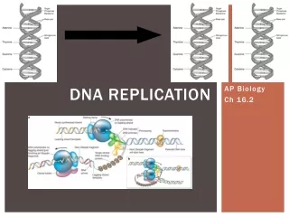
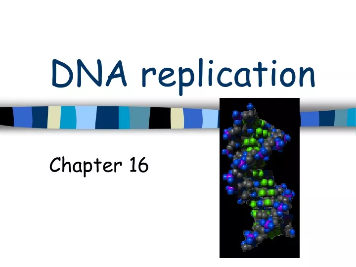
DNA replication Chapter 16
Summary of history • Griffith • Mice & Strep • Transformation • External DNA taken in by cell
Summary of history • Hershey-Chase • Bacteriophages • Supported heredity information was DNA
D:\Chapter_16\A_PowerPoint_Lectures\16_Lecture_Presentation\1604HersheyChaseExpA.htmlD:\Chapter_16\A_PowerPoint_Lectures\16_Lecture_Presentation\1604HersheyChaseExpA.html
Summary of history • Franklin • X-ray diffraction • Double helix • Watson-Crick • Double helix model
Nucleic acid structure • DNA deoxyribonucleic acid • RNA ribonucleic acid • Nucleotides
Nucleotide structure • 1. 5 carbon sugar (ribose) • 2. Phosphate • 3. Nitrogenous base
Nitrogenous base • Purines (2 rings) • Adenine(A) & Guanine(G) • Pyrimidines (1 ring) • Cytosine (C), Thymine (T) DNA only • Uracil (U) RNA only
Nucleic acids • 5’ Phosphate group (5’C) at one end • 3’ Hydroxyl group (3’C) at the other end • Sequence of bases is expressed in the 5’ to 3’ direction • GTCCAT 5’pGpTpCpCpApT---OH 3’
Double helix • Complementary • Sequence on one chain of DNA • Determines sequence of other chain • 5’-ATTGCAT-3’ • 3’-TAACGTA-5’
Double Helix • Complementary • Purines pair with pyrimidines • Diameter of base pairs are the same • Adenine (A) forms 2 hydrogen bonds with Thymine (T) • Guanine (G) forms 3 hydrogen bonds with cytosine (C)
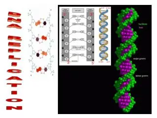
Double Helix • Sugar-phosphates are the backbone • Complementary • Phosphodiester bonds • Strands are antiparrellel • Bases extend into interior of helix • Base-pairs form to join the two strands
Fig. 16-7 5 end Hydrogen bond 3 end 1 nm 3.4 nm 3 end 0.34 nm 5 end (a) Key features of DNA structure (b) Partial chemical structure
Duplication • DNA unzips-breaks hydrogen bonds • New strand forms based on existing strand • Old strand is saved • Compliment of new strand • New DNA-one old strand & one new strand • Semiconservative replication
Fig. 16-9-3 A T A T A T A T C G C G C G C G A T A T A A T T T A T A T T A A C C G C G C G G (c) “Daughter” DNA molecules, each consisting of one parental strand and one new strand (b) Separation of strands (a) Parent molecule
Duplication study • Meselson and Stahl • Bacteria • 14N and 15N • Semiconservative method.
First replication Second replication Fig. 16-10 Parent cell (a) Conservative model (b) Semiconserva- tive model (c) Dispersive model
Summary G:\Chapter_16\A_PowerPoint_Lectures\16_Lecture_Presentation\1605DNAandRNAStructureA.html
Duplication Enzymes • DNA helicase: • Enzyme opens helix starts duplication • Separates parental strands • Single-strand binding protein: • Binds to unpaired DNA • After separation • Stabilizes DNA
Duplication Enzymes • DNA polymerases: • Help lengthen new strand of DNA • Adds new nucleotides strand • Synthesis occurs only one direction • 5’ to 3’ • Adding new nucleotides to the 3’OH
Duplication Enzymes • Primer: • Section of RNA • Complementary to the parental DNA • Synthesis occurs only one direction • 5’ to 3’ • DNA primase: • Enzyme creates the primer
Duplication Enzymes • Topoisomerase: • Relieves strain of unwinding DNA • DNA pol1: • Removes primers • Replaces with DNA nucleotides • DNA ligase: • Creates phosphodiester bonds between Okazaki fragments
Duplication • OriC • Origins of replication • Starting point in DNA synthesis • Replication is bidirectional • Proceeds in both directions from origin • 5’to 3’direction
Duplication • E coli (bacteria) • Circular DNA • One origin • Eurkaryotes • Multiple origins
Duplication • Replication bubble: • Separation of strands of DNA • Replication of DNA • Replication fork: • Y-shaped region • End of replication bubble • Site of active replication
Duplication • Leading strand: • DNA continuous 5’ to 3’ replication (towards fork) • Template is 3’ to 5’ • Lagging strand: • DNA duplicated in short segments (away from fork) • Okazaki fragments: • Short stretches of new DNA-lagging side
Duplication D:\Chapter_16\A_PowerPoint_Lectures\16_Lecture_Presentation\1609DNAReplicatOverviewA.html
Duplication • Unzips (helicase, single-strand binding protein, topoisomerase) • Primer • DNA polymerase (5’to3’) • DNA ligase
Fig. 16-14 New strand 5 end Template strand 3 end 5 end 3 end Sugar T A A T Base Phosphate C G C G G C G C DNA polymerase 3 end A A T T 3 end Pyrophosphate C C Nucleoside triphosphate 5 end 5 end
D:\Chapter_16\A_PowerPoint_Lectures\16_Lecture_Presentation\1615LeadingStrandA.htmlD:\Chapter_16\A_PowerPoint_Lectures\16_Lecture_Presentation\1615LeadingStrandA.html
Fig. 16-13 Primase Single-strand binding proteins 3 Topoisomerase 5 3 RNA primer 5 5 3 Helicase