Download
1 / 20
200 likes | 335 Views
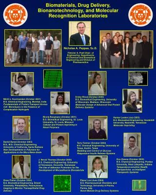
Intelligent Biomaterials Protein Delivery Molecular Imprinting and Micropatterning. Nicholas A. Peppas. Our Laboratories. 22 Researchers 12 Ph.D. students (8 ChEs, 4 BMEs) 3 Visiting scientists (Italy) 1 Technician 6 Undegraduate students About 3,800 sq.ft. facilities

E N D
Intelligent BiomaterialsProtein Delivery Molecular Imprinting and Micropatterning Nicholas A. Peppas
Our Laboratories • 22 Researchers • 12 Ph.D. students (8 ChEs, 4 BMEs) • 3 Visiting scientists (Italy) • 1 Technician • 6 Undegraduate students • About 3,800 sq.ft. facilities • Modern equipment including cellular facilities • Budget of about $ 2M • Grants from NIH, NSF, industry
The Changing World of Biomaterials, Drug Delivery and Biomolecular Engineering • Formation and fabrication of supramolecular assemblies comprising natural biological elements, structures or membranes. • Synthesis and preparation of modified biological molecules • Biomolecules as the basis of nanostructures, molecular adhesives • Micropatterned and microfabricated arrays
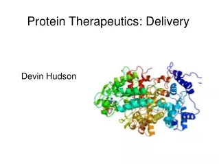
Oral Delivery of Proteins “Oral delivery of peptides and proteins has long been dubbed the ‘Holy Grail’ of drug delivery…” Why? • Increase patient compliance and comfort over other forms of drug delivery (i.e. injection) • Mimic physiologic delivery of proteins • Simple administration • Reduce costs • Potentially improve efficacy
Challenges of Oral Protein Delivery GI Tract is designed to digest proteins and food. • Protect the drug • Acidic environment in the stomach • Proteolytic enzymes in the GI tract • Improve bioavailability • Increase drug transport across intestinal epithelium • Localize drug at targeted site of absorption • Maintain biologically active and stable drug
Transport for Oral Drug Absorption • Transport Mechanism • Transcellular pathway • Paracellular pathway • Transcytosis and receptor-mediated endocytosis • Lymphatic absorption through M cells • P-glycoprotein efflux (not shown) • Factors Affecting Transport • Molecular mass of drug • Drug solubility
In Vivo Study with pH-Responsive Complexation Hydrogels • P(MAA-g-EG) microspheres loaded with insulin • Administered to diabetic rats 40% drop in blood glucose levels • Prior work done by Tony Lowman
Carrier Mediated Goal:Protect drug in the GI tract and be absorbed with drug by epithelial layer. • Biodegradable polymers, lectin modified carriers • Sites of uptake • M cells (majority of uptake) • Transcellular • Paracellular • Poor particle absorption Florence, A. T. The oral absorption of micro- and nanoparticulates: neither exceptional nor unusual. Pharm Res 1997, 3, 259-266.
Upper small Intestine Stomach Mucoadhesion Decomplexation Mucosa Mucosa
Blood Glucose Response in Healthy and Diabetic Wistar Rats
Advantages Spontaneously differentiate Produce enzymes Posses tight junctions Develop microvilii Transport of inorganic molecules correlates well with the in vivo absorption Disadvantages Do not produce mucus The properties are determined by the passage number Caco-2 Cells as GI Model
I I I I I I I I I I I I Nanodevices of Intelligent Gelsfor Protein Release Empty hydrogel absorbs glucose leading to gluconic acid production GOx GOx G GOx GlucA G G G GlucA GOx G G GlucA GlucA GOx G G G Decrease in pH leads to gel expansion which releases insulin GOx G GlucA
Targetting and Nanotechnology • Targeted delivery for cancer therapy • Gene delivery • Long term treatment of chronic diseases
Laser beam Polymer Silicon Change in pH, temperature, etc. hydrogel swells θ Laser beam φ φ > θ BioMEMS Sensor Platform • Pattern environmentally responsive hydrogels onto silicon microcantilevers to create a BioMEMS/MEMS sensor device.
Experimental Procedure • Surface Modification • Micropatterning
Micropatterned Hydrogel on Silicon Microcantilever • Volume shrunk as the polymerization proceeded • Polymer adhered to silicon surface and could not shrink at the interface, resulting in stress formation in the polymer film • This stress in the polymer film resulted in bending the microcantilever B) A) Top view images obtained utilizing an optical microscope in Nomarski mode showing a silicon microcantilever patterned with an environmentally responsive hydrogel. In A), the focus is on the substrate, while in B), the focus is on the microcantilever tip. Profilometry indicated that the thickness of the patterned hydrogel is approximately 2.2 mm.
50 m Confocal Images of MicroarraysAcrylamide-PEG200DMA with 67% Crosslinking Ratio 3D Projection of micropatterned recatangular array of a biorecognitive networks obtained utilizing a confocal microscope. Profilometry indicated that the thickness of the micropatterns are approximately 13 mm.
A) Polymer Silicon 50 m 25 m 25 m Optical and Confocal Images ofMicropatternsAcrylamide-PEG200DMA with 67% Crosslinking RatioMicrocantilever Shape B) C) Control Recognitive Images of micropatterned biorecognitive networks. In A), an optical image (Nomarski mode) of recognitive network patterned in shape of cantilever is demonstrated. In B) and C), a confocal microscope slice through middle cantilever pattern of a control and recognitive network, respectively, are shown. Profilometry indicated that the thickness of the micropatterns are approximately 13 mm.