Download

1 / 90
970 likes | 1.02k Views
Congenital Anomalies of the Kidney and Urinary Tract (CAKUT). Congenital anomalies of the kidney and urinary tract (CAKUT) constitute approximately 20 to 30 % of all anomalies identified in the prenatal period.

E N D
Congenital Anomalies of the Kidney and Urinary Tract (CAKUT)
Congenital anomalies of the kidney and urinary tract (CAKUT) constitute approximately 20 to 30 % of all anomalies identified in the prenatal period. • Defects can be bilateral or unilateral, and different defects often coexist in an individual child. • The overall rate of CAKUT in live and stillborn infants is 0.3 to 1.6 per 1000 .
The incidence is higher in women with a family history of CAKUT. • Of all antenatal renal anomalies, the most frequent abnormality is hydronephrosis, (ie, upper urinary tract dilatation). • Renal malformations are associated with non-renal congenital anomalies in about 30 % of cases
1-Renal hypoplasia : • A lower number of structurally normalnephrons, is a distinct entity separate from renal dysplasia • Unknown causes
The clinical diagnosis of renal hypoplasia is suggested when all of the following criteria are met : * Reduction of renal size by 2 standard deviations for the mean size by age * Exclusion of renal scarring by 99mTc–dimercaptosuccinic acid (DMSA) radionuclide scan مهمه * In cases of unilateral renal hypoplasia, compensatory hypertrophy of the contralateral kidney
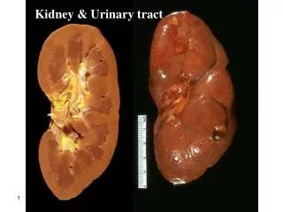
2-Renal dysplasia: • Renal dysplasia is characterized by the presence of malformed kidney tissue elements • Dysplastic kidneys are variable in size but most are smaller than normal. Size is often determined by the presence or absence of cysts. • Renal dysplasia may be unilateral or bilateral
Renal dysplasia may be discovered during routine antenatal screening or postnatally when renal ultrasonography is performed in a dysmorphic infant. • Bilateral dysplasia is likely to be diagnosed earlier than unilateral dysplasia especially if oligohydramnios is present.
Infants with bilateral dysplasia may have impaired renal function at birth and subsequent progressive renal failure may occur. • Associated urological findings include abnormalities of the renal pelvis and calyces (congenital hydronephrosis) and ureters (duplicating collecting system), megaureter, ureteralstenosis, and vesicoureteral reflux (VUR).
Because of the frequent association of renal dysplasia with a collecting system anomaly, voiding cystourethrography should be considered in all patients with renal dysplasia. • The prognosis of renal dysplasia depends on whether there is unilateral versus bilateral disease. In general, the long-term outcome of unilateral renal dysplasia is excellent, particularly if there is a normal contralateral kidney.
3-Multicystic dysplasia : • Multicystic dysplastic kidney (MCDK) is a nonfunctioning dysplastic kidney with multiple cysts, which is thought to arise from an alteration in renal parenchymal differentiation. MCDK consists of a nonreniform mass of cysts and connective tissue, and is most commonly detected by routine antenatal screening.
4-Renal agenesis: • Renal agenesis is defined as congenital absence of renal parenchymal tissue and results from major disruption of metanephric development at an early stage. • Unilateral RA accounts for 5 percent of renal malformations . • The incidence of renal agenesis is approximately 1 per 2900 births
Multiple factors are thought to be implicated in the pathogenesis of renal agenesis including mutations in genes important in renal development, and teratogenic and environmental agents (eg, retinoic acid and cocaine exposure)
Other urological abnormalities have been reported in up to 33 to 65 percent of unilateral cases • Vesicoureteral reflux (VUR) is the most commonly identified urological abnormality, • Nonrenal associated anomalies include cardiac anomalies (most commonly septal anomalies), genital tract, and gastrointestinal, respiratory, and skeletal malformations
5-Genetic cystic diseases : • Genetic cystic renal diseases are disorders of terminal epithelial differentiation A-Autosomal recessive polycystic kidney disease (ARPKD): ( more in children ) • It is caused by mutations in the PKHD1 gene, which codes for fibrocystin. • ARPKD is characterized by multiple microscopic cysts, principally involving the distal collecting ducts Of both kidneys • Kidneys are usually greatly enlarged and contain small cysts; renal failure is common in childhood.مهمه
The liver is enlarged and has periportal fibrosis and scattered cysts. • Fibrosis produces portal hypertension by age 5 to 10 yr. • Disease severity and progression vary. Severe disease may manifest prenatally or soon after birth or in early childhood with renal-related symptoms; less severely affected patients present in late childhood or adolescence with hepatic-related symptoms.
Severely affected neonates commonly have pulmonary hypoplasia secondary to the in utero effects of renal dysfunction and oligohydramnios. • If the patient presents in adolescence, nephromegaly is less marked, renal insufficiency may be mild to moderate, and the major symptoms are those related to portal hypertension.
Diagnosis may be difficult, especially without a family history. Ultrasonography may demonstrate renal or hepatic cysts; definitive diagnosis may require biopsy. • Ultrasonography in late pregnancy usually allows presumptive in utero diagnosis. • Clinical manifestations include oligohydramnios, pulmonary hypoplasia, hypertension, congestive cardiac failure, liver disease, and renal failure. • The perinatal prognosis depends on the pulmonary status.
B-Autosomal dominant polycystic kidney disease (ADPKD) ( more in adults ) • ADPKD is characterized by bilateral renal enlargement secondary to multiple cysts. • It is caused by mutations in either PKD1 (85 percent of patients) or PKD2 genes (15 percent) • There is a greater variability in clinical manifestations of ADPKD with most patients having significant clinical findings only in adulthood.
There are a subset of children who have an early onset of disease (in utero or in the first year of life) with symptoms similar to those with ARPKD. • These include gross or microscopic hematuria, hypertension, proteinuria, cyst infection, and renal insufficiency
RENAL ECTOPY: • Renal ectopy occurs when the kidney does not normally ascend to the retroperitoneal renal fossa (level of the second lumbar vertebra). • Simple congenital ectopy refers to a kidney that lies on the correct side of the body but lies in an abnormal position.
Crossed renal ectopia • Different forms of crossed renal ectopia 1- Fused: Ectopic kidney moves across the midline and fuses to the lower pole of the normally positioned contralateral kidney. 2-Nonfused: Ectopic kidney moves across the midline without fusion and positioned at the rim of the pelvis (pelvic kidney). 3-Bilateral: Both kidneys are ectopic and cross the midline with the ureters maintaining their normal bladder insertion.
RENAL FUSION: • Renal fusion occurs when a portion of one kidney is fused to the other. • The most common fusion anomaly is the horseshoe kidney, which involves abnormal migration of both kidneys (ectopy), resulting in fusion. • This differs from crossed fused renal ectopy, which usually involves abnormal movement of only one kidney across the midline with fusion of the contralateralnoncrossing kidney.
Horseshoe kidney can be a feature of many syndromes including genetic disorders such as Turner syndrome, Trisomy 13, 18 and 2 • Patients with a horseshoe kidney appear to have an increased risk for Wilms tumor.
Most patients with an ectopic or fused kidney(s) are asymptomatic and are diagnosed coincidentally, often by antenatal ultrasonography. • In patients diagnosed symptomatically with either anomaly, symptoms at presentation are generally related to associated complications including urinary tract infection (with or without VUR), obstruction, and renal calculi.
B-Ureter& Bladder تم الاستعانه بسلايدات من محاضرات الدكتور الحازمي لقله المعلومات هنا
Congenital anomalies of the urogenital system Most common of all organ system. مهمه اووووي 10% of population has some type of urogenitalanomaly. 14:1000 birth has antenatal diagnosis of urogenital anomaly. Antenatal ultrasound after 28 weeks gestation.
Congenital anomalies of the urogenital system… Antenatal Hydronephrosis Anomaly of position, number and rotation Cystic abnormalities Prune Belly Syndrome Hypospadiasabnormally located meatus ( which contin the urethral opening ) Epispadias Bladder e Exstrophy
اولا : Antenatal Hydronephrosis(ANH) Causes: Pelviureteric junction obstruction (41%) Ureterovesical junction obstruction (23%) Vesicouretericreflux(7%) Duplication anomalies (13%) Posterior urethral valves (10 %) MCDK Others (6%)
ANH ANH usually detected during pregnancy مهمه And it graded into 5 grads depend on the level of the dialitation
SFU Grading G0 G1 G2 G3 G4
causes of ANH : 1- Pelviureteric junction obstruction(PUJO)
Presentation: • Incidental in Neonates ( by US ) • Incidental in Children • Symptomatic: • UTI • Pain • Mass • Hematuria • Stone
2- Megaureter : narrowing of the ureter in the uretrovesceral junction
3- Duplication Anomalies Renal and ureteric duplication 1%, 1.6:1 F:M, 85% unilateral. ( more in F مهمه اوووي ) Either two urethral buds meeting the meta-nephros or one ureteric bud that bifurcates. مهمه جدا – انظر الصور للتوضيح Associated with: reflux 43%, renal dilatation 29%, ectopic insertion 3%, infections and ureterocele. Duplication per se is of no clinical significance, but the associated anomalies may require intervention
Type 1 : ( A ) : biforcation of the ureter after the generation Type1 : ( b ) : 2 separetureter generate and end at the same kidney للتوضيح اكثر انظر الصور الشريحهالقادمه
Weiger - Meyer Law : the ureter which drain the upper pole of the kidney end in the lower portion of the urinary bladder and vise versa يجب ان تعرف ان كل جزء من الكليه ( العلوي والسفلي ) له حوض خاص به لذلك يمكن القول انها كليتين في كليه – لذلك اذا حدث سدد في احد الحالبين فان الجزء من الكليه الذي يصب منه خذا الحالب هو وحده سيتمدد ولكن الجز الاخر سيظل سليم لان الحالب فيه سليم كما سيوضح بالصور لاحقا
4- Ureterocele : cystic dialatation of the terminal part of the ureter Ureterocele is always associated with Duplication Anomalies مهمه جدا
Ureterocele Sacculationof the terminal portion of the ureter. Has 2 types : مهمه اووووي 1- Orthotopic= intravesical=simple=adult type ureterocele. 2- Ectopic = extravesical=duplex system= infant type ureterocele. In ectopic ureterocele it involve the upper pole system. 7:1 F:M, 10% bilateral, ectopic: orthotopic 4:1 Commonest cause of urine retention in female infants. مهمه اوووووووي
Ureterocele Presentation: Antenatal (U/S) we use MCUG ( which is radiological study ) to confirm the diagnosis مهمه Urine retention Infection Calculus formation
5- Ectopic ureter Most commonly associated with duplex system and with ureterocele. مهمه اووووي Clinical picture depend on: associated anomalies, site and sex of the patient.