Download
1 / 37
380 likes | 796 Views
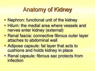
Kidney Development: One variation on the theme of organogenesis. Chris Campbell cc59@buffalo.edu 829-3462. Oct 29, 2013. Renal capsule. Cross section of a human kidney. Functions of the kidney. The functional unit of the kidney is the nephron. ~13,000 in mice ~1,000,000 in humans.

E N D
Kidney Development:One variation on the theme of organogenesis Chris Campbell cc59@buffalo.edu 829-3462 Oct 29, 2013
Renal capsule Cross section of a human kidney Functions of the kidney
The functional unit of the kidney is the nephron ~13,000 in mice ~1,000,000 in humans
The functional unit of the kidney is the nephron Quaggin (2008) Devt 135, 609 Human Anatomy, 3rd edition, 2001
Techniques to Study Development • Observation of normal development in animals of different species • Reporter genes or other markers (such as antibodies) to delineate particular cell types • Use classical genetics to identify genes in which mutations affect kidney development or cause kidney cancer or other kidney disease. (phenotype to genotype) eg. Wt1 • Loss or gain of function mutations (knockouts, transgenics, injection of sense, antisense or small inhibitory RNAs) • In vitro organ and cell culture
Similarities and Differences between Kidney Development and Lung Development • Similarities • Involves inductive interactions between mesenchymal and epithelial cells • Branching morphogenesis • Stem cell maintenance and differentiation • Differences • Involves sequential formation of three pairs of increasingly complex organs, • pronephros, mesonephros and metanephros, the first two of which are transient • Mesenchymal to epithelial transition
Specification of the intermediate mesoderm /Odd1 Dressler G R Development 2009;136:3863-3874
Development of the Kidney • The nephric duct and nephrogenic cord arise from intermediate mesoderm D > V • The nephric duct begins to elongate and undergo epithelialization in a rostral – caudal direction. Costantini (2010) Dev Cell 18, 698
Odd1/Osr1 is the earliest known marker of IM E7.5 E8.5 E9.5 But based on the expression of other early markers of IM in Odd1 k/o Odd1 isn’t required for the formation of the nephric duct or nephrogenic cord Wang (2005) Dev Biol 288, 582
Pax2 & Pax8 are required for the formation of the nephric duct ~E8.0 Laminin E-cadherin Bouchard Genes & Dev. 16,2958 (2002)
Development of the Pronephros As this duct extends caudally (eventually joining up with the cloaca), the anterior region of the duct induces the adjacent mesenchyme to form the simple tubule(s) of the pronephros . • In mouse the pronephros consists of nothing more than a few • cell condensates and is non functional but in amphibians • such as Xenopus it is a functional kidney. Chan (2006) Exp Neph 103 e81 • In mammals, pronephric tubules and the anterior portion of the nephric duct degenerate, but the more caudal portions of the duct persist and serve as the Wolffian duct.
Development of the Mesonephros • (B) As the pronephric tubules degenerate, the middle portion of the nephric duct induces a new set of kidney tubules in the mesenchyme constituting the mesonephros or mesonephric kidney. Sainio et al. (1997) Dev 124, 1293 Grote (2006) Dev 133, 53
Development of the Metanephros • (C) In male mammals, some of the mesonephric tubules persist as the vas deferens and efferent ducts of the testes but the remainder degenerates. • The metanephric kidney is initiated by the outgrowth of the ureteric bud from the Wolffian duct into themetanephricmesenchyme. ~E10.5 Bouchard (2004) Differentiation 72, 295
Development of the Metanephros • The development of the metanephros begins with the outgrowth of the ureteric bud from the Wolfian duct. • The ureteric bud grows out into the nephrogenic cord which then condenses around the bud to form the metanephric blastema or mesenchyme. • As this mesenchyme differentiates it induces the ureteric bud to branch and grow. • At the tips of the branches the mesenchyme undergoes epithelialization to form the structures of the nephron • The differentiated metanephric mesenchyme gives rise to the cells of the proximal and distal tubules, as well as the glomerular podocytes. Metanephric mesenchyme also gives rise to the renal stroma. • This process of branching of the UB and differentiation of the mesenchyme continues along a radial axis until ~P2-4 with the oldest nephrons located inside and the newest nephrons in the periphery or nephrogenic zone. • The ureteric bud gives rise to the ureter, the renal pelvis, and the collecting duct system. Shah et al (2004) Development 131, 1449
Ureteric bud tips Cap mesenchyme E17.5 cap mesenchyme in purple Renal vesicle Pretubular aggregate http://www.gudmap.org/Organ_Summaries/Metanephros/index.html
Comma shaped body S-shaped body Loop of Henley Distal tubule Renal corpuscle Proximal tubule
Mature nephron vasculature Cortical collecting ducts Medullary collecting ducts Renal capsule Renal interstitium
Kidney maturation involves reorientation within the body (Foxd1-/-) Levinson (2005) Dev 132, 529 Foxd1 Pax2 Hatini (1996) Gen&Dev 10, 1467
WT1 and Metanephric Development • WT1 was originally identified as a gene involved in Wilms tumor, a pediatric cancer in which kidney elements are incompletely differentiated and proliferate to form tumors. • Wt1 is first expressed in intermediate mesoderm prior to kidney development, and then in the nephrogenic cord and MM but not the WD or UB.
The Wt1-/- Mouse E14.5 no kidney kidney E11.5 Wt1+/+ Wt1-/- In the absence of WT1 the UB fails to grow out from the Wolfian duct. Kreidberg et al. (1993) Cell 74, 679
Using in vitro organ culture to determine where the defect in WT1-/- mice lies Explanted murine kidney buds will partially differentiate in culture. Ureteric bud and metanephric mesenchyme can be mechanically or enzymatically separated and then recombined. When separated both tissues undergo apoptosis but if recombined UB will branch and MM will differentiate into tubules
of MM differentiation The Wolffian duct in Wt1-/- mice is normal and can induce a wild type metanephric mesenchyme to differentiate normally but the mesenchyme in Wt1-/- mice can neither signal to the Wolffian duct to form a UB nor can it respond to signals from the UB. Donovan et al.(1999) Dev. Genet. 24, 252
At what stage in MM differentiaton are Wt1-/- mice defective ? Conclusion Even though Wt1 is expressed in the intermediate mesoderm prior to formation of the metanephric mesenchyme, the earliest stages of metanephric mesenchyme differentiation don’t require Wt1 expression (or contact with UB). Pax2 E11 -11.5 Donovan et al.(1999) Dev. Genet. 24, 252 Six2
Sall1 (MM) E11.5 Emx2 (UB) E11.5 Eya1 (MM) Xu (1999) Nat Gen 23, 113 Miyamoto (1997) Dev 124, 1653 Nishinakamura (2001) Dev 128, 3105 Six1 (MM) UB forms but fails to branch. Can be rescued in vitro with Grem1 (antagonist of BMP signaling). Nie (2011) Dev Bio 352, 141
Odd1 Ribes et al. J Am Soc Nephrol 14:S9, 2003
GDNF- Ret Signaling in Kidney Development Majumdar (2003) Dev 130, 3175 • Ret was initially identified based on the ability of an oncogenic variant of the gene to transform NIH3T3 cells. • Sequence homology with other proteins identified ret as a receptor tyrosine kinase • Ret is expressed in the Wolfian duct and the ureteric bud. By the time the bud has branched several times, expression is restricted to the tips of the branches.
Analyzing kidney development in Ret-/- Mice • The phenotype of Ret-/- kidneys is variable ranging from complete absence of both kidneys and ureters to presence of two very small kidneys and relatively normal looking ureters. E11.5 +/+ E11.5 -/- Schuchardt Nature 367,380 (1994) & Schuchardt (1996) Dev 122, 1919
Analysis of Ret-/- kidneys Conclusion (i)Mutant mesenchyme can signal wild type ureteric bud (ii) mutant ureteric bud cannot respond to wt mesenchyme. THE DEFECT IS IN THE URETERIC BUD. Schuchardt et al. (1996) Development 122, 1919
Identifying the ret receptor ligand GDNF was first identified as a factor capable of promoting growth of neurons in culture Following cloning of the gene, its expression pattern (expressed in MM but not UB) suggested it might play a role in kidney development. Mouse kidney organ culture + Conditioned medium from cells expressing GDNF control +aGDNF Ab +recombinant human GDNF Vega et al. (1996) PNAS 93, 10657
GDNF-/- mice have kidney defects similar to (but more severe than) Ret-/- Pichel et al. (1996 Nature 382, 73
Altering GDNF-ret signaling affects in any way affects kidney development Spry1-/- Davidson 2009 stembook.org Basson(2006) Dev Bio 299,466 Rozen (2009) JASN 20, 255
Wnt signaling and nephrogenesis Wnt4 E11.5 E12.5 Wnt4 E15 Wnt4-/- MM fails to undergo MET Stark (1994) Nature 372, 679 Isolated MM from either +/+ or Wnt4-/- mice can be induced to undergo tubulogenesis by Wnt4. Kispert (1998) Dev 125,4425
Wnt4 is required for MET of mesenchyme but Wnt4 is NOT expressed by UB. UB expresses a protein that induces Wnt4 expression in mesenchyme. + Wnt9b>Wnt4>tubulogenesis (UB) (MM) Carroll (2005) Dev Cell 9,283
Six2 and the identification & maintenance of nephron stem/progenitor cells (UB) (nephron) Wt1 (cap mesenchyme) E11.5 kidney explants Overexpression of Six2 prevents MM differentiation Self(2006) EMBO25,5214
Are Six2+ cells self renewing stem cells? Using FACS of GFP+ cells derived from Six2GCE/+ R26R embryonic kidneys to show Six2+ compartment increases 15 fold from E11.5 to E19.5 Cells that are expressing Six2 at the time of TAM injection will activate the lacZ gene and they and their progeny will express B-gal. Kobayashi (2008) Cell Stem Cell 3, 169
Can a single Six2+ cell give rise to multiple cell types? Higher magnification of D Kobayashi (2008) Cell Stem Cell 3, 169