Download
1 / 17
310 likes | 755 Views
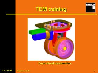
FEI TECNAI G2 F20 S-TWIN TEM Training SOP. NanoTech User Facility (NTUF) Center for Nanotechnology University of Washington June 2014 Contacts: primary: Ellen Lavoie 206-685-6775 lavoie@uw.edu Secondary: Scott Braswell 206-685-6774 sbras@uw.edu. TEM Pre-start. Tecnai User Interface (UI).

E N D
FEI TECNAI G2 F20 S-TWIN TEMTraining SOP NanoTech User Facility (NTUF) Center for Nanotechnology University of Washington June 2014 Contacts: primary: Ellen Lavoie 206-685-6775 lavoie@uw.edu Secondary: Scott Braswell 206-685-6774 sbras@uw.edu
TEM Pre-start Tecnai User Interface (UI) • Check that Microscope Control (Tecnai User Interface) & Digital Micrograph programs are open (in addition, be sure the CCD control box is on and the temp is -25). • Click on the Setup tab in UI and check that the high tension (HT) is on (yellow) and Operate is also yellow (if is not contact a staff member immediately…DO NOT turn on yourself). • In the “Setup” tab check the following: a. “Status: Col Valves” is red b. Pressure readings: Notify Staff if any indicators are abnormal Gun = 1 Column = 6 Camera = 31-ish • Check that the stage has been re-centered – double check both the numbers at the bottom of the software interface and in the Stage tab. • Cover the glass viewing chamber (both covers), then fill the nitrogen trap (anti contamination device or ACD). PPE is provided. Liquid nitrogen transfer dewars can be filled with the 50 L tank in the hallway near G44M entrance. Do not touch the SOOP Stage tab Viewing Chamber ACD
Single Tilt Sample HolderNote: Do not ever touch the rubber o-ring or the copper colored bits of the holder even with a gloved hand! • Remove the cap at the end of the holder stand. • Be careful to make sure TEM holder will not slip into the cover tube while you load the grid. • Fit the tool into the hole in front of the spring clamp. • Open the clamp to 90° and replace the tool in the stand. • Put the grid in the specimen carrier. Be careful that it fits flush into holder. • Carefully lower the spring clamp onto the grid with the tool. • Test that the grid is clamped firm and not loose in the carrier. • Remove the holder from the carrier and carefully check the rubber o-ring for any hairs or debris and clean gently with lens tissue (do not use tweezers to remove debris – you may damage the o-ring). O-ring Cover tube Lens tissue Holder Tool
Double Tilt Sample Holder – you must be specifically trained in order to use this holder!Important: beryllium is VERY toxic and special care must always be taken when working with this holder! Do not push down or apply force to the carrier at all or the pivots will break. The pivots are mounted on fragile crystals that calibrate the b-tilt. • Use the Hex Tool to unscrew the retaining nut and lift it gently out of the carrier (DO NOT use tweezers – use the hex tool). • Place grid in carrier and check alignment on the stereo microscope. • Put the washer on top of the grid with the tabs aligned if a thin grid or sample is being loaded. If you have a thicker sample, do not use the washer. Always store the washer when not being used in the small plastic box labeled for it. • Lift the retaining nut on the Hex Tool and carefully screw it into the carrier (again, DO NOT use tweezers – use the hex tool). • Test that the grid is clamped firm and not loose in the carrier. *attention…always use the vacuum tweezers when handling beryllium parts…this alleviates the possibility of contaminating tweezers with beryllium. If you touch beryllium with a gloved hand accidently, remove and throw away the glove without touching it on anything else. • Helpful Tips: • Keep small parts close to the table top in case they fall. • Don’t over tighten the nut. You should get about 1 full turn on the threads and the nut will be recessed into the carrier. • Take care not to insert the retaining nut upside down. There is a flange around the top of the nut. Hex Tool Washer
Insert sample holder • Before inserting go to “Stage” tab and ensure the stage has been reset. • Open “Setup” Tab and “Vacuum Overview” in the lower right hand bar of software. • Align pin to 4 o’clock position, and insert TEM holder straight into the airlock with a • slight clockwise turn until it sets in. • “Turbo On” button will turn from gray to Orange and a message will pop up below • for holder selection. Select holder type in message box and click Enter (arrow) button. • “Turbo On” button will change from Orange to Yellowand once it is yellow will go • through a series of pumping cycles for three minutes. Red LED on the • goniometer (stage) will light up. • Watch vacuum status change to “Airlock.” V41 & V8 will show as open on Vacuum Overview. • Once Red LED on the goniometer (stage) turns off and vacuum status changes back to • “Col Valves Closed”, the holder can be inserted further into the goniometer: • - Turn the holder counter-clockwiseto the stop, hold it (without pulling the holder or resisting), • and gently guide it in. DO NOT let go since it may slam against the goniometer and can cause • damage to the electronics behind the purple plate. • Turn Turbo off by clicking the Yellow “Turbo On” button. • Gloves can be removed at this point. Setup tab Vacuum status window
Getting a Beam • Wait for column pressure to drop below 10 Log. It may initially rise slightly after holder insertion…don’t panic but if it continues to rise, remove holder and find a staff member immediately. It should reach 10 Log within a few minutes. • Click the yellow “Col. Valves Closed” button to open the column valves. • In the Vacuum Overview V7 & V4 will open. • If no beam is visible in the chamber, check: Lower magnification on RH control panel to 8700X Move stage to one side with the joystick Ensure a large condenser aperture is in Check the spot size (typical is 3 or 2) • Load settings for saved alignment: “Gun” tab > “FEG Register” menu > select 200kV TEM > click “Set.” Setup tab Beam will be visible in chamber Left-hand control panel (LH) Right-hand control panel (RH)
TEM Alignment: Condenser • Choose spot size. • Select an appropriate C2 aperture by turning the dial with pin to a numbered position. For most samples in TEM mode use the 100 um aperture in position #3. • Increase the magnification to at least 38kX. • Turn the “Intensity” knob on the LH control panel in the counterclockwise position to focus the beam to a small spot. • Center the beam on the phosphor screen with the trackball on the LH control panel. • Turn the BRIGHTNESS knob clockwise until it fills the 4 cm ring on the phosphor screen. If the illumination shifts, re-center it with the condenser aperture X and Y knobs. • Repeat steps c, d, and e until the illumination no longer moves off center while turning “Intensity” knob in the crossover position. X-Align 4 cm ring on phosphor Y-Align Selector dial C2 Aperture
TEM Alignment: Condenser Stigmation • IF the area of illumination is not circular or the beam appears to stretch at the intensity cross-over then perform the following: • Go to “Tune” tab > “Stigmator” menu and click on “Condenser.” • Use the “Multifunction Knobs” (MF) to adjust the beam roundness in both the X and Y dimensions. Check that the beam does not stretch at the “Intensity” cross-over. • Click “None” after finishing. Tune tab Multi-Function Y Multi-Function X This legend at the lower-left corner identifies the buttons on the control panel
TEM Alignment: Eucentric Height and a-tilt Flap-out Arrow • Find an interesting large feature on the grid or sample and go to a mag of ~10kX with “Magnification” knob. • Press “Eucentric focus” on the RH control panel. • Go to “Stage” tab, click the flap-out arrow, and activate the “Wobbler.” Alternatively, use the short cut button if available. • Eucentric height is approx-100um with the single-tilt holder. • Adjust Z-height on the RH control panel to minimize image movement while wobbling the a-tilt. • Click “Wobbler” again to stop a-tilting. • Repeat a-tilt wobbler step at a higher mag of ~35kX. Stage Map Stage tab
TEM Alignment: Focus, Beam Tilt, Beam Shift, and Rotation Center Small phosphor screen lever Go to “Tune” tab and Direct Alignments: • Pull lever on the left side of the viewing chamber towards you to raise the small screen (this puts the phosphor image in the plane of view of the binocular system). Focus the binoculars by adjusting each eye piece while the pointer is in the center of the small screen. DO NOT FOCUS THE BINOCULARS ON THE ACTUAL SAMPLE. • Move away from sample and select “Beam tilt pp X” and focus “Intensity” to ~1” diameter. • Using MF knobs, merge the two beams into one spot. c. Select “Beam tilt pp Y” and adjust as for pp X. • Using MF knobs, merge the two beams into one spot. d. Select “Beam shift” • Lower magnification as needed to find beam. • Move beam to the center using MF knobs. • Bring the magnification back up to 125k and re-center beam. • Find a feature of interest on the sample and “Focus” on RH control panel to Gaussian focus (minimum contrast) (Obj ~91.9% at 200kV.) f. Select “Rotation center” • i. Position sample edge at the center of the screen with joystick. • ii. Watch the center of the image on the small phosphor screen. • Use MF knobs to minimize image shift at the center of the image. • Go back and forth between focusing and rotation center until it is correct. Misaligned beam tilt pivot point
Objective Aperture (Optional for Contrast) • Insert objective aperture by turning selector pin to 4, 3, 2, or 1. • Aperture 4 is smallest; it gives the most contrast and least resolution. Aperture 1 is largest and gives slightly more contrast than no aperture at all. • Press “Diffraction” on the RH panel. • Center the aperture on the screen with the two aperture screws (similar to centering the C2 aperture.) • Turn Focus knob to “D 1.65 m” and use the OA screws to center the aperture around the diffraction image. • Turn “Diffraction” off. • If the beam still looks ‘clipped’ then the aperture is too small or it’s off-center. • When finished working please remember to turn the OA selector back to position 7. This ensures no damage will be done to the aperture during holder removal and/or ACD cryo cycle.
Using the CCD camera NOTE: The CCD chip is sensitive to beam intensity so do not change magnification (up or down) with the camera exposed to the beam (screen raised). When adjusting intensity, be sure you have chosen FINE with the “Intensity” knob. • Choose a magnification then spread the beam intensity (clockwise) on LH panel until the beam is spread across the entire screen (the recommended “Exp Time” > 2.0 sec…but if there is a very dark feature on the screen this time could be invalid). • On “Camera” tab > flap out > “Settings” tab set “Search” integration time to 0.1 sec and “Acquire” integration to 2 sec. • Click “Insert” in “Camera” tab to insert the CCD camera (In the DM software the search and acquire can be controlled but not insertion). • Lift the big phosphor screen using “R1” on RH panel or the screen lift button. • Click “Search” to display live image or “Acquire” to capture. • Double-check the pixel intensity of the image < 3000. • If “Value” > 3000 quickly stop the camera, lower the phosphor screen, and spread the beam “Intensity” knob or the CCD could be damaged. Main phosphor screen up/down
TEM Alignment 5: Objective Stigmation and Focus • Go to “Process” tab in Digital Micrograph program and select Live > FFT. • IF the FFT image is not circular go to the “Tune” tab > “Stigmator” menu and click “Objective.” • Turn MF knobs to correct any objective astigmatism and make the FFT image circular. • Click “None” after finishing. • Adjust “Focus” to maximize the diameter of the rings which puts you near minimum contrast focus. • Defocus image counter-clockwise to make edges appear sharper. • Click “Acquire” to capture the focused micrograph. • Optional: at minimum contrast focus push “L1” on LH control panel to reset defocus reference point. Tune tab FFT in focus Out of focus (no rings) Astigmatic objective Astigmatism corrected
Save TEM Images in Digital Micrograph – Method 1 Follow the steps illustrated below to setup a sequential save. Pick the Tools icon in the bottom right of the Digital Micrograph application Highlight “Save Numbered” in the options menu of the Saving Dialogue. Click “Browse” and select Q: or M: from “My Computer.” Assign a file name ‘string.’ “Next Index” indicates the next numbered file. Save Display as DM3 file. After each image is acquired click Save123 icon. Make sure all micrographs are saved before you close them. After all files are transferred use ‘Batch Close’. Ctrl-Alt-Shift and “X” to close all frames. When finished for day, convert all images from DM to JPEG or TIFFs by doing a “batch convert”…see instructions on Method 2 for batch converting. Close all your images as a courtesy for the next user when you are finished.
Save TEM Images in Digital Micrograph – Method 2 Save each image individually in order to specifically name each: • Click on the drop down menu File: Save as: and save file as a DM initially. • Be sure to save in your file in either the M or Q drive. • When finished with you session convert all of your images from DM to a JPEG or TIFF file by: • File • Batch convert • Browse for the appropriate folder • Be sure to check the box “convert sub folders” • Under “save display folders”, choose the format you would like • Click “ok” In the folder there will be one copy of DM and one copy of your converted image. The original DM image also has a permanently attached record of all the parameters of the microscope. This is especially important in the case of forgetting to use a scale bar or to aid in reproducibility. Unfortunately a DM file can only be opened using DM software(free offline software can be found on the Gatan website). d. Close all your images as a courtesy for the next user when you are finished.
Changing Samples or Finishing • Be sure objective and selected area apertures removed. • Be sure the phosphor plate is down. • Switch back to “Bright Field” mode (if you were using STEM, Diffraction, or Nanoprobe Mode.) • “Camera” Tab > click “Insert” to remove CCD. • Go to “Stage” > Flap-out and click Reset: “Holder” button to zero the stage XYZ and holder tilt angles. Failure to “Reset: Holder” before removing it from the TEM is likely to damage the holder and stage. • Set magnification to 125kx. This is important for the stability of the objective lens system. • “Setup” Tab > Click “Col. Valves Closed” button. When closed, menu shows “Status: Col. Valves” in red. Failure to close column valves before removing the holder can damage the electron gun. • Take out the holder as shown at right. • IF the turbo starts after you remove the holder, click the orange turbo button to turn it off . • NOTE: IF YOU HAVE HAD A LONG SESSION AND THE NEXT USER IS NOT READY, PLEASE CHECK AND FILL LIQUID NITROGEN. THIS WILL ALLEVIATE THE ISSUE OF IT RUNNING “DRY” Be sure “Setup” and “Vacuum overview” tabs are visible during holder removal 1. Pull straight until stop 2. Turn clockwise until stop 3. Grab stem and pull holder out while thumb and forefinger are anchored on the purple plate Remove
Cryo Cycle If you are the last user of the day, please don’t forget to run the Cryo Cycle. • After 4pm, login in to the NTUF webpage at the end of your session and check the TEM schedule to see if you are the last person. • Remove liquid nitrogen dewar and pour remaining liquid carefully back into the transfer dewar. Put styrofoam cup underneath the copper coils. Place the Tecnai ACD dewar on the tabletop (not a top shelf). • Go to “Setup” > “Vacuum” menu > “Flapout” arrow > and click “Cryo Cycle.” • The Red LED on the stage will light and the turbo will run for 240 min to remove residual water from the column. NOTE: the cryo cycle can NOT be disrupted during the cycle…it must run the entire time. Have a nice evening!