Download
1 / 1
20 likes | 392 Views
Motoric abnormalities. Generation of Neural specific knockout. A. B. A.

E N D
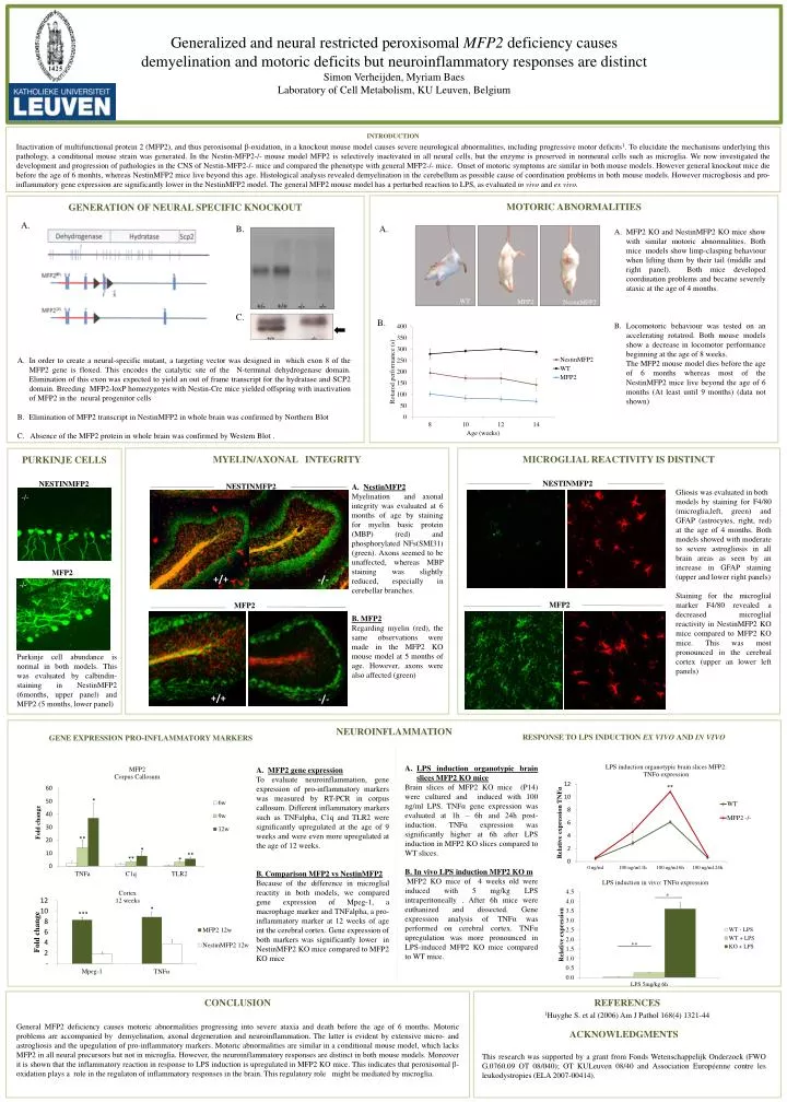
Motoric abnormalities Generation of Neural specific knockout A. B. A. MFP2 KO and NestinMFP2 KO mice show withsimilarmotoricabnormalities. Both mice models show limp-claspingbehaviourwhenliftingthembytheirtail (middle and right panel). Both micedevelopedcoordinationproblems and becameseverelyataxic at the age of 4 months. Locomotoricbehaviour was testedonanacceleratingrotatrod. Both mouse models show a decrease in locomotor performance beginning at the age of 8 weeks. The MFP2 mouse model dies before the age of 6 monthswhereas most of the NestinMFP2 mice live beyond the age of 6 months (At leastuntil 9 months) (data notshown) WT MFP2 NestinMFP2 C. B. Generalized and neural restricted peroxisomalMFP2 deficiency causesdemyelination and motoric deficits but neuroinflammatory responses are distinctSimon Verheijden, Myriam BaesLaboratory of CellMetabolism, KU Leuven, Belgium In order to create a neural-specific mutant, a targeting vector was designed in which exon 8 of the MFP2 gene is floxed. This encodes the catalytic site of the N-terminal dehydrogenase domain. Elimination of this exon was expected to yieldan out of frame transcript for the hydratase and SCP2 domain. Breeding MFP2-loxP homozygotes with Nestin-Cre mice yielded offspring with inactivation of MFP2 in the neural progenitor cells Elimination of MFP2 transcript in NestinMFP2 in whole brain was confirmedbyNorthernBlot C. Absence of the MFP2 protein in whole brain was confirmed by Western Blot . MYELIN/AXONAL INTEGRITY MICROGLIAL REACTIVITY IS DISTINCT PurkinjeCells NESTINMFP2 NestinMFP2 Myelination and axonalintegrity was evaluated at 6 months of agebystainingformyelin basic protein (MBP) (red) andphosphorylatedNFs(SMI31) (green). Axonsseemed to beunaffected, whereas MBP staining was slightlyreduced, especially in cerebellar branches. B. MFP2 Regardingmyelin (red), the sameobservationswere made in the MFP2 KO mouse model at 5 months of age. However, axons werealsoaffected (green) Gliosis was evaluated in both models bystainingfor F4/80 (microglia,left, green) and GFAP (astrocytes, right, red) at the age of 4 months. Both models showedwith moderate to severeastrogliosis in all brain areas as seenbyanincrease in GFAP staining (upper and lower right panels) Stainingfor the microglial marker F4/80 revealed a decreasedmicroglialreactivity in NestinMFP2 KO micecompared to MFP2 KO mice. This was most pronounced in the cerebral cortex (upper anlowerleft panels) -/- MFP2 MFP2 MFP2 +/+ -/- -/- MBP - blue SMI31 – green KO INTRODUCTION Inactivation of multifunctional protein 2 (MFP2), and thus peroxisomalβ-oxidation, in a knockout mouse model causes severe neurological abnormalities, including progressive motor deficits1. To elucidate the mechanisms underlying this pathology, a conditional mouse strain was generated. In the Nestin-MFP2-/- mouse model MFP2 is selectively inactivated in all neural cells, but the enzyme is preserved in nonneural cells such as microglia. We now investigated the development and progression of pathologies in the CNS of Nestin-MFP2-/- mice and compared the phenotype with general MFP2-/- mice. Onset of motoric symptoms are similar in both mouse models. However general knockout mice die before the age of 6 monhts, whereas NestinMFP2 mice live beyond this age. Histological analysis revealed demyelination in the cerebellum as possible cause of coordination problems in both mouse models. However microgliosis and pro-inflammatory gene expression are significantly lower in the NestinMFP2 model. The general MFP2 mouse model has a perturbed reaction to LPS, as evaluated in vivo and ex vivo. NESTINMFP2 NESTINMFP2 Purkinjecellabundance is normal in both models. This was evaluatedbycalbindin-staining in NestinMFP2 (6months, upper panel) and MFP2 (5 months, lower panel) +/+ -/- NEUROINFLAMMATION Response to LPS induction EX VIVO and IN VIVO Gene expression pro-inflammatory markers * LPS inductionorganotypicbrain slices MFP2 KO mice Brain slices of MFP2 KO mice (P14) werecultured and inducedwith 100 ng/ml LPS. TNFα gene expression was evaluated at 1h – 6h and 24h post-induction. TNFαexpression was significantlyhigher at 6h after LPS induction in MFP2 KO slices compared to WT slices. B. In vivo LPS induction MFP2 KO m MFP2 KO mice of 4 weeks oldwereinducedwith 5 mg/kg LPS intraperitoneally . After 6h micewereeuthanized and dissected. Gene expressionanalysis of TNFα was performedoncerebral cortex. TNFαupregulation was more pronounced in LPS-induced MFP2 KO micecompared to WT mice. MFP2 gene expression Toevaluateneuroinflammation, gene expression of pro-inflammatory markers was measuredby RT-PCR in corpus callosum. Different inflammatory markers such as TNFalpha, C1q and TLR2 weresignificantlyupregulated at the age of 9 weeks and were even more upregulated at the age of 12 weeks. B. Comparison MFP2 vs NestinMFP2 Because of the difference in microglialreactity in both models, we compared gene expression of Mpeg-1, a macrophage marker and TNFalpha, a pro-inflammatory marker at 12 weeks of age int the cerebral cortex. Gene expression of both markers was significantlylower in NestinMFP2 KO micecompared to MFP2 KO mice *** * ** ** * ** ** * * ** CONCLUSION General MFP2 deficiencycausesmotoricabnormalitiesprogressinginto severe ataxiaanddeathbefore the age of 6 months.Motoricproblems are accompaniedbydemyelination, axonaldegenerationandneuroinflammation. The latter is evident byextensive micro- andastrogliosisand the upegulation of pro-inflammatorymarkers. Motoricabnormalities are similar in a conditional mouse model, whichlacks MFP2 in allneural precursors but not in microglia. However, the neuroinflammatory responses are distinct in both mouse models. Moreoverit is shownthat the inflammatoryreaction in response to LPS induction is upregulated in MFP2 KO mice. Thisindicatesthatperoxisomalβ-oxidationplays a role in the regulaton of inflammatory responses in the brain. Thisregulatoryrolemightbemediatedbymicroglia. REFERENCES 1Huyghe S. et al (2006) Am J Pathol 168(4) 1321-44 ACKNOWLEDGMENTS This researchwassupported by a grantfrom Fonds WetenschappelijkOnderzoek (FWO G.0760.09 OT 08/040); OT KULeuven 08/40 andAssociationEuropéennecontre les leukodystropies (ELA 2007-00414).