Download
1 / 57
570 likes | 649 Views
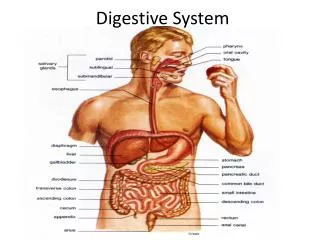
Digestive System. AKA. G-I Tract Alimentary Canal. Overview. Consists of Mouth, pharynx, esophagus, stomach, small intestine, large intestine, anus About 30’ in length Accessory Organs Teeth, tongue, gall bladder, salivary glands, liver and pancreas

E N D
AKA • G-I Tract • Alimentary Canal
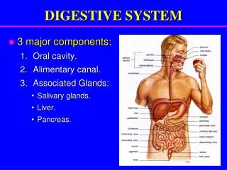
Overview • Consists of • Mouth, pharynx, esophagus, stomach, small intestine, large intestine, anus • About 30’ in length • Accessory Organs • Teeth, tongue, gall bladder, salivary glands, liver and pancreas • Glands secrete saliva, bile and enzymes
Process Includes • Ingestion • Mechanical – chewing, churning • Propulsion – swallowing and peristalsis • Chemical – breakdown via enzymes • Absorption – transport of end products into blood • Defecation – elimination as feces
Peritoneum and cavity • Parietal peritoneum surrounds cavity, lines body wall • Visceral surrounds organ • Serous membrane allows organs to glide/expand • Retroperitoneal refers to organs in the dorsal region • Peritonitis = inflammation of peritoneum • Perforation = infection
Mesentery • Double layer of peritoneum, holds organs in place • Omentums - protection • Lesser = fatty skin, superficial near the stomach • Greater = deeper, made of connective tissue, significant fat • Three layers of fascia – Skin, Fascia, mesentary
Histology • Smooth muscle - peristalsis • Glands with ducts • Nerves, arteries and veins
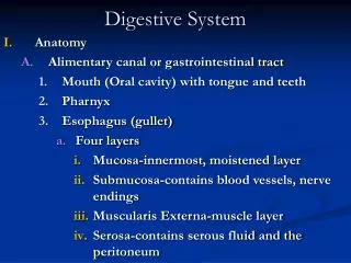
MouthPoint of Origin • Oral cavity • Tongue • Soft and hard palate • Uvula • Oropharynx • Epiglottis • Larynx/pharynx • Esophagus
Salivary glands • Produce saliva, a mixture of water, ions, mucous, and enzymes • Dissolve food • Wets food to help bind it to become a bolus • Neutralizes acids, helps growth of beneficial bacteria • Intrinsic –in tongue, palate, lips and cheeks • Extrinsic = outside mouth • Parotids • Submandibular • Sublingual
Pharynx • Oso • Laryngopharynx • Lined with epithelial cells for protection
Esophagus • Muscular tube • Propels swallowed food to stomach • Passes through diaphragm (esophageal hiatus) into abdomen • Join the stomach at cardiac orifice • Cardiac sphincter prevents reflux or regurgitation of acid
Stomach • Food churned into chyme; a paste • Secretes pepsin – a protein digesting enzyme and HCl • Waters, electrolytes, some drugs absorbed through stomach • Anatomy • Cardiac orifice, fundus, lesser and greater curvature, pylorus • Rugae = numerous longitudinal folds of mucosa which flatten as stomach fills, allows expand
Small Intestine • Longest part of alimentary canal • Most enzymes involved in small intestine come from pancreas • Three divisions – each approximately • Duodenum – 5% • Jejunum – 40% • Ileum – almost 50%
Duodenum • Receives digestive enzymes from pancreas • Bile from gall bladder and liver • Almost all nutrients are absorbed in small intestine • Large surface area, great length
Gall Bladder • Cystic duct • Bile duct – empties into small cystic duct • Secrete bile for duodenum digestion • Bile – Right and Left hepatic ducts to common cystic duct to Gall bladder for storage • From GB to bile duct to duodenum
Pancreas • Exocrine gland – produces most enzymes for digestion in small intestine • Endocrine function = produce hormones that regulate levels of sugar in the blood • Main pancreatic duct
Large Intestine • Most material has been digested by the time it reaches LI • 12-24 hours in large bowel • Little breakdown • Performs some absorption, especially water • Components • Ascending colon • Transverse colon • Descending colon • Sigmoid to rectum and anus
Cecum and appendix • Cecum is a small sac (blind pouch) • Valve prevents a back up of fecal matter to ileum • Appendix • Lymph tissue neutralizes bacteria • Diverticulosus – a small outward herniation of colon, especially sigmoid • Diverticulitis – infection with leaks into peritoneal cavity may lead to peritonitis
Anal Canal • ANS balance between defecation or not • PNS = increase movement • SNS = decrease • Voluntary control via external sphincter muscles