Download

1 / 45
470 likes | 849 Views
The brain stem. Liu jiao Binzhou Medical University Department Of Anatomy. The brain. Telencephalon Diencephalon Cerebellum Brain stem. Names of cranial nerves. Ⅰ Olfactory nerve Ⅱ Optic nerve Ⅲ Oculomotor nerve Ⅳ Trochlear nerve Ⅴ Trigeminal nerve

E N D
The brain stem Liu jiao Binzhou Medical University Department Of Anatomy
The brain • Telencephalon • Diencephalon • Cerebellum • Brain stem
Names of cranial nerves • Ⅰ Olfactory nerve • Ⅱ Optic nerve • Ⅲ Oculomotor nerve • Ⅳ Trochlear nerve • Ⅴ Trigeminal nerve • Ⅵ Abducent nerve • Ⅶ Facial nerve • Ⅷ Vestibulocochlear nerve • Ⅸ Glossopharyngeal nerve • Ⅹ Vagus nerve • Ⅺ Accessory nerve • Ⅻ Hypoglossal nerve
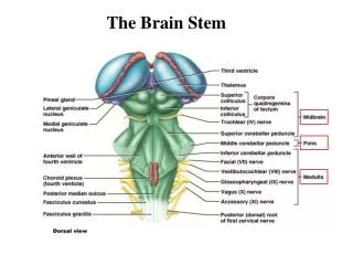
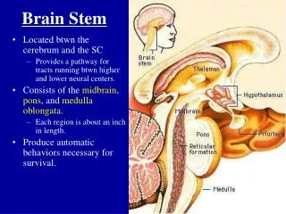
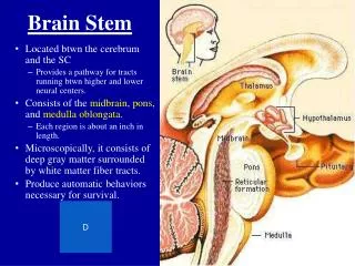
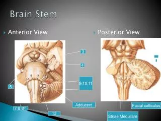
The brain stem Consists of • Midbrain • Pons • Medulla oblongata
Medulla oblongata Ventral surface • Pyramid: contain pyramidal tract (corticospinal tract) • Decussation of pyramid: formed by crossing fibers of corticospinal tract • Olive: produced by underlying inferior olivary nucleus • Anterolateral sulcus: rootlets of hypoglossal nerve emerge from it • Retroolivary sulcus: rootlets of glossopharyngeal, vagus and accessory nerves emerge from it
Medulla oblongata Dorsal surface • Lower portion • Gracile tubercle:produced by underlying gracile nucleus • Cuneate tubercle: marks the site of cuneate nucleus • Inferior cerebellar peduncle • Obex • Upper portion: forms the lower half of rhomboid fossa
Pons Ventral surface • Basilar part • Basilar sulcus • Bulbopontine sulcus : from medial to lateral, the abducent, facial and vestibulocochlear nerves appear • Middle cerebellar peduncle • Trigeminal nerve • Pontocerebellar trigone : the junction of medulla, pons and cerebellum
Pons Dorsal surface • Superior cerebellar peduncle • Superior medullary velum • Trochlear nerve
Midbrain Ventral surface • Crus cerebri • Interpeduncular fossa oculomotor nerves emerge from medial of crus cerebri • Posterior perforated substance
Midbrain Dorsal surface • Superior colliculus constitute centers for visual reflexes • Inferior colliculus associated with auditory pathway • Brachium of superiorcolliculi • Brachium of inferior colliculi
Fourth ventricle Central canal →fourth ventricle →mesencephalic aqueduct→third ventricle Position • Situated ventral to cerebellum, and dorsal to pons and cranial half of medulla
Boundaries • Inferolateral: gracile and cuneate tubercles, inferior cerebellar peduncle • Superolateral: superior cerebellar peduncle • Lateral recess
Features • Median sulcus • Sulcus limitans • Vestibular area • overlies vestibular nuclei • Acoustic tubercle • overlyingdorsal cochlear nucleus • Medial eminence • Striae medullares
Facial colliculus: • overlies nucleus of abducent n. and genu of facial nerve • Hypoglossal triangle: verlyinghypoglossal nucleus • Vagal triangle: • overlies dorsal nucleus of vagus nerve • Funiculus separans • Area postrema • Locus ceruleus
Roof • Anterior part: formed by superior cerebellar peduncle and superior medullary velum • Posterior part: formed by inferior medullary velum and choroid plexus of fourth ventricle • Three apertures • Median aperture of fourth ventricle • Two lateral apertures of fourth ventricle
Internal structures Gray matter • Cranial nerve nuclei • Relay nuclei
General somatic motor nuclei • Nucleus of oculomotor n. • Nucleus of trochlear n. • Nucleus of abducent n. • Nucleus of hypoglossal n.
Special visceral motor nuclei • Motor nucleus of trigeminal n. • Nucleus of facial n. • Nucleus ambiguus • Accessory nucleus
General visceral motor nuclei • Accessory oculomotor nucleus • Superior salivatory nucleus • Inferior salivertory nucleus • Dorsal nucleus of vagus n.
Visceral sensory nuclei ( general and special ) • Nucleus of solitary tract
General somatic sensory nuclei • Mesencephalic nucleus of trigeminal n. • Pontine nucleus of trigeminal n. • Spinal nucleus of trigeminal n.
Special somatic sensory nuclei • Cochlear nuclei • Vestibular nuclei
Relay nuclei • Gracile nucleus • Cuneate nucleus • Inferior olivary nucleus • Superior olivery nucleus • Pontine nucleus • Nucleus of inferior colliculus • Gray matter layers of superior colliculus • Red nucleus • Substantia nigra • Pretectal area
White matter Ascending tracts • Medial lemniscus • Spinal lemniscus • Trigeminal lemniscus • Lateral lemniscus
Descending tracts • Corticospinal tract • Rubrospinal tract • Tectospinal tract • Vestibulospinal tract • Reticulospinal tract
Reticular formation of brain stem • Ascending reticular activating system (ARAS) • Motor central and vital centres • Reticulospinal tract • Cardiovascular center and respiratory center • Serotonergic rapheal nuclei
Medulla oblongata Lower part (closed part) • Two decussations– • Decussations of medial lemniscus • Decussations of pyramid
Medulla oblongata Upper part (open part) • Appearance of inferior olivary nuculeus and inferior cerebellar peduncle • Enlargement of central canal to form the fourth ventricle floor
Pons • Tegmentum of pons • directed upward continuation of medulla oblongata • Basilar part • contain both longitudinal and transverse fibers intermixed with pontine nuclei
Midbrain • Rectum of midbrain: includes superior and inferior colliculi • Cerebral peduncle • Tegmentum : contain ascending tracts, central gray matter, recticular formation and so on • Substentia nigra • Crus cerebri : • Pyramidal tract • middle three-fifths of the crus: • Frontopontine tract • medial one-fifth: • pariatotempopontine tract • lateral one-fifth