Download
1 / 40
440 likes | 1.55k Views
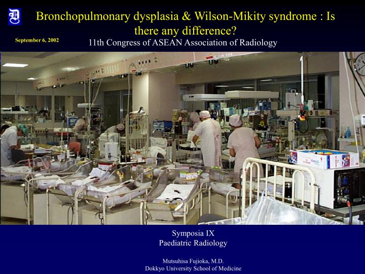
Bronchopulmonary dysplasia & Wilson-Mikity syndrome : Is there any difference?. September 6, 2002. 11 th Congress of ASEAN Association of Radiology. Symposia IX Paediatric Radiology Mutsuhisa Fujioka, M.D. Dokkyo University School of Medicine. What is this?.

E N D
Bronchopulmonary dysplasia & Wilson-Mikity syndrome : Is there any difference? September 6, 2002 11th Congress of ASEAN Association of Radiology Symposia IX Paediatric Radiology Mutsuhisa Fujioka, M.D. Dokkyo University School of Medicine
What is this? Bronchopulmonary dysplasia(BPD) or chronic lung disease(CLD) 80 day-old-baby
Why is it so important ? Griscom NT: Caldwell Lecture. Respiratory problems of early life now allowing survival into adult hood: concepts for radiologists. AJR 1993 Sep; 161(3) : 574-5 Survivors of bronchopulmonary dysplasia have decreased exercise capacity, wheezing, and recurrent pneumonia, although their chest radiographs may become normal or almost normal.
Aquino Sl, Schechter MS, Chiles C, Ablin DS et al :High-resolution inspiratory and expiratory CT in older children and adults with bronchopulmonary dysplasia.AJR 1999 0ct. 173(4):963-7 Massachusetts General Hospital, USA Abnormal findings on inspiratory and expiratory high-resolution CT of older children with bronchopulmonary dysplasia include scarring and air trapping with architectural distortion. The correlation between these findings and physiologic evidence of air trapping and obstructive lung disease was statistically significant.
What is BPD(bronchopulmonary dysplasia)? Originally(1967): a new chronic pulmonary syndrome that is associated with the use of intermittent positive pressure respirators(IPPR) and high oxygen for longer than 150 hours. Currently(1992): chronic pulmonary syndrome occurring in prematurely born infants treated with positive pressure ventilation and oxygen supplementation for respiratory insufficiency. Northway WH. Bronchopulmonary dysplasia: Twenty-five Years Later. Pediatrics (1992) 89:969-973
BPD or CLD ??? In 1989, the Bureau of Maternal and Child Health and Resources Development put forward the following diagnostic criteria for BPD (1 ) Positive pressure ventilation during the first 2 weeks of life for a minimum of 3 days (2) Clinical signs of respiratory compromise persisting longer than 28 days of age (3) Requirement for supplemental oxygen longer than 28 days of age to maintain a PaO2 above 500mmHg (4) Chest radiograph with findings characteristic of BPD Northway WH. Bronchopulmonary dysplasia: Twenty-five Years Later. Pediatrics (1992) 89:969-973
Day 1 Day 2
Day 3 Day 4
Day 5 Day 6
Day 14 Day 20
Day 25 Day 30
Radiographic findings of BPD Chest radiograph: Original four-stage radiographic progression to the severly hyperinflated cystic appearance of the lungs in chronic BPD is still seen in severe cases. More commonly, the radiographic changes are more subtle and the progression may be more prolonged. Northway WH. Bronchopulmonary dysplasia: Twenty-five Years Later. Pediatrics (1992) 89:969-973
Radiographic findings of BPD Complete opacification of the lungs, originally described as stage II, is now uncommon. Northway WH. Bronchopulmonary dysplasia: Twenty-five Years Later. Pediatrics (1992) 89:969-973
Radiographic findings of BPD The characteristic spectrum of chest radiographic findings of chronic BPD are bilateral diffuse interstitial thickening of mild to very severe degree with normal to increased expansion of the lungs gradual onset with little change over time. Northway WH. Bronchopulmonary dysplasia: Twenty-five Years Later. Pediatrics (1992) 89:969-973
The broadening of the spectrum of radiologic findings characteristic of BPD has resulted in the use of terms such as chronic lung disease. Northway WH. Bronchopulmonary dysplasia: Twenty-five Years Later. Pediatrics (1992) 89:969-973
Definition of CLD(2) Clinical signs of respiratory compromise persisting longer than 28 days of age(3) Requirement for supplemental oxygen longer than 28 days of age to maintain a PaO2 above 500mmHg Northway WH. Bronchopulmonary dysplasia: Twenty-five Years Later. Pediatrics (1992) 89:969-973
Prevention 1 technologic approarch: decreasing pulmonary oxygen toxicity and barotrauma, high –frequency ventilation, extracorporeal membrane oxygenation Northway WH. Bronchopulmonary dysplasia: Twenty-five Years Later. Pediatrics (1992) 89:969-973
Prevention 2 molecular-biologic approarch: exogenous surfactants(bovine, porcine, human, synthetic); prenatal induction of pulmonary surfactant and antioxidant enzyme activity by maternal corticosteroid treatment and use of exogenous antioxidants as well as steroid and nonsteroid inhibitors of the pulmonary inflammatory cascade. Northway WH. Bronchopulmonary dysplasia: Twenty-five Years Later. Pediatrics (1992) 89:969-973
Prevention 3 socioeconomic-political approarch: Decreasing incidence of premature birth by socioeconomic-political action Northway WH. Bronchopulmonary dysplasia: Twenty-five Years Later. Pediatrics (1992) 89:969-973
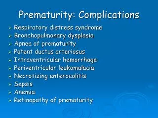
Pathogenesis and Risk factors 1respiratory distress or failure, 2 premature birth, 3 oxygen supplementation, and 4 intermittent positive pressure ventilation Northway WH. Bronchopulmonary dysplasia: Twenty-five Years Later. Pediatrics (1992) 89:969-973
Pathogenesis and Risk factors Causes of respiratory distress are now recognized to include not only RDS but also meconium aspiration pneumonia, neonatal pneumonia, congestive heart failure, Wilson-Mikity syndrome, and prematurity. Pulmonary air leaks(pulmonary interstitial emphysema, pneumomediastinu, and pneumothorax), pulmonary edema and pulmonary infection, all of which prolong the need for mechanical ventilation and supplemental oxygen therapy,. Increase the incidence of BPD. Northway WH. Bronchopulmonary dysplasia: Twenty-five Years Later. Pediatrics (1992) 89:969-973
Day 1 Day 2
A cystic or reticular chest radiographic change is occasionally seen in the first week of life in low birth weight prematurely born infants being mechanically ventilated with low concentrations of supplemental oxygen or room air. This radiographic change has been suggested by some to represent a new form of BPD but more likely represents a reappearance of the Wilson-Mikity syndrome. Northway WH. Bronchopulmonary dysplasia: Twenty-five Years Later. Pediatrics (1992) 89:969-973
Chest radiographic changes of this type have been described as early as the first day of life in the Wilson-Mikity syndrome. Since infants with the Wilson-Mikity syndrome generally do not die, pathologic confirmation of this clinical diagnosis is usually not possible. The pathology of the Wilson-Mikity syndrome and that of BPD are quite different. Northway WH. Bronchopulmonary dysplasia: Twenty-five Years Later. Pediatrics (1992) 89:969-973
Fujimura M, Takeuchi T, Kitajima H, Nakayama M : Elevated serum IgM of the neonate and chronic inflammation of the placenta with subsequent development of Wilson-Mikity syndrome. Biol Neonate., 47 : 251-251, 1985 大出集、勝又大助、小幡一夫、名越廉、猪谷泰史、大野勉、新津直樹、藤岡睦久: 慢性肺疾患32例の臨床的検討 新生児学会誌 (1988) 24(2):464-472 Ohide S et al: Clinical evaluation in 32 cases with chronic lung disease. Acta Neonatologica Japonica (1988) 24(2):464-472
Fujimura M et al: Increased leukocyte elastase of the tracheal as pirate at birth and neonatal pulmonary emphysema. Pediatrics. 92(4):564-569, 1993 Oct. The level of PMN elastase-alpha 1-Pl (polymorphonuclear leukocyte elastase-alpha 1-proteinase inhibitor complex ) was increased in the tracheal aspirates of newborns in whom pulmonary emphysema developed. Intrauterine inflammation may increase the level of PMN elastase in the fetal respiratory tract. This increase in PMN elastase-alpha 1-Pl in fetal lung tissue may cause lung injury in utero, resulting in postnatal pulmonary empysema consisitent with the Wilson-Mikity syndrome following ventiration
Ogawa Y:Chronic lung disease of the very low birth weight infant – is it preventable?Turkish Jorunal of Pediatrics. 40(1):33-44, 1998 Jan-Mar CLD(chronic lung disease) An oxygen requirement greater than that obtainable in room air at 28 days of age, with symptoms of persistent respirator distress and a hazy or emphysematous appearance upon chest x-ray 4964 infants weighing less than 1500g at birth and born in 1990 admitted to and cared for at level II and III neonatal care centers in Japan. A total 4293(86.3%) survived at 28 days after birth.
Six types classification according to their preceding illnesses and chest x ray findings Type I and II: CLD following the acute stage respiratory distress syndrome(RDS). Type I with typical radiographic findings for classical BPD. Type II with atypical radiographic findings, only diffuse haziness without typical emphysema and fibrosis. Type III has a history of intrauterine inflammation with typical bubling and cystic appearance identical to those described with Wilson-Mikity syndrome(neonatal pulmonary emphysema in the very low birth weight infant). Type IV does not have a history of either intrauterine inflammation or RDS but shows typical emphysematous and fibrous appearance upon chest x-ray. Type V includes those with atypical chest x-ray appearance similar to Type II but without history of RDS and intrauterine inflammation.
The prevention of Type I and II CLD or CLD following RDS, should be accomplished by successful prophylactic surfactant replacement therapy with additional application of high frequency oscillatory ventilation(HFOV) in the acute stage of RDS or at the time of stabilization right after birth.
Type III and III’ CLD present the most difficult challenge for prevention strategy because the disease process already started before birth. At the moment there are no effective measures for prevention.
The strategy for the prevention of Type IV and V CLD may reside in the early detection and treatment of patent ductus arteriosus, sepsis and airway infection including pneumonia.
Management of premature infant with respiratory distress Prenatal steroid: inducing pulmonary maturity, reduction in the incidence of intraventricular hemorrhage and necrotizing enterocolitis Surfactant: no longer confined to premature babies with RDS. Conditions such as infection, meconium aspiration and diaphragmatic hernia Postnatal steroid : short time therapy, long term treatment Nitric Oxide:inhalated nitric oxide providing effective, non toxic, local vasodilatation to treat secondary persistent pulmonary hypertension(PPHN) High frequency ventilation: delivering small tidal volumes at very rapid rate with lower proximal airway pressures, most useful in situations with air leak or as an option or a bridge to ECMO when more conventional ventilation methods fail. ECMO : severe reversible respiratory failure and pulmonary hypertension most often related to sepsis, meconium aspiration or congenital diaphragmatic hernia. Because of the need for anticoagulation, ECMO is contraindicated in infants with prexsisting intraventricular hemorrhage(IVH) greater than grade 1-2 or premature infants with a high risk of IVH.
Conclusion Wilson-Mikity syndrome is a specific entity in premature infants caused by prenatal infection from chorioamnionitis and may result in bronchopulmonary dysplasia or chronic lung disease.
Conclusion Bronchopulmonary dysplasia is a condition of infants with chronic lung disease showing characteristic radiographic findings.
Thank you for your attention! Department of RADIOLOGY