Download
1 / 47
760 likes | 2.56k Views
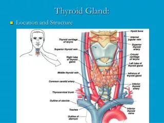
Trachea, bronchi, LUNGS & bronchopulmonary segmentS. Dr. Mujahid Khan. Trachea. The trachea is a mobile cartilaginous and membranous tube It begins in the neck as a continuation of the larynx at the lower border of the cricoid cartilage at the level of the sixth cervical vertebra

E N D
Trachea, bronchi, LUNGS & bronchopulmonary segmentS Dr. Mujahid Khan
Trachea • The trachea is a mobile cartilaginous and membranous tube • It begins in the neck as a continuation of the larynx at the lower border of the cricoid cartilage at the level of the sixth cervical vertebra • It descends in the midline of the neck
Trachea • In the thorax the trachea ends below at the carina by dividing into right and left principal (main) bronchi • During expiration the bifurcation rises by about one vertebral level • During deep inspiration may be lowered as far as the sixth thoracic vertebra
Trachea • In adults the trachea is about 4½ in. (11.25 cm) long and 1 in. (2.5 cm) in diameter • The fibroelastic tube is kept patent by the presence of U-shaped rings of hyaline cartilage embedded in its wall • The posterior free ends of the cartilage are connected by smooth muscle, the trachealis muscle
Relation • Anteriorly: The sternum, the thymus, the left brachiocephalic vein, the origins of the brachiocephalic and left common carotid arteries, and the arch of the aorta • Posteriorly: The esophagus and the left recurrent laryngeal nerve
Relation • Right side: The azygos vein, the right vagus nerve, and the pleura • Left side: The arch of the aorta, the left common carotid and left subclavian arteries, the left vagus and left phrenic nerves, and the pleura
Blood Supply • The upper two thirds are supplied by the inferior thyroid arteries • The lower third is supplied by the bronchial arteries
Lymph Drainage • The lymph drains into the pretracheal and paratracheal lymph nodes and the deep cervical nodes
Nerve Supply • The sensory nerve supply is from the vagi and the recurrent laryngeal nerves • Sympathetic nerves supply the trachealis muscle
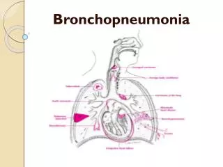
Bronchi • The trachea bifurcates behind the arch of the aorta into the right and left principal (primary, or main) bronchi • The bronchi divide into several million terminal bronchioles that terminate in one or more respiratory bronchioles
Bronchi • Each respiratory bronchiole divides into 2 to 11 alveolar ducts that enter the alveolar sacs • The alveoli arise from the walls of the sacs as diverticula
Right Principal Bronchus • Is wider, shorter, and more vertical than the left • Is about 1 in. (2.5 cm) long • Before entering the hilum of the right lung, it gives off the superior lobar bronchus • On entering the hilum, it divides into a middle and an inferior lobar bronchus
Left Principal Bronchus • Is narrower, longer, and more horizontal than the right and is about 2 in. (5 cm) long • It passes to the left below the arch of the aorta and in front of the esophagus • On entering the hilum of the left lung, the principal bronchus divides into a superior and an inferior lobar bronchus
Lungs • Two lungs are soft, spongy and elastic • In the child, they are pink, but with age, they become dark and mottled because of the inhalation of dust particles • These particles are trapped in the phagocytes of the lung • The lungs are situated so that one lies on each side of the mediastinum
Lungs • Two lungs are separated from each other by the heart and great vessels and other structures in the mediastinum • Each lung is conical, covered with visceral pleura and suspended free in its own pleural cavity • Attached to the mediastinum only by its root
Lungs • A blunt apex projects upward into the neck for about 1 in. (2.5 cm) above the clavicle • A concave base that sits on the diaphragm • A convex costal surface, which corresponds to the concave chest wall • A concave mediastinal surface, which is molded to the pericardium and other mediastinal structures
Lungs • Hilum is a depression in which the bronchi, vessels, and nerves that form the root enter and leave the lung • The anterior border is thin and overlaps the heart • It is here on the left lung that the cardiac notch is found • The posterior border is thick and lies beside the vertebral column
Lobes and Fissures of Rt. Lung • The right lung is slightly larger than the left • Divided by the oblique and horizontal fissures into three lobes: upper, middle, and lower • The oblique fissure runs from the inferior border upward and backward across the medial and costal surfaces until it cuts the posterior border about 2.5 in. (6.25 cm) below the apex
Lobes and Fissures of Rt. Lung • The left lung is divided by a similar oblique fissure into two lobes: upper and lower • There is no horizontal fissure in the left lung
Bronchopulmonary Segment • The bronchopulmonary segments are the anatomic, functional, and surgical units of the lungs • Each lobar (secondary) bronchus gives off branches called segmental (tertiary) bronchi • Each segmental bronchus passes to a structurally and functionally independent unit of a lung lobe called a bronchopulmonary segment, surrounded by connective tissue
Bronchopulmonary Segment • The segmental bronchus is accompanied by a branch of the pulmonary artery • Each segment has its own lymphatic vessels and autonomic nerve supply • On entering a bronchopulmonary segment, each segmental bronchus divides repeatedly
Bronchopulmonary Segment • As the bronchi become smaller, the U-shaped bars of cartilage found in the trachea are gradually replaced by irregular plates of cartilage • The smallest bronchi divide and give rise to bronchioles, which are less than 1 mm in diameter
Bronchopulmonary Segment • Bronchioles possess no cartilage in their walls and are lined with columnar ciliated epithelium • The submucosa possesses a complete layer of circularly arranged smooth muscle fibers
Bronchopulmonary Segment • The bronchioles then divide and give rise to terminal bronchioles which show delicate outpouchings from their walls • Gaseous exchange between blood and air takes place in the walls of these outpouchings, which explains the name respiratory bronchiole
Bronchopulmonary Segment • The diameter of a respiratory bronchiole is about 0.5 mm • The respiratory bronchioles end by branching into alveolar ducts, which lead into tubular passages with numerous thin-walled outpouchings called alveolar sacs
Bronchopulmonary Segment • The alveolar sacs consist of several alveoli opening into a single chamber • Each alveolus is surrounded by a rich network of blood capillaries • Gaseous exchange takes place between the air in the alveolar lumen through the alveolar wall into the blood within the surrounding capillaries
Characteristics of Bronchopulmonary Segment • It is a subdivision of a lung lobe • It is pyramid shaped, with its apex toward the lung root • It is surrounded by connective tissue
Characteristics of Bronchopulmonary Segment • It has a segmental bronchus, a segmental artery, lymph vessels, and autonomic nerves • The segmental vein lies in the connective tissue between adjacent bronchopulmonary segments • Because it is a structural unit, a diseased segment can be removed surgically
Right lung • Superior lobe: Apical, posterior, anterior • Middle lobe: Lateral, medial • Inferior lobe: Superior (apical), medial basal, anterior basal, lateral basal, posterior basal
Left lung • Superior lobe: Apical, posterior, anterior, superior lingular, inferior lingular • Inferior lobe: Superior (apical), medial basal, anterior basal, lateral basal, posterior basal
Blood Supply • The bronchi and the connective tissue of the lung receive their blood supply from the bronchial arteries, which are branches of the descending aorta • The bronchial veins communicate with the pulmonary veins and drain into the azygos and hemiazygos veins
Blood Supply • The alveoli receive deoxygenated blood from the terminal branches of the pulmonary arteries • The oxygenated blood leaving the alveolar capillaries drains into the tributaries of the pulmonary veins • Two pulmonary veins leave each lung root to empty into the left atrium of the heart
Lymph Drainage • The lymph vessels originate in superficial and deep plexuses • They are not present in the alveolar walls • The superficial (subpleural) plexus lies beneath the visceral pleura and drains over the surface of the lung toward the hilum, where the lymph vessels enter the bronchopulmonary nodes
Lymph Drainage • The deep plexus travels to the hilum of the lung and enters the bronchopulmonary nodes • All the lymph from the lung leaves the hilum and drains into the tracheobronchial nodes and then into the bronchomediastinal lymph trunks
Nerve Supply • At the root of each lung is a pulmonary plexus composed of efferent and afferent autonomic nerve fibers • The plexus is formed from branches of the sympathetic trunk and receives parasympathetic fibers from the vagus nerve • The sympathetic efferent fibers produce bronchodilatation and vasoconstriction • The parasympathetic efferent fibers produce bronchoconstriction, vasodilatation, and increased glandular secretion