Download
1 / 56
750 likes | 1.86k Views
Chapter 23. Microbial Diseases of the Digestive System. Structures of the Digestive System. Digestive system structures divided into two groups Gastrointestinal tract (GI tract) The pathway from the mouth to the anus Most organs of the GI tract protected by the peritoneum

E N D
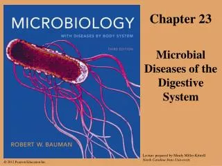
Chapter 23 Microbial Diseases of the Digestive System
Structures of the Digestive System Digestive system structures divided into two groups Gastrointestinal tract (GI tract) The pathway from the mouth to the anus Most organs of the GI tract protected by the peritoneum Accessory digestive organs Organs involved in grinding food or providing digestive secretions © 2012 Pearson Education Inc.
Structures of the Digestive System The Gastrointestinal Tract Digests food, absorbs nutrients and water into the blood, and eliminates waste Components of the gastrointestinal tract Mouth Esophagus Stomach Small intestine Large intestine (colon) Rectum and anus © 2012 Pearson Education Inc.
Uvula Tongue Teeth Salivaryglands Figure 23.1 Major structures of the digestive system Pharynx Mouth Esophagus Liver Stomach Pancreas Gallbladder Transversecolon Duodenum Ascendingcolon Largeintestine Smallintestine Jejunum Descendingcolon Ileum Sigmoid colon Rectum Anus
Structures of the Digestive System The Accessory Digestive Organs Tongue and teeth Salivary glands Liver Gallbladder Pancreas © 2012 Pearson Education Inc.
Bacteria Enamel Figure 23.2 Detailed structure of teeth and socket Dentin Pulp Gingiva(gum) Bone Branches ofblood vesselsand nervein root canal
Normal Microbiota of the Digestive System Esophagus, Stomach, Duodenum These regions are almost free of microbes Peristalsis and rapid transport of food helps prevent microbial colonization Tongue and teeth Viridans streptococci are most prevalent in this region Lower small intestine and colon Microbiota here are microbial antagonists Mucous membrane prevents microbes entering the bloodstream © 2012 Pearson Education Inc.
Bacterial Diseases of the Digestive System Dental Caries, Gingivitis, and Periodontal Disease Signs and symptoms Caries Appear as holes or pits in the teeth Periodontal disease Gums that are swollen, tender, bright red, or bleeding Pathogen and virulence factors, and pathogenesis Streptococcus mutans is a frequent cause of caries Dextran and pili allow biofilm formation on the tooth Porphyromonas gingivalis causes periodontal disease Proteases break down gingival tissue © 2012 Pearson Education Inc.
Figure 23.3 The process of tooth decay Plaque (biofilm)
Bacterial Diseases of the Digestive System Dental Caries, Gingivitis, and Periodontal Disease Epidemiology Most adults have experienced dental caries Diets high in sucrose increase the risk of decay Diagnosis, treatment, and prevention Caries Diagnosed by visual inspection Treat by filling cavities if caught early Gingivitis Diagnosed by inspection of gums Treat by scaling and use of antibacterial rinses Prevention involves good oral hygiene © 2012 Pearson Education Inc.
Bacterial Diseases of the Digestive System Peptic Ulcers Signs and symptoms Abdominal pain is main symptom Pathogen and virulence factors Caused by Helicobacter pylori Numerous virulence factors Flagella enable burrowing through stomach lining Adhesins facilitate attachment to gastric cells Urease neutralizes stomach acid © 2012 Pearson Education Inc.
Figure 23.4 The role of Helicobacter pylori in the formation of ulcers Helicobacter pylori(neutralizes stomachacid) Layer of mucus Acidic gastric juice Nucleus Neutrophil Lymphocyte Epithelial cellin stomach lining Mucus-secreting cell Ulcer Red bloodcells in capillaries Bacteria invade mucus and attach togastric epithelial cells. Helicobacter, its toxins, andinflammation cause the layer ofmucus to become thin. Gastric acid destroys epithelial cellsand underlying tissue.
Bacterial Diseases of the Digestive System Peptic Ulcers Epidemiology Fecal-oral transmission is likely Stress may worsen ulcer symptoms Diagnosis, treatment, and prevention Diagnosis based on X-ray exam to identify ulcers and presence of H. pylori in clinical specimens Treat with antimicrobials and drugs that inhibit stomach acid Prevent by avoidance of fecal-oral transmission © 2012 Pearson Education Inc.
Bacterial Diseases of the Digestive System Bacterial Gastroenteritis Inflammation of stomach or intestines caused by bacteria Associated with contaminated food or water and poor living conditions General features Similar manifestations despite different causative agents Nausea, vomiting, diarrhea, abdominal pain, and cramps Dysentery produces loose, frequent stool containing mucus and blood © 2012 Pearson Education Inc.
Bacterial Diseases of the Digestive System Bacterial Gastroenteritis: Cholera Pathogen and virulence factors Caused by Vibrio cholerae Most important virulence factor is production of cholera toxin Pathogenesis and epidemiology Pandemics have occurred throughout history © 2012 Pearson Education Inc.
Intestinal lumen A Water followselectrolytes intolumen. Cholera toxinbinds tomembraneof epithelialcell. B Figure 23.5 The action of cholera toxin in intestinal epithelial cells Epithelialcell Portion of toxin(part of A)enters cell. Cyclic AMP stimulatescell tosecrete Cl,Na, andotherelectrolytes. A1 A1 is anenzyme thatactivatesadenylatecyclase. Adenylatemakescyclic AMP(cAMP).
Figure 23.6 Cholera pandemic Initial epidemicsJanuary 1991 August 1991 February 1992 November 1994
Bacterial Diseases of the Digestive System Bacterial Gastroenteritis: Cholera Diagnosis, treatment, and prevention Diagnosis based on presence of “rice-water stool” Treat with supportive care and administration of tetracycline Available vaccine provides only short-lived immunity Proper hygiene is an important preventive measure © 2012 Pearson Education Inc.
Bacterial Diseases of the Digestive System Bacterial Gastroenteritis: Shigellosis Pathogen and virulence factors Caused by four species of Shigella Virulence factors include type III secretion systems and enterotoxins Pathogenesis and epidemiology Pathogen colonizes cells of the small, then large intestine Diagnosis, treatment, and prevention Diagnose by symptoms and presence of Shigella in stool Supportive treatment and administration of antimicrobials © 2012 Pearson Education Inc.
Shigella Shigella attaches toepithelial cell of colon. Epithelial cell Nucleus Figure 23.7 The events in shigellosis Shigella triggersendocytosis. Shigella multipliesin cytosol. Actin fibers Shigella invadesneighboring epithelialcells, thus avoidingimmune defenses. Mucosal abscess An abscess forms asepithelial cells are killedby the infection. Blood vessel Phagocyte Shigella thatenters the bloodis quicklyphagocytizedand destroyed.No bacteremia.
Bacterial Diseases of the Digestive System Bacterial Gastroenteritis: Traveler’s Diarrhea Pathogen and virulence factors Caused by Escherichia coli Virulence factors include adhesins, fimbriae, and toxins Pathogenesis and epidemiology Diarrhea mediated by enterotoxins Diagnosis, treatment, and prevention Diagnosis based on signs and symptoms Treatment based on fluid and electrolyte replacement Antidiarrheal drugs prolong the symptoms © 2012 Pearson Education Inc.
Bacterial Diseases of the Digestive System Bacterial Gastroenteritis: Campylobacter Diarrhea Pathogen and virulence factors Caused by Campylobacter jejuni Virulence factors include adhesins, cytotoxins, endotoxin Pathogenesis and epidemiology Virulence factors cause bleeding lesions and inflammation Diagnosis, treatment, and prevention Diagnosis based on signs and symptoms Most cases resolve without treatment Prevent with proper hygiene after handling raw poultry © 2012 Pearson Education Inc.
Figure 23.8 Campylobacter jejuni, the most common cause of bacterial gastroenteritis in the U.S.
Bacterial Diseases of the Digestive System Bacterial Gastroenteritis: Antimicrobial-Associated Diarrhea Signs and symptoms Pseudomembranous colitis occurs in severe cases Pathogen and virulence factors Caused by Clostridium difficile Antimicrobial use facilitates overgrowth of C. difficile C. difficile produces two toxins Pathogenesis Toxins mediate inflammation and pseudomembrane formation © 2012 Pearson Education Inc.
Lesions Figure 23.9 Pseudomembranous colitis
Bacterial Diseases of the Digestive System Bacterial Gastroenteritis: Antimicrobial-Associated Diarrhea Epidemiology By-product of modern medicine Any antimicrobial can trigger the disease Diagnosis, treatment, and prevention Diagnosis based on presence of bacterial toxin in stool Treat with antimicrobials Avoid unnecessary use of antimicrobials © 2012 Pearson Education Inc.
Bacterial Diseases of the Digestive System Bacterial Gastroenteritis: Salmonellosis and Typhoid Fever Pathogen and virulence factors Caused by Salmonella enterica serotypes Serotypes Typhi and Paratyphi cause typhoid fever Serotypes Enteritidis and Typhimurium cause salmonellosis Bacteria tolerate acidity of stomach and pass to the intestine Toxins disrupt numerous cellular activities Pathogenesis and epidemiology Typhoid fever acquired by contaminated food or water Salmonellosis often acquired by consuming contaminated eggs © 2012 Pearson Education Inc.
Salmonella Salmonella attaches toepithelial cells liningthe small intestine. Epithelial cell Figure 23.10 The events in salmonellosis Nucleus Salmonella triggersendocytosis. Salmonella multiplieswithin food vesicle. Salmonella kills hostcell, inducing fever,cramps, and diarrhea. Capillary (blood vessel) Bacteremia:Salmonellamoves intobloodstream.
Bacterial Diseases of the Digestive System Bacterial Gastroenteritis: Salmonellosis and Typhoid Fever Diagnosis, treatment, and prevention Diagnosis made by finding Salmonella in stool Salmonellosis is usually self-limiting Typhoid fever can be treated with antimicrobial drugs Prevented with proper hygiene © 2012 Pearson Education Inc.
Bacterial Diseases of the Digestive System Bacterial Food Poisoning (Intoxication) Signs and symptoms Nausea, vomiting, diarrhea, cramping Pathogen and virulence factors Caused by Staphylococcus aureus Virulence factors include five enterotoxins Pathogenesis and epidemiology Outbreaks associated with social functions Diagnosis, treatment, and prevention Diagnosis based on signs and symptoms Treated with fluid and electrolyte replacement Proper hygiene can reduce incidence © 2012 Pearson Education Inc.
Viral Diseases of the Digestive System Oral Herpes Signs and symptoms Presence of cold sores Pathogen and pathogenesis Caused by human herpesvirus 1 (HHV-1) Virions avoid host’s immune system by forming syncytia Epidemiology Infections occur by casual contact in childhood Diagnosis, treatment, and prevention Diagnosis based on characteristic lesions Topical acyclovir limits duration of lesions Avoid direct contact with infected individuals © 2012 Pearson Education Inc.
Figure 23.12 Latency and reactivation of oral herpesviruses Trigeminal nerve Trigeminal (V)nerve ganglion,site of virallatency • Ophthalmic branch • Maxillary branch • Mandibular branch Fever blister
Viral Diseases of the Digestive System Mumps Caused by the mumps virus Humans are the only natural host Once a very common childhood disease Nearly nonexistent in developed countries due to immunization No specific treatment for mumps Infected individuals develop lifelong immunity © 2012 Pearson Education Inc.
Viral Diseases of the Digestive System Viral Gastroenteritis Signs and symptoms Similar to bacterial gastroenteritis Pathogens and pathogenesis Caused by caliciviruses, astroviruses, and rotaviruses Epidemiology More cases occur in winter Diagnosis, treatment, and prevention Serological test distinguishes among viruses Treatment is based on fluid and electrolyte replacement Vaccine for rotavirus exists © 2012 Pearson Education Inc.
Figure 23.14 Deaths from rotaviral diarrhea are most common in developing countries 1000 deaths
Viral Diseases of the Digestive System Viral Hepatitis Signs and symptoms Jaundice, abdominal pain, fatigue, vomiting, appetite loss Symptoms may occur years after initial infection Host immune response causes much of the liver damage Pathogen and pathogenesis Hepatitis A virus (HAV) Hepatitis B virus (HBV) Hepatitis C virus (HCV) Hepatitis delta virus (HDV) Hepatitis E virus (HEV) © 2012 Pearson Education Inc.
Viral Diseases of the Digestive System Viral Hepatitis Diagnosis, treatment, and prevention Initial diagnosis made by observation of jaundice, enlarged liver, or fluid in the abdomen Serological testing can identify viral antigens HBV diagnosed by presence of viral proteins in body fluids Supportive care for symptoms Prevent with good hygiene and protected sex or abstinence Vaccines are available against HAV and HBV © 2012 Pearson Education Inc.
Figure 23.15 The three types of viral particles produced by hepatitis B viruses-overview
Protozoan Diseases of the Intestinal Tract Giardiasis Signs and symptoms Often asymptomatic Diarrhea and associated symptoms can last up to four weeks Pathogen and pathogenesis Caused by Giardia intestinalis G. intestinalis interferes with intestinal absorption, causing flatus © 2012 Pearson Education Inc.
Mark left byadhesive disk Ventral adhesivedisk Figure 23.16 Trophozoites of Giardia intestinalis Intestinal villi Dorsal surface
Protozoan Diseases of the Intestinal Tract Giardiasis Epidemiology Infection results from ingesting cysts in contaminated water Hikers and campers are at particular risk Diagnosis, treatment, and prevention Diagnosed by microscopic observation of Giardia in stool Treat with metronidazole (adults) or furazolidone (children) © 2012 Pearson Education Inc.
Protozoan Diseases of the Intestinal Tract Cryptosporidiosis Signs and symptoms Severe watery diarrhea with potentially serious complications Pathogen and pathogenesis Caused by Cryptosporidium parvum Pathogenicity of C. parvum unclear Epidemiology Infection results from drinking contaminated water Diagnosis, treatment, and prevention Treated with fluid and electrolyte replacement Prevented with proper hygiene © 2012 Pearson Education Inc.
Oocysts Figure 23.17 Oocysts of Cryptosporidum parvum in feces
Protozoan Diseases of the Intestinal Tract Amebiasis Signs and symptoms Luminal amebiasis is asymptomatic Invasive amebic dysentery causes severe diarrhea, colitis, appendicitis Invasive extraintestinal amebiasis causes necrotic lesions in various organs Pathogen, virulence factors, and pathogenesis Caused by Entamoeba histolytica Trophozoites in the peritoneal cavity or blood cause symptoms © 2012 Pearson Education Inc.
Protozoan Diseases of the Intestinal Tract Amebiasis Epidemiology Transmitted by consumption of contaminated food or water, from contaminated hands, or oral-anal intercourse Majority of individuals develop luminal amebiasis Diagnosis, treatment, and prevention Diagnosed by microscopic observation of Entamoeba in stool or intestinal biopsy Treat with oral rehydration therapy and antiamebic drugs Prevent with proper hygiene and safe sex practices © 2012 Pearson Education Inc.
Helminthic Infestations of the Intestinal Tract Helminths are macroscopic, multicellular worms Helminths can infest the GI tract as non-disease-causing parasites Tapeworm is the common name for a cestode Flat, segmented, parasitic helminth Tapeworms are intestinal parasites that lack their own digestive system © 2012 Pearson Education Inc.