Download
1 / 17
810 likes | 2.92k Views
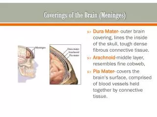
The Meninges. Head & Neck Unit – Lecture 3 د. حيدر جليل الأعسم. The Meninges. Three protective membranes that surround the brain, cerebellum and the spinal cord . The D ura mater (tough) The Arachnoid mater (delicate) The P ia mater (thin but firmly attached). Dura Mater.

E N D
The Meninges Head & Neck Unit – Lecture 3 د. حيدر جليل الأعسم
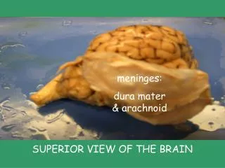
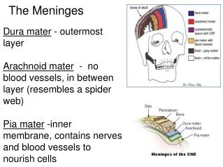
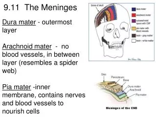
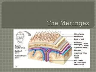
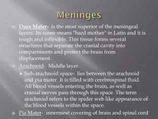
The Meninges Three protective membranes that surround the brain, cerebellum and the spinal cord. • The Dura mater (tough) • The Arachnoid mater (delicate) • The Piamater (thin but firmly attached)
Dura Mater The Dura Mater: • fibrous layer that is divided into two layers (except@ venous sinuses). • Endosteallayer: (periosteum) • Do not extend beyond the skull. • Fuse with periosteum of the skull outside. • Fuse with suturalligaments. • Meningeal layer: (dura mater proper) • Extend beyond the skull @ foramen magnum. • Fuse with epineurium of cranial nerves. • Sends inward septa to form cranial partitions.
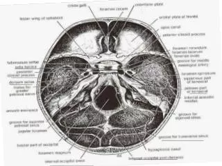
Dura Partitions • Falx Cerebri • Tentorium Cerebelli • Falx Cerebelli • The diaphragma sellae
Dura Partitions – Falx Cerebri Falx Cerebri: • Sickle-shaped fold of dura mater. • Its ant end is attached to the internal frontal crest and the crista galli. Its posterior end blends with the upper surface of the tentorium cerebelli. • The superior sagittal sinus runs in its upper fixed margin • The inferior sagittal sinus in its lower free margin • The straight sinus runs along its attachment to the tentorium cerebelli.
Dura Partitions – Tentorium Cerebelli Tentorium Cerebelli • crescent-shaped fold of dura mater. • anteriorly, the tentorial notch for the midbrain. • its outer border is attached to the posterior clinoid processes, the superior borders of the petrous bones, and the margins of the grooves for the transverse sinuses on the occipital bone. • It inner free border crosses the attached border, and is attached to the anterior clinoid process.
Dura Partitions – Falx Cerebelli Falx Cerebelli: • is a small, sickle-shaped fold of dura mater. • Its fixed posterior margin is attached to the internal occipital crest and contain the occipital sinus. • Its free border anteriorly separate the two cerebellar hemispheres.
Dura Partitions - The diaphragmasellae The diaphragma sellae • is a small circular fold of dura mater • it forms the roof for the sella turcica. • It has a small opening in its center for stalk of the pituitary gland.
Innervation of the Dura • Dura is sensitive to ?? --► headache • Dura of Posterior cranial fossa by branches from cervical spinal nerves (C1, C2 & C3) • Dura of ACF, MCF, Falx cerebri & Tentorium Cerebelli by Trigeminal nerve: 1- Anterior meningeal nerves 2- Tentorial nerve 3- Meningeal branches of maxillary and Mandibular divisions of Trigeminal nerve
Arterial Supply of Dura • Anterior meningeal artery (branches of ethmoidal arteries which are branches of maxillary artery (ECA). • Middle meningeal artery and accessary meningeal artery: branches of Maxillary artery (ECA) • Posterior meningeal artery (terminal branch of ascending pharyngeal artery (ECA) & other meningeal branches from: 1- Ascending pharyngeal artery 2- Occipital artery 3- Vertebral artery
Venous Drainage • Venous drainage of dura usually follow the arterial arrangement of meningeal arteries: 1- anterior Meningeal veins 2- middle Meningeal veins 3- Posterior Meningeal veins
Arachnoid mater • Delicate avascular layer (lies against but not firmly attached to dura mater). It sends numerous trabeculaetoward the pia mater. • Subarachnoid space?? CSF? • Arachnoid cisternae. • All cerebral arteries and veins lie in subarachnoid space. • Arachnoid villi: numerous projections to the venous sinus. • Arachnoid granulations: aggregations of these villi at which CSF diffuses to venous sinuses. • Arachnoid mater fuses with epineurium of nerves at the exit foramina except for optic nerve.
Pia mater thin vascular layer that is adherent to the brain surface. It extends with cerebral sulci and cover gyri and fuses with epineurium of cranial nerves at their exit foramina.
Venous Sinuses • Blood-filled spaces situated between the layers of the dura mater. • The sinuses have no valves. • They receive tributaries from the brain, the diplo of the skull, the orbit, and the internal ear. • The superior sagittal sinus- receives the superior cerebral veins. • The inferior sagittal sinus - joins the great cerebral vein to form the straight sinus. It receives cerebral veins from the medial surface of the cerebral hemisphere. • The straight sinus- it drains into the left transverse sinus. • The left transverse sinus is a continuation of the straight sinus & end on each side by becoming the sigmoid sinus. • The right transverse sinus is as a continuation of the superior sagittal sinus. • The sigmoid sinuses - leave through jugular foramen --►internal jugular vein. • The occipital sinus- communicates with the vertebral veins through the foramen. • The cavernous sinusAnteriorly, the sinus receives the inferior ophthalmic vein and the central vein of the retina & drains posteriorly into the transverse sinus through the superior petrosal sinus. • Intercavernous sinuses connect the two cavernous sinuses through the sellaturcica.