Download
1 / 81
820 likes | 1.09k Views
Case Report. A 44-year-old African-American woman presented with complaints of worsening dyspnea and chest discomfort for several months Review of systems revealed long-standing dyspnea on exertion, orthopnea, and paroxysmal nocturnal dyspnea.

E N D
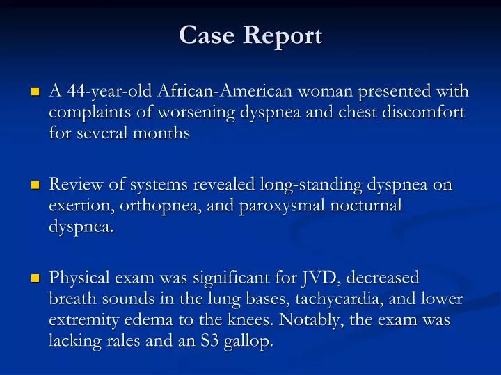
Case Report • A 44-year-old African-American woman presented with complaints of worsening dyspnea and chest discomfort for several months • Review of systems revealed long-standing dyspnea on exertion, orthopnea, and paroxysmal nocturnal dyspnea. • Physical exam was significant for JVD, decreased breath sounds in the lung bases, tachycardia, and lower extremity edema to the knees. Notably, the exam was lacking rales and an S3 gallop.
Chest radiograph revealed an enlarged cardiac sillouette and bilateral pleural effusions (Figure 1).
Bedside echocardiography revealed a pericardial effusion (Figure 2).
Data • Laboratory findings included a macrocytic anemia (MCV 114.6 fL, HCT 22.2%) with a high corrected reticulocyte count (14.5) and normal B12 and folate levels. • Auto-immune hemolytic anemia was confirmed by a decreased haptoglobin (<14 mg/dL), an elevated LDH (1411 U/L), and the detection of a warm IgG auto-antibody. • ANA testing revealed a 160 titer and an anti-DNA DS level of 101 IU. Right heart catheterization revealed tamponade physiology.
Discussion • This case represents a rare presentation in a patient with SLE. • The patient's symptoms were secondary to both tamponade and hemolytic anemia, perhaps two of the most morbid of the diagnostic criteria. • The eventual diagnosis may have been delayed were it not for careful attention to the physical exam and prompt diagnostic testing to validate those findings. • Standard treatment for congestive heart failure based solely on reported symptoms would have greatly increased her morbidity, and could have been fatal. • This case emphasizes the importance that our history AND physical examinations guide our diagnostic and therapeutic measures.
Systemic Lupus ErythematosusJuly 13, 2009 Julie Schwartzman, MD Associate Program Director, Rheumatology Director of Arthritis and Lupus Clinics
SLE: Subsets • Discoid LE • Drug Induced SLE • Neonatal SLE • Antiphospholipid Syndrome • SLE • Benign, incomplete • Subacute cutaneous, ANA negative, Ro + lupus • DNA positive, complement fixing, hypocomplementic
Drug Induced SLE • Hydralazine • Procainamide • Minocycline (ANCA+) • Chlorpromazine • Isoniazid • Penicillamine • Methyldopa • Interferon-alpha
SLE: Demographics • Affects .5 million (.2%) vs 1.5 million (.6%) of US population (epidemiologic vs LFA random digit dialing telephone survey) • Female:Male ratio of 10:1 • Closer to 2:1 during childhood and after menopause, suggesting hormonal influence • Disease in males is often more severe • 70% of SLE: females between ages 15-45 • African American to Caucasian ratio 3:1 • Highest prevalence in Afro-Caribbean females 1:250
Demographics • Genetic factors HLA-A1, B8, Dr3 - C4A null genes - Fc receptor polymorphisms -gene linked to chromosome 1 • Environmental factors - Concordance for monozygotic twins is 30% (70% of genetically identical twins will not share the disease) • Child of SLE mother risk of SLE 1:15 (7%) • ANA positive in 5-20% of population. 10 times more likely to have false positive ANA than disease
Null alleles that cause a deficiency of one of the early complement components — C1q, C2, or C4 — are a strong risk factor for lupus. • Family studies have identified genes that are more likely to occur in patients with lupus than in their healthy relatives. • Many of these genes encode components of the immune system.
SLE: ETIOLOGY • AUTOANTIBODY PRODUCTION • GENERATION OF CIRCULATING IMMUNE COMPLEXES • EPISODIC COMPLEMENT ACTIVATION
SLE: Pathobiology • Autoantibodies (AIHA, AITP, Anti-neuronal antibody, APS) • Immune complex disease (microangiitis and vasculitis) • Neutrophil and endothelial cell adhesive interaction with leukoaggregation • Thrombophilia: Antibody mediated thrombosis in secondary APLS with micro and macrovascular non-inflammatory occlusion
SEROLOGY • ANA (Titer and pattern: diffuse, speckled, rim, nucleolar, centromere) • double stranded-DNA- highly specific, correlates with disease activity • Sm- highly specific for SLE, no correlation with disease activity • RNP- also seen in MTCD/UCTD • Ro (SS-A)/La (SS-B)- neonatal lupus, SS, subacute cutaneous lupus, photosensitivity (Ro) • C3 • C4
POSITIVE ANA • SLE • Non SLE CTD (RA, SS, PSS, CREST, DM/PM) • DRUG-INDUCED • NORMALS (FALSE POSTIVE) • LYMPHOPROLIFERATIVE DISORDER • CHRONIC INFECTION (HIV, Leprosy)
Malar rash Discoid rash Photosensitivity Oral ulcers Arthritis Serositis Renal disorder Neurologic disorder Hematologic disorder Immunologic disorder Anti-nuclear antibody 1982 ACR Classification Criteria Require four simultaneously or serially
Mucocutaneous Manifestations • * Malar rash • * Discoid rash • * Photosensitivity • *Oral ulcers • Vasculitis • Periungual erythema • Alopecia (*classification criteria)
“Butterfly” or Malar Rash 1. Malar rash: Fixed erythema, flat or raised, over the malar eminences, tending to spare the nasolabial folds
Discoid Rash 2. Discoid rash: Erythematous raised patches with adherent keratotic scaling and follicular plugging; atrophic scarring may occur in older lesions
Photosensitivity “allergic to the sun” 3. Photosensitivity: Skin rash as a result of unusual reaction to sunlight, by patient history or physician observation
Oral Ulcers 4. Oral ulcers: Oral or nasopharyngeal ulceration, usually painless
Subacute Cutaneous Lupus Erythematosus Left, Papulosquamous lesions are characterized by erythematous scaling papules and plaques that resemble psoriasis. The distribution in light-exposed areas suggests photosensitivity. Right, The annular polycyclic lesions have an erythematous, slightly scaling border with central clearing.
Arthritis • 5. Arthritis: Non-erosive arthritis involving 2 or more peripheral joints, characterized by tenderness, swelling, or effusion • 80% • Single or multiple joints • Reducible deformities • Pain may be out of proportion with appearance
Serositis • 6. Serositis a) Pleuritis--convincing history of pleuritic pain or rubbing heard by a physician or evidence of pleural effusion • OR b) Pericarditis--documented by ECG or rub or evidence of pericardial effusion • Peritonitis - diffuse abdominal pain, nausea and vomiting, ascites is rare
Lupus Serositis Pleuritis Pericarditis Chest X-ray Chest X-ray Peritonitis Abdominal X-ray
Renal 7. Renal disorder a) Persistent proteinuria greater than 0.5 grams per day OR b) Cellular casts--may be red cell, hemoglobin, granular, tubular, or mixed
Lupus Nephritis (WHO Classification) • I Normal glomeruli a) Nil by all techniques b) Normal by light but deposits on EM or IF • II Mesangial nephritis • III Focal glomerulonephritis • IV Diffuse proliferative glomerulonephritis • V Membranous nephritis • VI Membranoproliferative glomerulonephritis • VII Advanced sclerosing glomerulonephritis
Lupus Nephritis (light microscopy) Mesangial nephritis Focal glomerulonephritis Diffuse proliferative glomerulonephritis Membranous glomerulonephritis
Lupus Nephritis (immunofluorescent staining) Staining with anti-Ig FITC
Lupus Nephritis (Electron Microscopy) A – subepithelial deposits B – intramembranous deposits C – basement membrane D – epithelial foot processes
Neurologic disorder 8. Neurologic disorder a) Seizures--in the absence of offending drugs or known metabolic derangements; e.g., uremia, ketoacidosis, or electrolyte imbalance OR b) Psychosis--in the absence of offending drugs or known metabolic derangements, e.g., uremia, ketoacidosis, or electrolyte imbalance
Seizures Psychosis Headache Coma Dementia Aseptic meningitis Chorea Ataxia Depression Cranial neuropathy Peripheral neuropathy Mononeuritis multiplex Stroke syndrome Transverse myelitis Neuropsychiatric Lupus
SLE Brain gross specimen photomicrograph fibrin thrombus leukoaggregation Left, Gross specimen reveals multiple cerebral infarctions. Right, Photomicrograph demonstrates occlusion of small vessels by both leukoaggregation (A) and a fibrin thrombus (B) as the causes of the infarctions. Vasculitis was not found
Hematologic Manifestations 9. Hematologic disorder a) Hemolytic anemia--with reticulocytosis OR b) Leukopenia--less than 4,000/mm on 2 or more occasions OR c) Lymphopenia--less than 1,500/mm on 2 or more occasions OR d) Thrombocytopenia--less than 100,000/mm in the absence of offending drugs
10. Immunologic disorder a) Anti-DNA OR b) Anti-Sm OR c) Positive finding of antiphospholipid antibodies 11. ANA: in the absence of drugs known to be associated with "drug-induced lupus" syndrome
Classification Criteria • 4/11 criteria >90% sensitivity and specificity • Criteria designed for classification, especially for entrance into laboratory studies and clinical trials but not for diagnosis • For mild and early disease may not be as sensitive • Examples: patient with malar rash and +ANA patient with GN, +ANA and elevated dsDNA Abs
Mortality • 90% survive 5 years, 80% 10 years • Worse if renal disease • AA more aggressive and treatment resistant disease • Bimodal distribution of etiology Early: disease activity and infection Late: disease activity, ESRD, arteriosclerotic and thromboembolic
CAD in SLE • Risk is 10-times increased in SLE patients, 50-times increased in SLE pts 35-44 yrs old • Factors contributing to increase: • Steroid therapy • Hyperlipidemia • HTN • Smoking • Coagulation abnormalities • Homocystinemia • Obesity • Vasculopathy from Immune Injury>>increased circulating endothelial cells activation *All modifiable risk factors are important targets of intervention by both PMD and Rheum MD
APLS • The anti-phospholipid Ab syndrome (APS) is vascular thrombosis and/or pregnancy morbidity developing in persistently anti-phospholipid antibody (aPL)-positive individuals. • The most commonly used aPL tests are the lupus anticoagulant test, the anti-cardiolipin antibody ELISA, and/or the anti- b2-glycoprotein I ELISA. • APS is present if more than 1 clinical and 1 lab criteria met
ANTIPHOSPHOLIPID SYNDROMESapporo Criteria, Sydney revisionMiyakis, J Thromb Haemost. 2006 4:295-306 • Clinical • Thrombosis: one or more confirmed episodes: venous, arterial or small vessel (exclude other causes, male 55+ female 65+) • Confirmed by imaging or Doppler or histopathology and without evidence of inflammation in vessel wall on histopathologic confirmation
ANTIPHOSPHOLIPID SYNDROMESapporo Criteria, Sydney revisionMiyakis, J Thromb Haemost. 2006 4:295-306 • Clinical • Pregnancy: • one or more unexplained deaths >10 wk • one or more pre-eclampsia/placental insufficiency < 34 wk • 3 or more unexplained consecutive spontaneous abortions <10 wk • exclude other causes
ANTIPHOSPHOLIPID SYNDROMESapporo Criteria, Sydney revisionMiyakis, J Thromb Haemost. 2006 4:295-306 • Laboratory • Medium/high IgG or IgM aCL, b2GP1 dependent on 2+ occasions 12 wk apart • LAC 2+ occasions 12 wk apart • prolonged PL-dependent screening test • failure to correct with mixing • shortening with excess PL • exclusion of other coagulopathies
ANTIPHOSPHOLIPID SYNDROMESapporo Criteria, Sydney revisionMiyakis, J Thromb Haemost. 2006 4:295-306 • aPL-associated findings (individual diagnosis) • cardiac valve disease • livedo reticularis • thrombocytopenia • nephropathy
Current Recommendations Erkan, Lockshin, Rheum Dis Clin North Am 2006;32:129-48 Lim, Crowther, Eikelboom, JAMA 2006;295:1050-1057