Download
1 / 22
220 likes | 391 Views
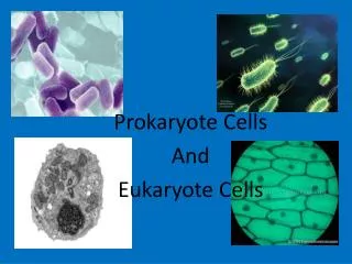
Life and Cells. What is Life? Can grow, i.e. increase in size. Can reproduce. Responsive to environment. Metabolism: can acquire and utilize energy. Schwann and Schleiden: cells basic unit of life Prokaryotes and eukaryotes from microscopy. Our focus: prokaryotic cells.

E N D
Life and Cells • What is Life? • Can grow, i.e. increase in size. • Can reproduce. • Responsive to environment. • Metabolism: can acquire and utilize energy. • Schwann and Schleiden: cells basic unit of life • Prokaryotes and eukaryotes from microscopy. • Our focus: prokaryotic cells. • Eubacteria and Archaebacteria
The Essential Cell Separates inside from environ. Genetic blueprint Needed for protein synthesis http://web.jjay.cuny.edu/~acarpi/NSC/images/cell.gif
Bacterial Appearance • Size • 0.2 µm – 0.1 mm • Most 0.5 – 5.0 µm • Shape • Coccus (cocci); rod (bacillus, bacilli); spiral shapes (spirochetes; spirillum, spirilla); filamentous; various odd shapes. • Arrangement • Clusters, tetrads, sarcina, pairs, chains http://www.cellsalive.com/howbig.htm http://www.ionizers.org/Sizes-of-Bacteria.html http://smccd.net/accounts/case/biol230/ex3/bact.jpeg
Overview of prokaryotic cell.
From Membrane Out:lecture order • Examination of layers of bacterial cell • Starting at cell membrane, working to outside • A look at how cells move • Examination of inside of bacterial cell • A look at how things get into cells • Brief review of eukaryotic cell structure.
Structure of phospholipids http://biyoloji_genetik.sitemynet.com/genel_biyoloji/genel_biyoloji_logos/phospholipids.gif
How phospholipids work Polar head groups associate with water but hydrophobic tails associate with each other to avoid water. When placed in water, phospholipids associate spontaneously side by side and tail to tail to form membranes. http://users.rcn.com/jkimball.ma.ultranet/BiologyPages/L/LipidBilayer.gif
Cell Membranes • 50/50 lipids and proteins • Fluid mosaic model • Effective barrier to large and hydrophilic molecules • O2, CO2, H2O, lipid substances can pass through • Salts, sugars, amino acids, polymers, cannot. • Proteins can be on inner, outer surfaces (peripheral) or transmembrane (integral) • Involved primarily with transport • Degradation and biosynthesis • Site of ATP synthesis
Membrane structure http://www.slic2.wsu.edu:82/hurlbert/micro101/images/cytomemb.gif
Outside the cell membrane:the Cell Wall Animal cells do not have a cell wall outside the cell membrane. Plant cells and fungal cells do. So do most prokaryotic cells, providing structural support and influencing the shape of the cell.
Gram stain Gram stain invented by Hans Christian Gram Divides Eubacteria into two main groups based on stain. Correlates with two types of cell wall architecture.
Division of the Eubacteria:Gram Negative and Gram Positive • Stain is valuable in identification. • Gram positives stain purple; Gram negatives stain pink. • Architecture: • Gram positives have a thick peptidoglycan layer in the cell wall; • Gram negatives have a thin peptidoglycan layer and an outer membrane. • BUT When we say Gram positive… • Cells stain purple? Or have a particular structure?
Gram Negative Gram Positive http://www.conceptdraw.com/sampletour/medical/GramNegativeEnvelope.gif http://www.conceptdraw.com/sampletour/medical/GramPositiveEnvelope.gif
Function and Structure of peptidoglycan • Provides shape and structural support to cell • Resists damage due to osmotic pressure • Provides some degree of resistance to diffusion of molecules • Single bag-like, seamless molecule • Composed of polysaccharide chains cross linked with short chains of amino acids: “peptido” and “glycan”.
Monomers of peptidoglycan Units added to PG as a pair. NAG:N-acetyl glucosamine NAM: N-acetyl muramic acid (NAG + lactic acid)
Glycan chains cross-linked with amino acids • G- and G+ vary w/ DAP vs. lysine and at the interbridge. • Note the presence of unusual “D” amino acids. • Peptides attached to NAM.
Peptidoglycan is a 3D molecule Cross links are both horizontal and vertical between glycan chains stacked atop one another. http://www.sp.uconn.edu/~terry/images/other/peptidoglycan.gif; http://www.alps.com.tw/cht/img/anti-allergy_002.jpg
Teichoic acid and lipoteichoic acid Found in G+ cell wall
Teichoic acid and lipoteichoic acidStructure and Function • Polymer of phosphate and ribitol or glycerol R: sugar or amino acid • Lipoteichoic acid covalently attached to membrane lipids. • Major contributor to negative charge of cell exterior. • Appears to function in Ca++ binding http://www.bact.wisc.edu/Microtextbook/images/textbook/structure/TAcid.gif;http://www.palaeos.com/Kingdoms/Prokaryotes/Images/GramPosCellEnvelope.gif
Effect of osmotic pressure on cells • Hypotonic: water rushes in; PG prevents cell rupture. • Hypertonic: water leaves cell, membrane pulls away from cell wall. plasmolysis
Bacteria and Osmotic pressure • Bacteria typically face hypotonic environments • Insides of bacteria filled with proteins, salts, etc. • Water wants to rush in, explode cell. • Protection from hypertonic environments is different, discussed later. • Peptidoglycan provides support • Limits expansion of cell membrane • Lysozyme found in secretions cuts peptidoglycan • Antibiotic penicillin prevents cross linking • Cells in isotonic medium are not harmed
Cell Wall Exceptions • Mycobacterium and relatives • Wall contains lots of waxy mycolic acids • Attached covalently to PG • Mycoplasma: no cell wall • Parasites of animals, little osmotic stress • Archaea, the 3rd domain • Pseudomurein and other chemically different wall materials (murein another name for PG)