Download
1 / 24
270 likes | 909 Views
The Value of Amplitude Integrated EEG in Monitoring Neonates at Risk for Seizures. Eilon Shany , Sonia Khvatskin, Agneta Golan , Michael Karplus . Neonatal Unit, Soroka University Medical Center, Ben-Gurion University, Beer Sheva , Israel. Background. Seizures. Common problem.

E N D
The Value of Amplitude Integrated EEG in Monitoring Neonates at Risk for Seizures. Eilon Shany, Sonia Khvatskin, Agneta Golan, Michael Karplus. Neonatal Unit, Soroka University Medical Center, Ben-Gurion University, Beer Sheva, Israel.
Seizures • Common problem. • Variable clinical appearances. • Etiology: • Hypoxic ischemic encephalopathy. • Metabolic, infectious, traumatic, hemorrhagic, structural. • Diagnosis: • Electroencephalography.
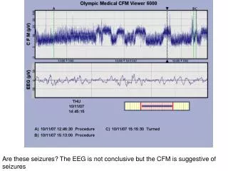
Cerebral Function Monitor (CFM) • Amplitude integrated EEG. • DE Maynard & PF Prior. • Single parietal channel. • Different filters. • Output: • Maximum and minimum amplitudes of EEG wave, on semilogarithmic scale, at a rate of 6 cm per hour.
EEG vs CFM Correlation • Simultaneous recording : • 10 infants with ictal EEG, 8 with clear ictal CFM, 1 suspected. • Sensitivity 80%, specificity 100%, PPV 100%, NPV 92%. • CFM has a good correlation with EEG for monitoring ictal activity. Toet et al, pediatrics 2002; 109(5): 772-779.
Seizure in CFM Normal CFM
Objective • To evaluate the clinical usefulness of amplitude integrated electroencephalogram (aEEG) using the cerebral function monitor (CFM) to diagnose seizures in a high risk neonates.
Inclusion Criteria • Infants born between December 1995 and August 2002. • Infants monitored with CFM due to: • Suspected asphyxia. • Suspected seizures.
Study Design • Records reviewed retrospectively by two investigators for presence of: • Clinical overt seizures. • Clinical subtle seizures. • CFM clear seizures. • CFM suspected seizures.
Suspected Seizures Clear Seizures
Data • 124 infants met inclusion criteria. • 1.5 hour to 10 days continuous recording. • 88 (71%) term, 36 (29%) preterm. • 101 (81%) asphyxia, 23 (19%) convulsions.
Interobserver ReliabilityCFM Tracings • Initial kappa: 74%. • Kappa after joined consult: 91%. • When no agreement was reached the more severe diagnosis was considered.
38 (95%) 2 (5%) 0 (0%) 9 (56%) 4 (25%) 3 (19%) 11 (16%) 22 (32%) 35 (52%) Table 1 CFM Clin Seizures Clear Seizures n=58 Suspected Seizures n=28 Normal n=38 Overt n=40 Subtle n=16 No Seizures n=68
CFM and Seizures • Correlates well with overt clinical seizures. • Helps to differentiate between abnormal movements and subtle seizures. • Identifies silent seizures. • Rules out seizures.
Cerebral Function Monitor is Useful for Diagnosing Seizures in the NICU.
CFM Links • CFAM Brain Monitors • Instruction Manual • Training Manual • Personal CFM page
Thank You Dr Eilon Shany