Download
1 / 33
360 likes | 420 Views
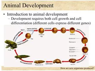
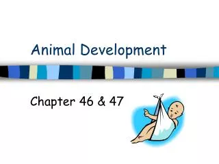
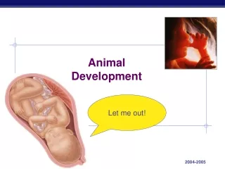
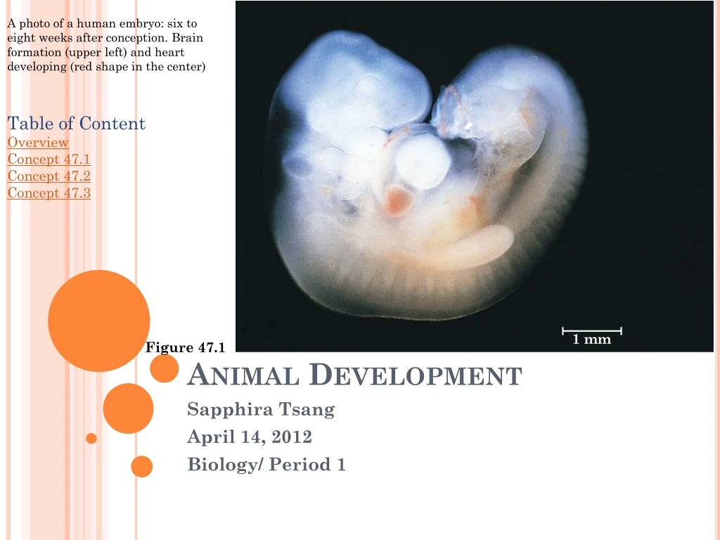
1 mm. Figure 47.1. A photo of a human embryo: six to eight weeks after conception. Brain formation (upper left) and heart developing (red shape in the center). Table of Content Overview Concept 47.1 Concept 47.2 Concept 47.3. Animal Development. Sapphira Tsang April 14, 2012

E N D
1 mm Figure 47.1 A photo of a human embryo: six to eight weeks after conception. Brain formation (upper left) and heart developing (red shape in the center) Table of Content Overview Concept 47.1 Concept 47.2 Concept 47.3 Animal Development Sapphira Tsang April 14, 2012 Biology/ Period 1
1 m Figure 47.2 Home Overview • Question: How does a zygote become an egg? • Theories: • 18th century—people believed that the answer was preformation • Preformation was the idea that the egg or sperm already had a “mini human” (called a homunculus) that grows and develops into a larger adult version • Aristotle proposed his theory of epigenesis • Epigenesis was the belief that an animal’s conformation is formed from a shapeless egg • Answer: • Genome of the zygote and differences between early embryonic cells are factors that change the way an organism develops • Cell differentiation: differences between the functions, structures, and roles of cells • Morphogenesis: a process wherein an animal takes shape and the location of the cells are already determined
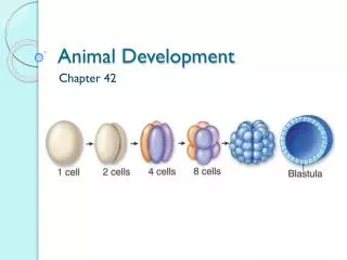
1 m Home Concept 47.1: Fertilization and three body structuring stages • Regulation of development occurs during fertilization • Three stages occur that starts building animal’s body: • Cleavage: cell divides from the zygote; creates a hollow ball of cells called a blastula • Gastrulation: production of a three-layered embryo called the gastrula • Organogenesis: basic organs are forming which eventually grows into adult structures
1 m Home Fertilization • Fertilization is when the sperm and egg, the gametes, unite • Fertilization combines the haploid sets of chromosomes into a diploid cell (called the zygote) • When the sperm comes in contact with the egg’s surface, metabolic reactions are triggered within the egg which activates the development of the embryo • Scientists study fertilization using sea urchins • Two reactions occur during fertilization: • Acrosomal reaction • Cortical reaction
1 m Home Acrosomal Reaction during sea urchin fertilization • Head of the sperm, acrosome, comes in contact with the egg, activating acrosomal reaction. • Acrosome releases hydrolytic enzymesdigest the jelly coat surrounding the egg. • Sperm to elongates a structure called the acrosomal process, made up of actin filaments,which will fully penetrate through the jelly coat. The acrosomal process contains molecules which bind to receptor proteins that are rooted into the vitelline layer. • The hole in the vitelline layer allows sperm membrane to fuse with egg membrane. The combined membranes are depolarized which provides a fast block to polyspermy. Polyspermy (fertilization of egg by more than one sperm) can lead to an abnormal number of chromosomes in the zygote. • Sperm enters and travels to cell’s nucleus. • Cortical reaction occurs. 4 5 3 2 1 6
1 m Home Cortical Reaction during sea urchin fertilization • When sperm binds to egg’s surface, signal transduction pathway activates calcium is released into cytosol. • High concentration of calcium initiates cortical reaction wherein fusion with the egg’s plasma membrane of vesicles located in the egg’s cortex (area beneath the cell’s membrane) occurs • Cortical granules are formed during oogenesis • Cortical granules release their contents into periveritelline space (area between the vitilline layer and the plasma membrane) • Vitelline layer detaches from plasma membrane; an osmotic gradient forces water into the perivitelline space, pushing it away from the membrane • The vitelline layer becomes a fertilization envelope, preventing other sperms from entering by a slow block to polyspermy A picture of calcium spreading out over the cell.
Sperm binds to the egg 1 Acrosomal reaction occurs 2 3 4 6 Seconds 8 Calcium level increases 10 20 Cortical reaction occurs 30 40 50 Fertilization envelope forms 1 2 pH level increases 3 4 5 Protein synthesis increases Minutes 10 The sperm is fused to the nuclei of the egg 20 30 1 m DNA synthesis occurs 40 60 Cell divides for the first time Home 90 Events of fertilization/ activation of a sea urchin egg • The rate of cell respiration and protein synthesis may increase as a result of the rise of calcium
Follicle cell Sperm basal body Zona pellucida Cortical granules Sperm nucleus Egg plasma membrane 1 m Acrosomal vesicle EGG CYTOPLASM Home Fertilization in mammals • Fertilization in mammals is internal but it is external for sea urchins • Mammalian eggs are surrounded by follicle cells • Follicle cells and the egg are released during ovulation • Sperm travels through follicle cells in order to get to the zonapellucida, the egg’s extracellular matrixsperm binds to receptor molecules in the zonapellucida. • Binding stimulates acrosomal reaction; sperm release hydrolytic enzymes • Zonapellucida is broken down by enzymes; sperm membrane can fuse with the plasma membrane of egg by receptors. • Sperm nucleus enters egg 4 3 2 1
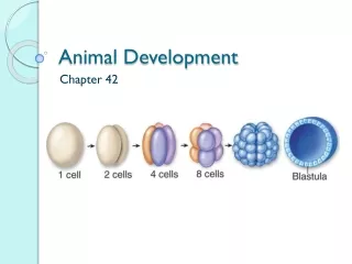
1 m Home Cleavage • After fertilization, cell division occurs • Process of cleavage takes place; cells undergo S (DNA synthesis) and M (mitosis) phase • Skip over G1 and G2 phases (no protein synthesis is occurring) • Embryo doesn’t enlarge; cytoplasm divides into smaller cells called blastomeres • Each has its own nucleus • First 5-7 divisions: cluster of cells is known as a morula • Blastocoel(fluid filled cavity) begins to form within morula • it is fully formed in the blastula (a hollow ball of cells) • In animals, the distribution of yolk (stored nutrients) affects pattern of cleavage • Vegetal pole: one pole of the egg where yolk is most abundant • Animal pole: opposite end of the egg where yolk concentration is significantly less • Gray crescent: a light gray area of the cytoplasm that helps the cell mark the dorsal side • Many zygotes and eggs of animals have a definite polarity. However, mammals do not. • Polarity: distribution of yolk to the vegetal pole, having most yolk, and animal pole, having less yolk
1 m Home Cleavage in echinoderm embryo The egg is fertilized and this photo shows the zygote prior to cleavage division. This is a picture of post-second cleavage division. The cell has divided and is at the four cell stage. The morula has formed and the blastocoel is beginning to form. A fully formed blastula is present and the embryo will later hatch from the fertilization envelope.
1 m Home Polarity determines body axes in an amphibian A picture of the body axes of a fully developed tadpole embryo. • Polarity of the egg helps with the determination of the anterior and posterior ends of an animal. • 2. Gray crescent on the cytoplasm marks the future dorsal site of the organism. • 3. Cleavage division begins; left-right axis is defined after the dorsal-ventral and anterior-posterior ends are defined.
Cleavage in a frog embryo 1 m Home Cleavage in a chick embryo • Zygote- cell is mostly made out of yolk. A small disk is located on the area of the animal pole. Egg white provides extra nutrients. • Four cell stage- early cell divisions are meroblastic(or incomplete). Holoblastic cleavage occurs when eggs containing very little yolk divides completely. The cleavage furrow forms through the cytoplasm and not through the yolk. • Blastoderm- cleavage divisions occur; a blastoderm is produced which is a group of cells covering the top of the yolk. • Blastoderm cells make up two layers: the epiblast and hypoblast, which enfold the blastocoel.
1 m Home Gastrulation • Cells of blastula rearranges • Gastrulation produces embryonic tissues/ embryonic germ layers. • “Gastrula” is the three layered embryo • Process is determined by: • Changes in cell motility • Changes in cell shape • Changes in cellular adhesion to other cells • Cells near the blastula surface will move into the interior regions and start forming three cell layers
1 m Home Gastrulation in sea urchin embryo • Gastrulation starts at vegetal poleCells from blastula wall travels into blastocoel; they are referred to as “mesenchyme cells” in the blastocoel; the cells still in blastula wall make up the “vegetal plate”. • Vegetal plate folds into itself, a process called invagination starts to form an archenteron, a primitive gut. The open end of archenteron becomes the anus. • Endoderm cells make up archenteron. The mesenchyme cells send thin extensions called filopodia towards the ectoderm cells of the blastocoel wall. • Filopodia contracts and pulls archenteron across the blastocoel space. • Archenteron combines with the blastocoel wall, forming digestive tube.
1 m Home Gastrulation in frog embryo Gastrulation starts on dorsal side of blastula. Invagination starts at gray crescent region, and becomes the dorsal lip. Involution: process that occurs when cells roll over the lip of the blastospore and goes into the embryoforms the endoderm and mesoderm. In the animal pole, ectoderm transforms and covers entire surface. Blastopore lip grows and invagination is still taking place. Once lips meet on either side, blastopore transforms into a circle which shrinks when the ectoderm starts covering surfacearchenteron forms, endoderm and mesoderm continues to expand by involution and blastocoel shrinks. Archenteron is replaced by blastocoel; all three germ layers are formed; the blastospore encompasses a group of yolk-filled cells.
Epiblast Future ectoderm Primitive streak Migrating cells (mesoderm) Endoderm Hypoblast YOLK 1 m Figure 47.13 Home Gastrulation in chick embryo • Some cells of the epiblast travel towards the inside of the embryo producing a primitive streak (a bunch of moving into the middle of the blastoderm). • Some cells form the endoderm; some form the mesoderm. • The cells that stay on the embryo’s surface becomes the ectoderm.
1 m Home Organogenesis Organogenesis in frog embryo • the three germ layers develop into basic organs • Somites- • neural tube formed • lateral mesoderm separates, making the coelom • mesoderm forms the somites • somites form axial skeleton muscles • Neural plate forms- • notochord grows from dorsal mesoderm • notochord signals for dorsal ectoderm to form neural plate Neural tube forms- neural plate folds into itself and detaches itself, resulting in neural tubeneural tube becomes the central nervous system.
1 m Home Organogenesis in chick • Organogenesis in a chick is similar to organogenesis in a frog
1 m Home Structures formed from the three embryonic germ layers in vertebrates Endoderm Ectoderm Mesoderm • Digestive tract lining • Respiratory system lining • Urethra, urinary bladder, and reproductive system lining • Liver • Pancreas • Thymus • Thyroid and parathyroid glands • Notochord • Skeletal system • Muscular system • Muscle that make up the stomach, intestines, etc • Excretory system • Circulatory and lymphatic system • Reproductive system (except for germ cells) • Dermis of skin • Body cavity lining • Adrenal cortex • Sweat glands • Hair follicles • Epidermis of skin • Lining of mouth and rectum • Sensory receptors • Eye cornea and lens • Nervous system • Adrenal medulla (part of the adrenal gland which secrete hormones) • Tooth enamel • Epithelium of pineal and pituitary glands
1 m Home Developmental adaptations of amniotes Amnion Allantois • Terms to know: • Amnion: serves as protection and cushion for embryo; prevents dehydration • Allantois: basically a garbage canstores embryo’s waste; functions with chorion as a “lung” • Chorion: exchange gases with allantois; provide embryo with oxygen and carbon dioxide • Yolk Sac: stores nutrients and feeds the embryo • Amniotes: animals that develop as embryos in fluid-filled sacs in eggs or a uterus Chorion Yolk sac • All vertebrates develop in aqueous environments • Evolution of animal movement onto land requires: • Shelled eggs • Uterus of marsupial and placental mammales
1 m Home Mammalian Development • Mammalian egg cell and zygote: • Do not contain polarity in their cytoplasm contents • Have yolk-lacking, holoblastic zygote cleavages • Have small eggs • Gastrulation and organogensis are similar to those processes in birds and reptiles Embryo development in early stages • Cleavage formed • Embryo traveled down oviduct to the uterus • Inner cell mass: a group of cells at one end of the cavity • Inner cell mass develops into embryo proper and add to all extra embryonic membranes • Implantation occurs • Trophoblast (outer epithelium of blastocyst) initiates implantation when it secretes enzymes that break down endometrium molecules (uterus lining) • Blastocyst can then enter the endometrium • Trophoblast thickens and it extends fingerlike projections into maternal tissues rich in blood vessels • Invasion by trophoblast results in erosion of capillaries in endometrium the blood spills out and covers trophoblast tissue • Gastrulation starts • Implantation completed • Cells from epiblast move inward through the primitive streak, forming the mesoderm and endoderm • Germ layers are formed • Four extraembryonic membranes also form: allantois, amnion, chorion, yolk sac
Microtubules elongate neural plate cells Ectoderm Neural plate Microfilaments, located on dorsal side, contracts, changing cell shape into a wedge Cell wedge in opposite direction can form a hinge. 1 2 3 4 Neural plate pinches off, forming neural tube 1 m Home Concept 47.2: Morphogenesis in animals involves specific changes in cell shape, position, and adhesion • Only in animals, morphogenesis involves movement of cells • Movement can determine cell shape or allow cells to travel within embryo • When the cytoskeleton changes, so does the cell shape Neural tube formation in vertebrates
Convergence Extension Figure 47.20 1 m Home Cell Crawling • Cell crawling: the movement of cells to other places • Convergent extension: type of morphogenetic movement wherein tissue layer cells arrange as a thin, long sheet • Cell crawling is involved in convergent extension
1 m Home Extracellular Matrix (ECM) and Cell Adhesion Molecules • Roles of ECM fibers: • Guide cells in morphogenetic movements • Function as tracks to direct migrating cells • Migrating cells moving along specific paths have receptor proteins that receive direction signals • Signals can direct the orientation of the cytoskeleton so that it can move the cell forward • Cell adhesion molecules (CAMs): located on cell surface and binds to other CAMs on neighboring cells • Help regulate movements and tissue building due to differences in amount of CAMs and chemical identity • Cadherins: require calcium for work • Gene for cadherins is differentiated by their location at certain times during embryo development
1 m Home Cell migration using fibronection Frog blastula formation through cadherin • Fibronecton provides anchorage for cells
1 m Home Concept 47.3: The developmental fate of cells depends on their history and inductive signals • Two principles of differentiation during embryonic development: • In early cleavage divisions, embryonic cells must become different from each other. • After asymmetries are determined, interactions between embryonic cells determine fate by causing changes in gene expression Fate Mapping • Fate map: territorial diagrams of embryonic development • Scientists studied fate maps while manipulating embryo parts to see whether a cell’s fate can be changed by moving it • Two conclusions were made: • “Founder cells” give rise to specific tissues in older embryos • As development proceeds, cell’s development potential becomes restricted • Developmental potential: range of structures it can form
1 m Home Fate mapping
1 m Home Establishing Cellular Asymmetries • In nonamniotic vertebrates, body axes determination are made early during oogenesis or fertilization • Example: locations of melanin and yolk in the unfertilized egg of a frog determines the vegetal hemispheres • In amniotes, body axes are not determined until later • Environmental factors determine the axes • Gravity establishes anterior-posterior axis of chicks in the eggs • pH differences between blastoderm cells determine the dorsal-ventral axis Restrictions of cellular potency • Totipotent: zygote is capable of developing into all adult cell types • Only the zygote is totipotent • Mammalian embryo cells remain totipotent until 16-cell stage; this is when they arrange into precursors of trophoblast and inner cell mass of blastocyst • Location determines cell fate • At 8-cell stage, each blastomeres can develop an embryo if isolated
1 m Home Cell Fate determination and pattern formation by inductive signals • Embryonic cell division creates cells that differ cells influence each other’s fates by induction • Inductive signals affect pattern formation • Pattern formation: development of spatial organization in an animal • Positional information: signals the cell its position in the animal’s body axes and determines how the cell will respond to molecular signals • Signal molecules: • Affect gene expression in receiving cells • Result in differentiation • Can develop certain structures
1 m Home Spemann and Mangold’s Experiment Spemann and Mangold’s experiment concluded that the dorsal lip of the blastopore acts as an organizer of the embryo
1 m Home A chick’s wing and legs start off as limb buds. Vertebrate Limb Development • Limp bud provides a model of pattern formation • Made up of a core mesodermal tissue surrounded by ectoderm layer • Two organizer regions in limp bud of vertebrate limbs: • Apical ectodermal ridge (AER): thick region of ectoderm at tip of the bud; produces secreted protein signals that promote limb-bud outgrowth • Zone of polarizing activity (ZPA): a block of mesodermal tissue underneath ectoderm; needed for proper pattern formation and produces posterior structures
1 m Home Tissue Tranplantation experiment • Tissue transplantation experiment supports the idea that ZPA produces an inductive signal message with information for posterior positions
Works Cited • www.campbellbiology.com • Campbell Biology textbook • Pictures from: www.campbellbiology.com